Ann Hepatobiliary Pancreat Surg.
2021 May;25(2):299-306. 10.14701/ahbps.2021.25.2.299.
Third retransplantation using a whole liver graft for late graft failure from hepatic vein stent stenosis in a pediatric patient who underwent split liver retransplantation
- Affiliations
-
- 1Departments of Surgery, Asan Medical Center, University of Ulsan College of Medicine, Seoul, Korea
- 2Departments of Pediatrics, Asan Medical Center, University of Ulsan College of Medicine, Seoul, Korea
- KMID: 2516499
- DOI: http://doi.org/10.14701/ahbps.2021.25.2.299
Abstract
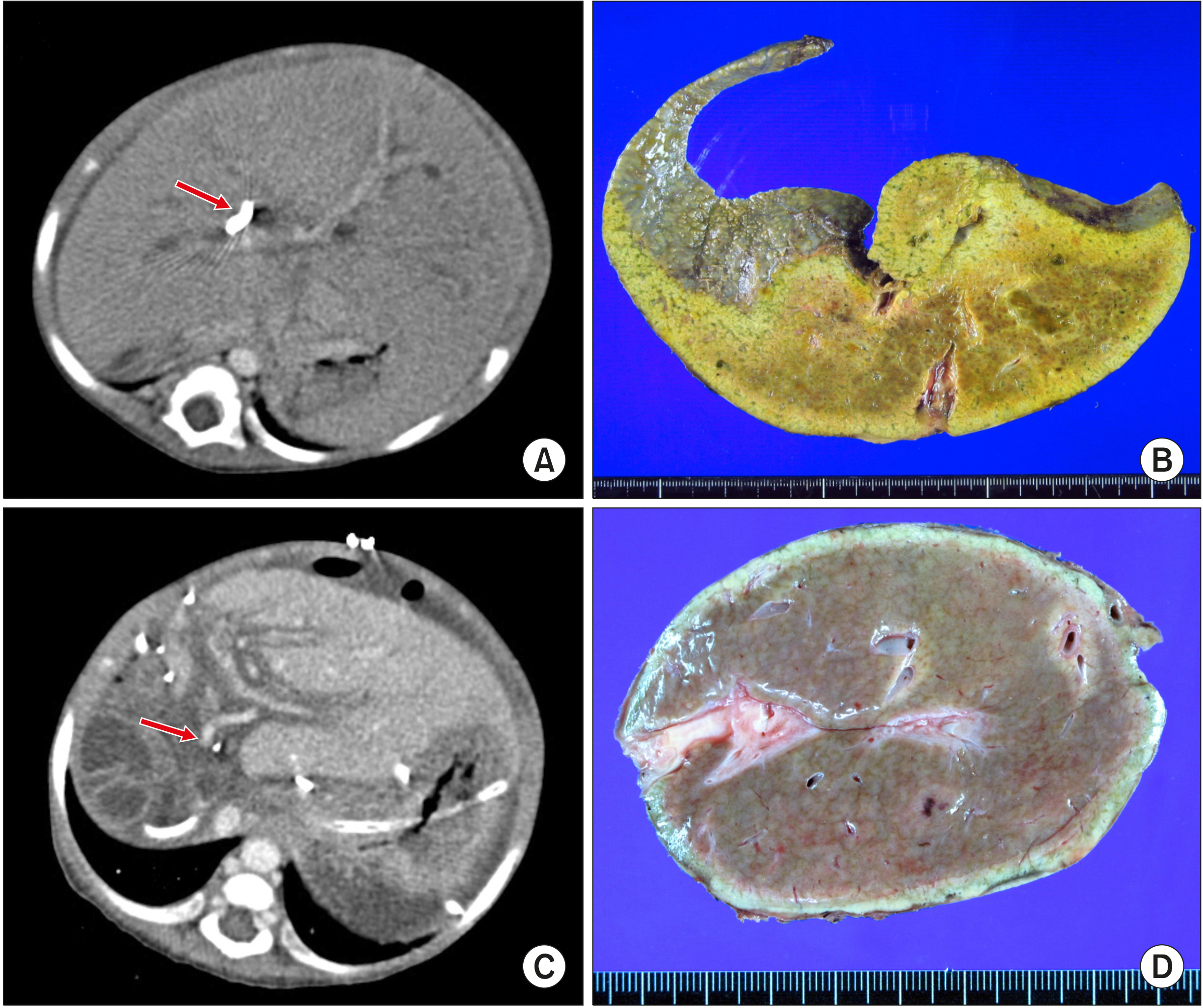
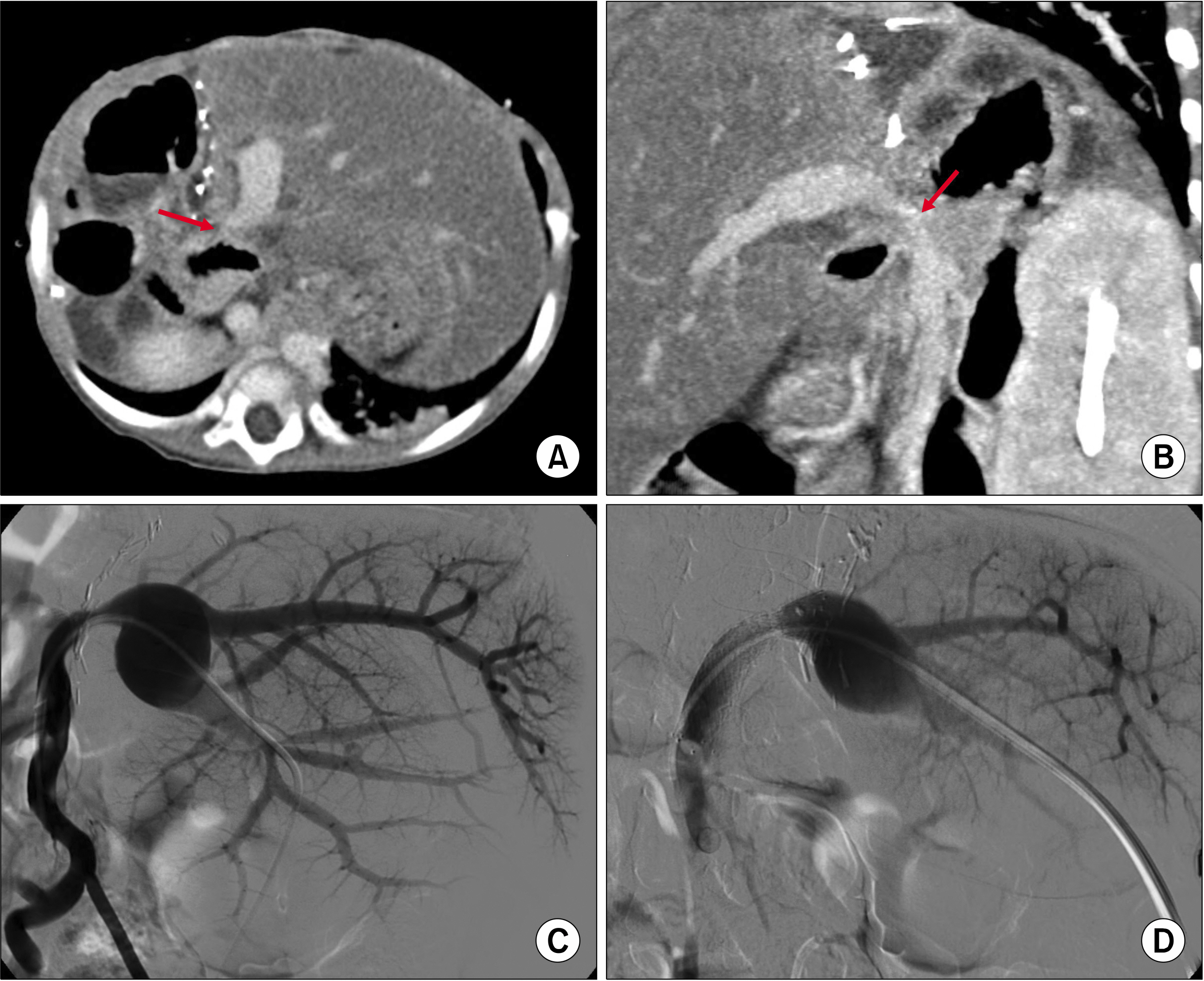
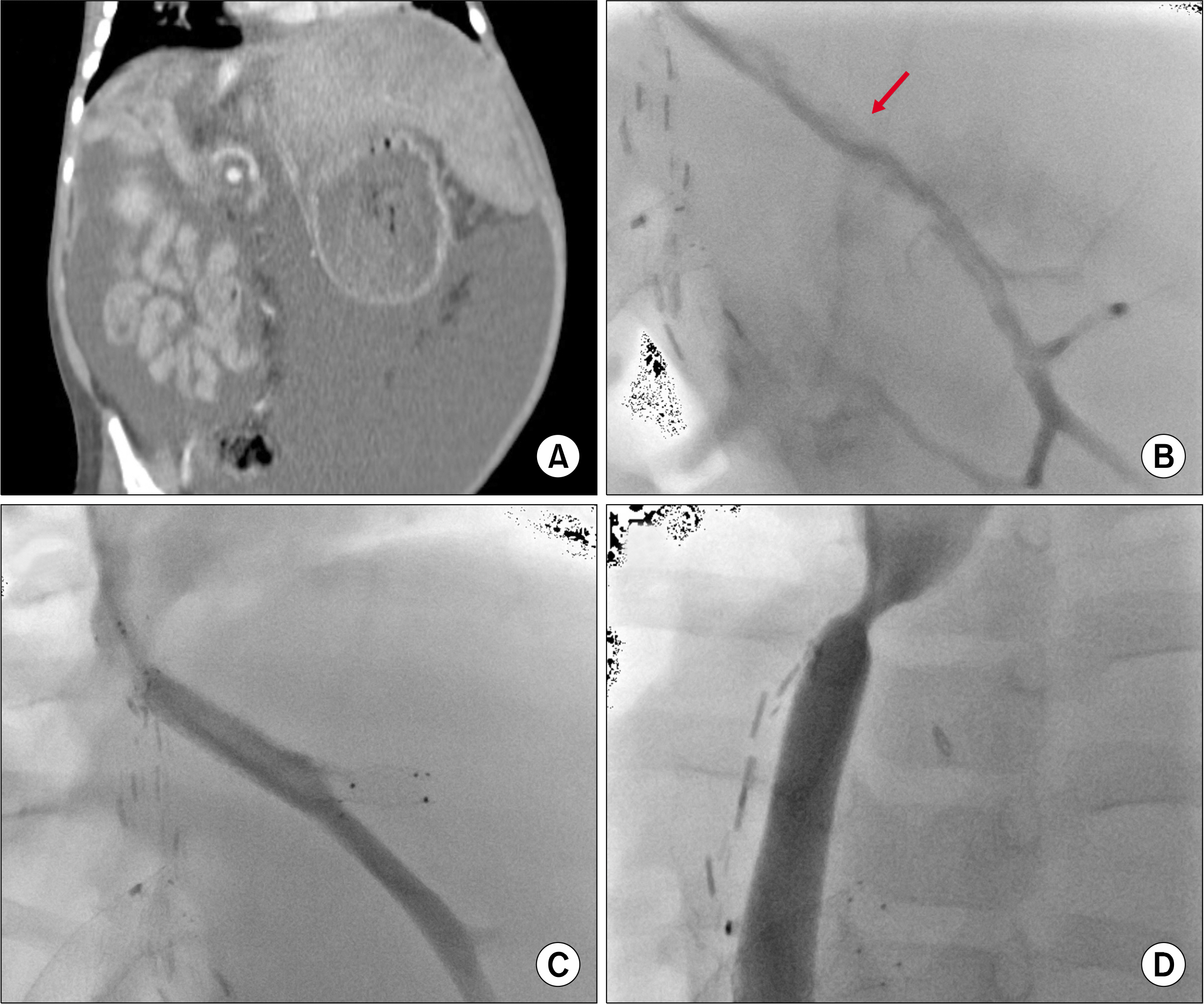
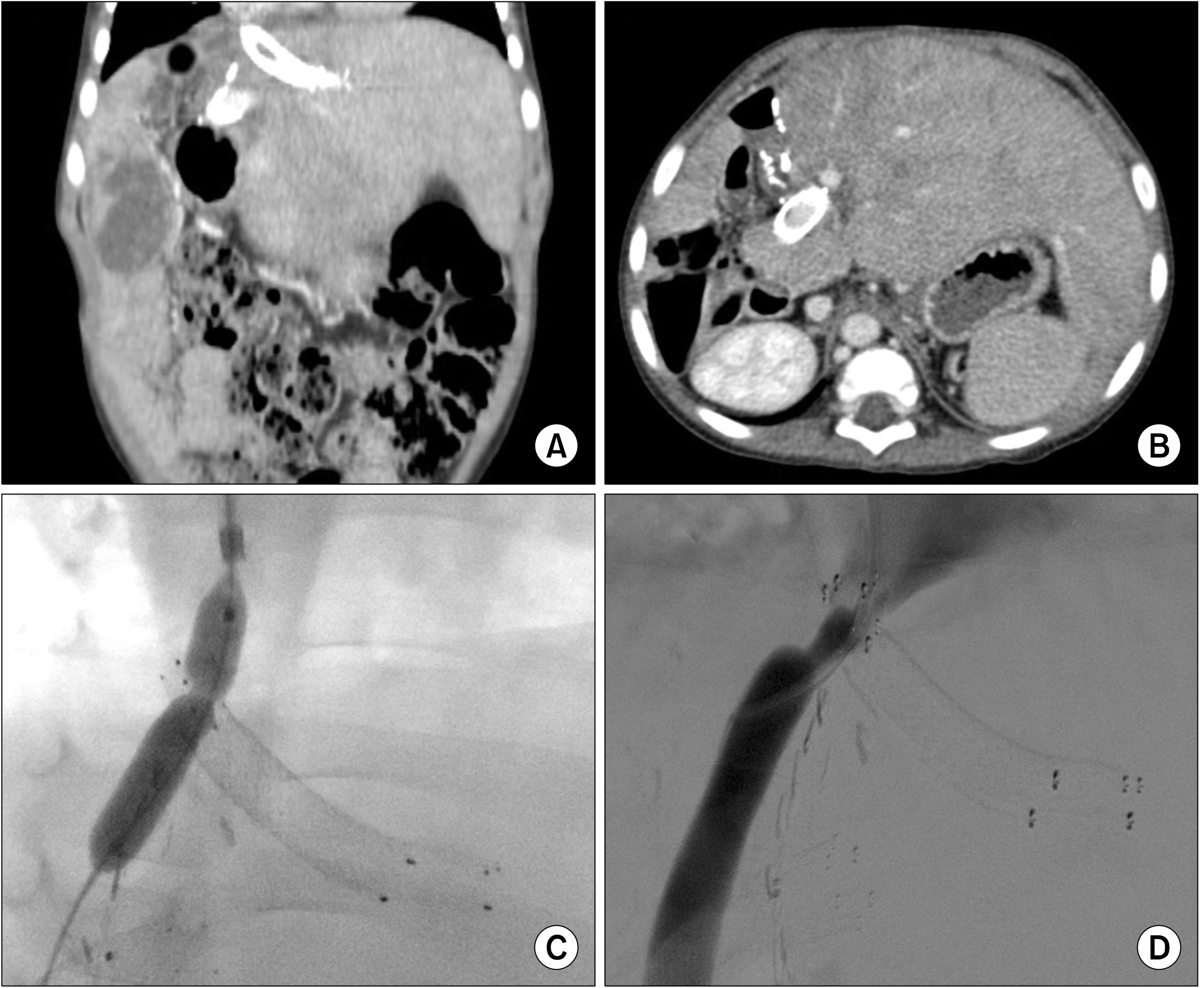
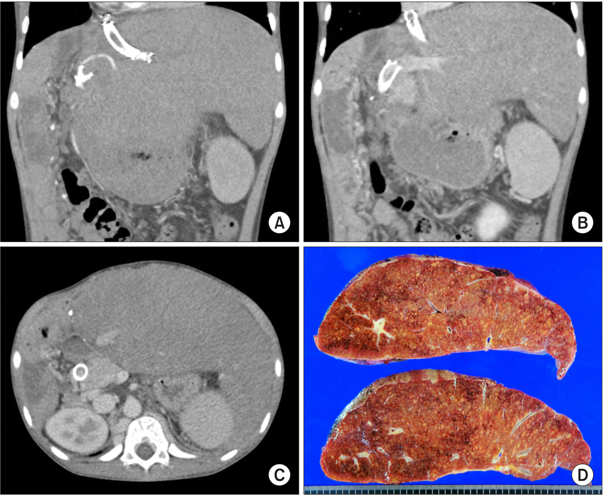
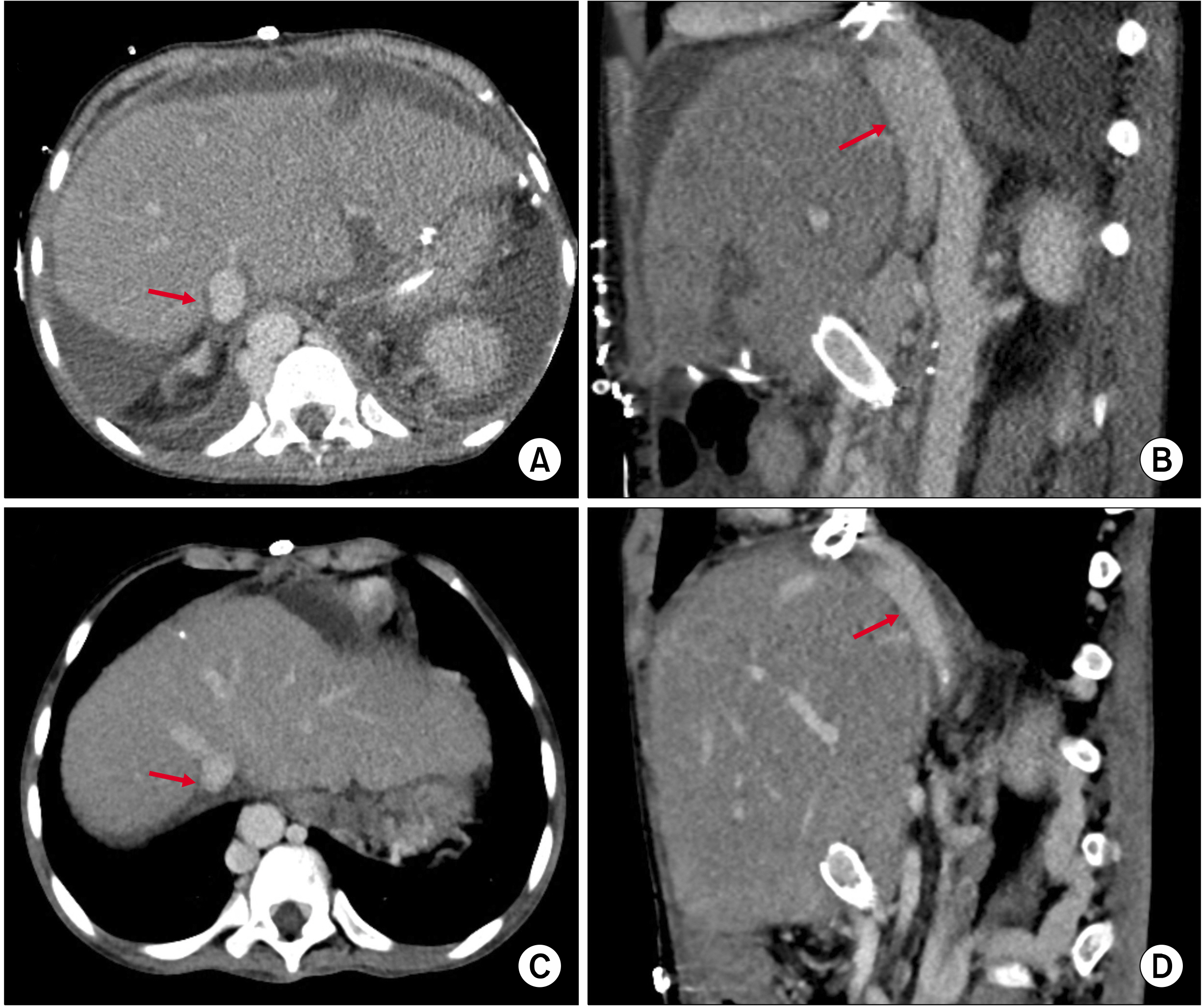
- We present a case of third retransplantation using a whole liver graft in a 13-year-old girl who suffered graft failure and hepatopulmonary syndrome following split liver retransplantation with endovascular stenting of the hepatic and portal veins as an infant. She was diagnosed with biliary atresia-polysplenia syndrome, and thus underwent living donor liver transplantation from her mother at 9 months of age. The first liver graft failed due to stenosis of the portal vein. She underwent the second liver transplantation with a split left lateral section graft. Endovascular stenting was performed to the portal vein stenosis 2 months and hepatic vein stenosis 9 months after transplantation. During the next 9 years, 11 sessions of balloon angioplasty for hepatic vein stent stenosis were performed. Ten years after the second transplantation, she underwent third transplantation using a whole liver graft recovered from a 12-year-old-girl. The double inferior vena cava technique was used for outflow vein reconstruction. The graft portal vein was anastomosed with the stent-containing portal vein stump because it was not possible to remove the stent and the inner diameter of the portal vein stent was large enough. An aorto-hepatic jump graft was used for arterial reconstruction. The patient recovered slowly and is doing well for 6 months posttransplant. In conclusion, because stenting of the hepatic vein or portal vein can induce graft failure leading to late retransplantation, we emphasize secure vascular reconstruction to prevent endovascular stenting during LT in infants.
Keyword
Figure
Cited by 1 articles
-
Endovascular stenting for late-onset stricture of interposed portal vein conduit following pediatric liver transplantation
Jung-Man Namgoong, Shin Hwang, Gi-Young Ko, Gil-Chun Park, Kyung Mo Kim, Seak Hee Oh
Ann Liver Transplant. 2023;3(1):29-34. doi: 10.52604/alt.23.0007.
Reference
-
1. Yeh YT, Chen CY, Tseng HS, Wang HK, Tsai HL, Lin NC, et al. 2017; Enlarging vascular stents after pediatric liver transplantation. J Pediatr Surg. 52:1934–1939. DOI: 10.1016/j.jpedsurg.2017.08.060. PMID: 28927979.
Article2. Galloux A, Pace E, Franchi-Abella S, Branchereau S, Gonzales E, Pariente D. 2018; Diagnosis, treatment and outcome of hepatic venous outflow obstruction in paediatric liver transplantation: 24- year experience at a single centre. Pediatr Radiol. 48:667–679. DOI: 10.1007/s00247-018-4079-y. PMID: 29468367.3. Katano T, Sanada Y, Hirata Y, Yamada N, Okada N, Onishi Y, et al. 2019; Endovascular stent placement for venous complications following pediatric liver transplantation: outcomes and indications. Pediatr Surg Int. 35:1185–1195. DOI: 10.1007/s00383-019-04551-9. PMID: 31535198.
Article4. Zhang ZY, Jin L, Chen G, Su TH, Zhu ZJ, Sun LY, et al. 2017; Balloon dilatation for treatment of hepatic venous outflow obstruction following pediatric liver transplantation. World J Gastroenterol. 23:8227–8234. DOI: 10.3748/wjg.v23.i46.8227. PMID: 29290659. PMCID: PMC5739929.
Article5. Lu KT, Cheng YF, Chen TY, Tsang LC, Ou HY, Yu CY, et al. 2018; Efficiency of transluminal angioplasty of hepatic venous outflow obstruction in pediatric liver transplantation. Transplant Proc. 50:2715–2717. DOI: 10.1016/j.transproceed.2018.04.022. PMID: 30401383.
Article6. Kawano Y, Mizuta K, Sanada Y, Urahashi T, Ihara Y, Okada N, et al. 2016; Complementary indicators for diagnosis of hepatic vein stenosis after pediatric living-donor liver transplantation. Transplant Proc. 48:1156–1161. DOI: 10.1016/j.transproceed.2015.12.114. PMID: 27320577.
Article7. Namgoong JM, Hwang S, Park GC, Kwon H, Kwon YJ, Kim SH. 2020; Graft outflow vein unification venoplasty with superficial left hepatic vein branch in pediatric living donor liver transplantation using a left lateral section graft. Ann Hepatobiliary Pancreat Surg. 24:326–332. DOI: 10.14701/ahbps.2020.24.3.326. PMID: 32843600. PMCID: PMC7452796.
Article8. Namgoong JM, Hwang S, Song GW, Kim DY, Ha TY, Jung DH, et al. 2020; Pediatric liver transplantation with hyperreduced left lateral segment graft. Ann Hepatobiliary Pancreat Surg. 24:503–512. DOI: 10.14701/ahbps.2020.24.4.503. PMID: 33234754. PMCID: PMC7691208.
Article9. Davenport M, Tizzard SA, Underhill J, Mieli-Vergani G, Portmann B, Hadzić N. 2006; The biliary atresia splenic malformation syndrome: a 28-year single-center retrospective study. J Pediatr. 149:393–400. DOI: 10.1016/j.jpeds.2006.05.030. PMID: 16939755.
Article10. Broniszczak D, Apanasiewicz A, Czubkowski P, Kaliciński P, Ismail H, Ostoja-Chyzynska A, et al. 2011; Liver transplantation in children with biliary atresia and polysplenia syndrome. Ann Transplant. 16:14–17.11. Nio M, Wada M, Sasaki H, Tanaka H, Watanabe T. 2015; Long-term outcomes of biliary atresia with splenic malformation. J Pediatr Surg. 50:2124–2127. DOI: 10.1016/j.jpedsurg.2015.08.040. PMID: 26613836.
Article12. Namgoong JM, Hwang S, Kim DY, Ha TY, Song GW, Jung DH, et al. 2020; Pediatric split liver transplantation in a patient with biliary atresia polysplenia syndrome and agenesis of inferior vena cava. Korean J Transplant. 34:286–292. DOI: 10.4285/kjt.20.0023.
Article
- Full Text Links
- Actions
-
Cited
- CITED
-
- Close
- Share
- Similar articles
-
- Living donor liver transplantation-associated retransplantation in adult patients
- Liver retransplantation for adult recipients
- A Case of Successful Hepatic Retransplantation
- Graft-recipient-weight ratio and lowered immunosuppression is important for the success of adult liver retransplantation: 25-year single center experience
- Portal vein reconstruction in pediatric liver transplantation using end-to-side jump graft: A case report

