Endoscopic Band Ligation in Endoscopic Retrograde Cholangiopancreatography Related Duodenal Perforation
- Affiliations
-
- 1Division of Gastroenterology and Hepatology, Department of Internal Medicine, Konyang University College of Medicine, Daejeon, Korea
- KMID: 2514090
- DOI: http://doi.org/10.4166/kjg.2020.164
Abstract
- Although ERCP is a therapeutic endoscopic procedure in pacreatico-biliary diseases, its rare complications, including pancreatitis, duodenal perforation, and bleeding, can be fatal. An 87-year-old woman with a history of gallbladder cancer presented with jaundice and general weakness. Her skin color was yellowish and epigastric tenderness was confirmed on a physical examination. On abdomen CT, the gallbladder cancer directly invaded the duodenum, common bile duct, and liver parenchyma. Enlarged portocaval lymph nodes obstructed the extrahepatic bile duct. ERCP was performed for bile duct decompression. When shortening of endoscopy was achieved, the duodenal lateral wall was perforated because of the endoscopic tip pressure. After inserting endoscopic retrograde biliary drainage and endoscopic nasobiliary drainage, endoclips were placed evenly around the defect, and a detachable snare was tightened around the endoclips. Three days later, the duodenal wall was not sealed on the abdomen CT scan. Repeat endoscopy was achieved, and the endoscopic nasobiliary drainage, endoscopic retrograde biliary drainage, endoclips, and detachable snare were removed. From the distal margin of the perforation, band ligation was performed, and a detachable snare was applied. The patient's condition improved after the second procedure. A percutaneous biliary stent was inserted, and she was discharged. This case highlights the successful endoscopic management of ERCP-related duodenal perforation.
Figure
-
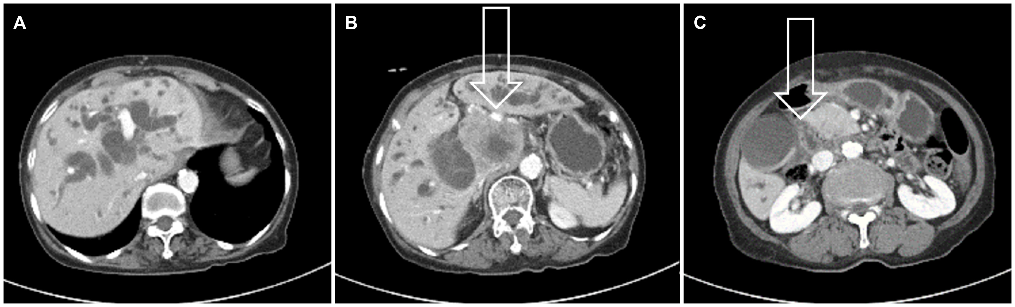
Fig. 1 Abdominal computed tomography (CT) findings. (A) CT showed a dilatated intrahepatic bile duct. (B) Enlarged lymph nodes (arrow) at the portocaval space (approximately 9 cm). (C) Tumor invasion of the duodenal bulb (arrow).
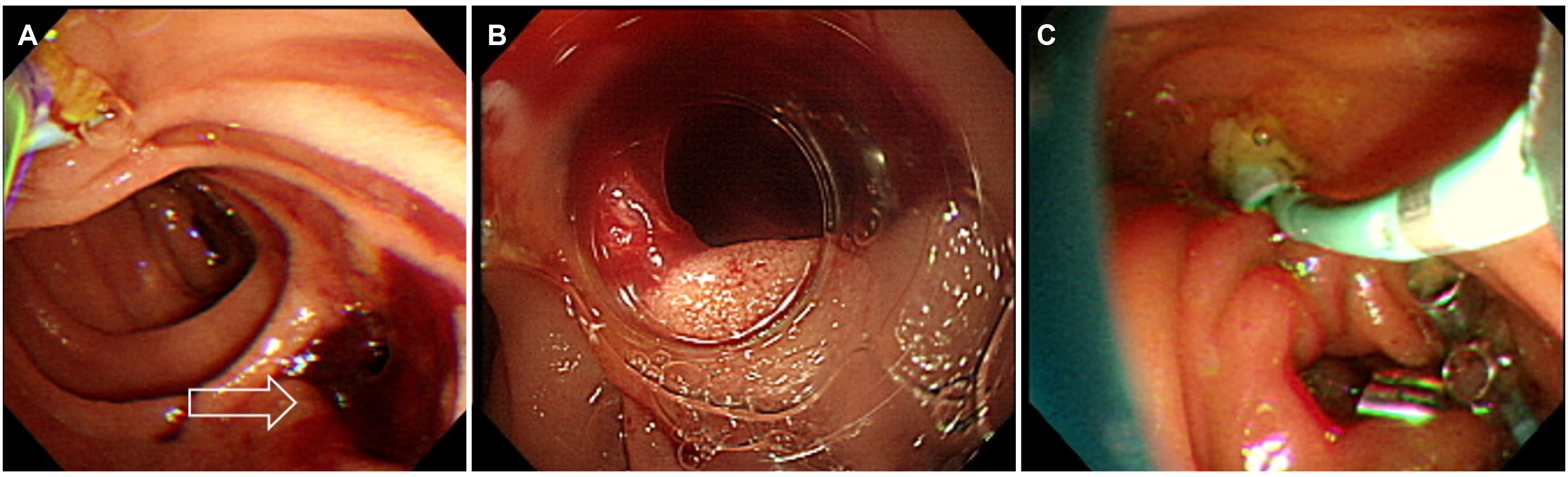
Fig. 2 Side-viewing endoscopic and gastroscopic findings. (A) Perforation (arrow) of the duodenal lateral wall. (B) Gastroscopic view through the transparent cap of perforation. (C) Endoscopic retrograde biliary drainage and endoscopic nasobiliary drainage were inserted, and endoclips and a detachable snare were applied.

Fig. 3 Abdominal X-ray and abdominal computed tomography findings. (A) Abdominal X-ray shows a retroperitoneal free air outlining the psoas muscle. (B) Anterior pararenal fluid collection (arrow) despite endoscopic retrograde biliary drainage and endoscopic nasobiliary drainage. (C) Small pneumoretroperitoneum (arrow) around 2nd-3rd duodenum junction, Rt. pararenal and perirenal space.
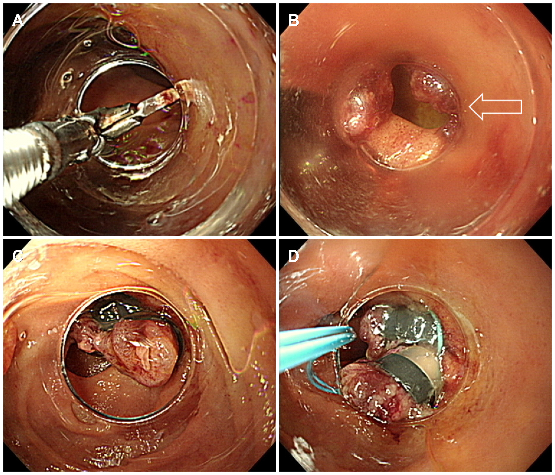
Fig. 4 Gastroscopic findings. (A) Endoclips were removed using rat tooth forceps. (B) An unsealed duodenal perforation site (arrow). (C, D) From the distal margin of the perforation, band ligation was done, and a detachable snare was applied.
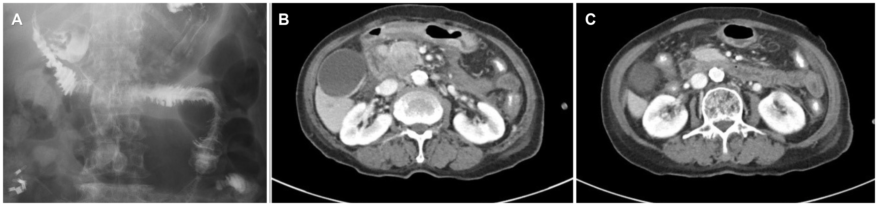
Fig. 5 Upper gastrointestinal (UGI) series and abdominal computed tomography findings. (A) No contrast leakage in UGI series. (B) Fluid collection was decreased. (C) Duodenal perforation site was sealed with band ligation.
Reference
-
1. Paspatis GA, Dumonceau JM, Barthet M, et al. 2014; Diagnosis and management of iatrogenic endoscopic perforations: European Society of Gastrointestinal Endoscopy (ESGE) Position Statement. Endoscopy. 46:693–711. DOI: 10.1055/s-0034-1377531. PMID: 25046348.
Article2. Vezakis A, Fragulidis G, Polydorou A. 2015; Endoscopic retrograde cholangiopancreatography-related perforations: diagnosis and management. World J Gastrointest Endosc. 7:1135–1141. DOI: 10.4253/wjge.v7.i14.1135. PMID: 26468337. PMCID: PMC4600179.
Article3. Stapfer M, Selby RR, Stain SC, et al. 2000; Management of duodenal perforation after endoscopic retrograde cholangiopancreatography and sphincterotomy. Ann Surg. 232:191–198. DOI: 10.1097/00000658-200008000-00007. PMID: 10903596. PMCID: PMC1421129.
Article4. Wu HM, Dixon E, May GR, Sutherland FR. 2006; Management of perforation after endoscopic retrograde cholangiopancreatography (ERCP): a population-based review. HPB (Oxford). 8:393–399. DOI: 10.1080/13651820600700617. PMID: 18333093. PMCID: PMC2020744.
Article5. Lee TH, Bang BW, Jeong JI, et al. 2010; Primary endoscopic approximation suture under cap-assisted endoscopy of an ERCP-induced duodenal perforation. World J Gastroenterol. 16:2305–2310. DOI: 10.3748/wjg.v16.i18.2305. PMID: 20458771. PMCID: PMC2868227.
Article6. Li G, Chen Y, Zhou X, Lv N. 2012; Early management experience of perforation after ERCP. Gastroenterol Res Pract. 2012:657418. DOI: 10.1155/2012/657418. PMID: 22899906. PMCID: PMC3412108.
Article7. Seibert DG. 2003; Use of an endoscopic clipping device to repair a duodenal perforation. Endoscopy. 35:189. DOI: 10.1055/s-2003-37004. PMID: 12561015.
Article8. Yang HY, Chen JH. 2015; Endoscopic fibrin sealant closure of duodenal perforation after endoscopic retrograde cholangiopancreatography. World J Gastroenterol. 21:12976–12980. DOI: 10.3748/wjg.v21.i45.12976. PMID: 26668519. PMCID: PMC4671050.
Article9. Li Y, Han Z, Zhang W, et al. 2014; Successful closure of lateral duodenal perforation by endoscopic band ligation after endoscopic clipping failure. Am J Gastroenterol. 109:293–295. DOI: 10.1038/ajg.2013.415. PMID: 24496428.
Article10. Buffoli F, Grassia R, Iiritano E, Bianchi G, Dizioli P, Staiano T. 2012; Endoscopic "retroperitoneal fatpexy" of a large ERCP-related jejunal perforation by using a new over-the-scope clip device in Billroth II anatomy (with video). Gastrointest Endosc. 75:1115–1117. DOI: 10.1016/j.gie.2011.05.029. PMID: 21820111.
Article11. Han JH, Park S, Youn S. 2011; Endoscopic closure of colon perforation with band ligation; salvage technique after endoclip failure. Clin Gastroenterol Hepatol. 9:e54–e55. DOI: 10.1016/j.cgh.2010.12.026. PMID: 21195795.
Article12. Vezakis A, Fragulidis G, Nastos C, Yiallourou A, Polydorou A, Voros D. 2011; Closure of a persistent sphincterotomy-related duodenal perforation by placement of a covered self-expandable metallic biliary stent. World J Gastroenterol. 17:4539–4541. DOI: 10.3748/wjg.v17.i40.4539. PMID: 22110286. PMCID: PMC3218146.
Article13. Park WY, Cho KB, Kim ES, Park KS. 2012; A case of ampullary perforation treated with a temporally covered metal stent. Clin Endosc. 45:177–180. DOI: 10.5946/ce.2012.45.2.177. PMID: 22866262. PMCID: PMC3401625.
Article14. Loperfido S, Angelini G, Benedetti G, et al. 1998; Major early complications from diagnostic and therapeutic ERCP: a prospective multicenter study. Gastrointest Endosc. 48:1–10. DOI: 10.1016/S0016-5107(98)70121-X. PMID: 9684657.
Article
- Full Text Links
-
- Actions
-
Cited
- CITED
-
- Close
- Share
-
- Similar articles
-
- Repair of an Endoscopic Retrograde Cholangiopancreatography-Related Large Duodenal Perforation Using Double Endoscopic Band Ligation and Endoclipping
- The Management of Endoscopic Retrograde Cholangiopancreatography-Related Duodenal Perforation
- Recent Advanced Endoscopic Management of Endoscopic Retrograde Cholangiopancreatography Related Duodenal Perforations
- Endoscopic Treatments of Endoscopic Retrograde Cholangiopancreatography-Related Duodenal Perforations
- Four Cases of Guidewire Induced Periampullary Perforation During Endoscopic Retrograde Cholangiopancreatography
