Clin Endosc.
2016 Jul;49(4):376-382. 10.5946/ce.2016.088.
Recent Advanced Endoscopic Management of Endoscopic Retrograde Cholangiopancreatography Related Duodenal Perforations
- Affiliations
-
- 1Department of Internal Medicine, Chungbuk National University College of Medicine, Cheongju, Korea. smpark@chungbuk.ac.kr
- KMID: 2348258
- DOI: http://doi.org/10.5946/ce.2016.088
Abstract
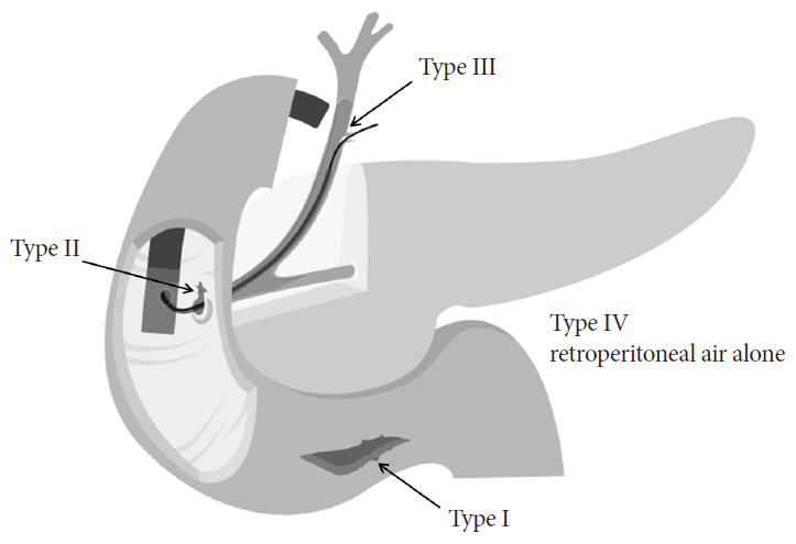
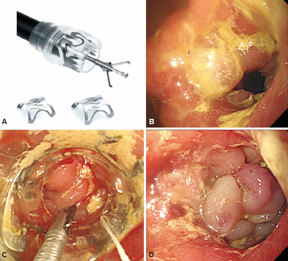
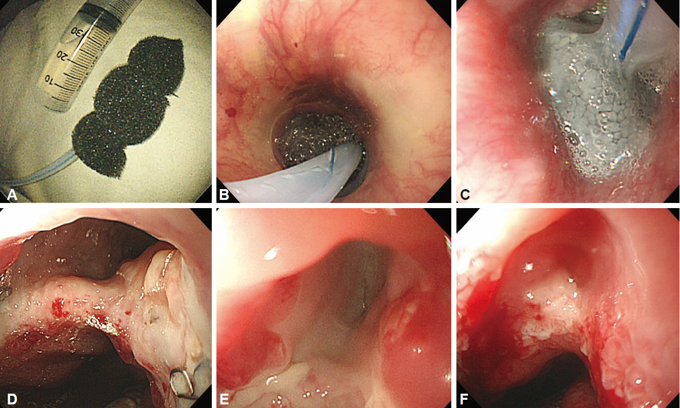
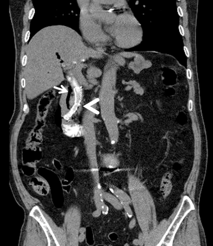
- The management strategy for endoscopic retrograde cholangiopancreatography-related duodenal perforation can be determined based on the site and extent of injury, the patient's condition, and time to diagnosis. Most cases of perivaterian or bile duct perforation can be managed with a biliary stent or nasobiliary drainage. Duodenal wall perforations had been treated with immediate surgical repair. However, with the development of endoscopic devices and techniques, endoscopic closure has been reported to be a safe and effective treatment that uses through-the-scope clips, ligation band, fibrin glue, endoclips and endoloops, an over-the-scope clipping device, suturing devices, covering luminal stents, and open-pore film drainage. Endoscopic therapy could be instituted in selected patients in whom perforation was identified early or during the procedure. Early diagnosis, proper conservative management, and effective endoscopic closure are required for favorable outcomes of non-surgical management. If endoscopic treatment fails, or in the cases of clinical deterioration, prompt surgical management should be considered.
MeSH Terms
Figure
Reference
-
1. Machado NO. Management of duodenal perforation post-endoscopic retrograde cholangiopancreatography: when and whom to operate and what factors determine the outcome? A review article. JOP. 2012; 13:18–25.2. Paspatis GA, Dumonceau JM, Barthet M, et al. Diagnosis and management of iatrogenic endoscopic perforations: European Society of Gastrointestinal Endoscopy (ESGE) Position Statement. Endoscopy. 2014; 46:693–711.
Article3. Andriulli A, Loperfido S, Napolitano G, et al. Incidence rates of post-ERCP complications: a systematic survey of prospective studies. Am J Gastroenterol. 2007; 102:1781–1788.
Article4. Vezakis A, Fragulidis G, Polydorou A. Endoscopic retrograde cholangiopancreatography-related perforations: diagnosis and management. World J Gastrointest Endosc. 2015; 7:1135–1141.
Article5. Enns R, Eloubeidi MA, Mergener K, et al. ERCP-related perforations: risk factors and management. Endoscopy. 2002; 34:293–298.
Article6. Verlaan T, Voermans RP, van Berge Henegouwen MI, Bemelman WA, Fockens P. Endoscopic closure of acute perforations of the GI tract: a systematic review of the literature. Gastrointest Endosc. 2015; 82:618–628.
Article7. Stapfer M, Selby RR, Stain SC, et al. Management of duodenal perforation after endoscopic retrograde cholangiopancreatography and sphincterotomy. Ann Surg. 2000; 232:191–198.
Article8. Freeman ML, Nelson DB, Sherman S, et al. Complications of endoscopic biliary sphincterotomy. N Engl J Med. 1996; 335:909–918.
Article9. Wu HM, Dixon E, May GR, Sutherland FR. Management of perforation after endoscopic retrograde cholangiopancreatography (ERCP): a population-based review. HPB (Oxford). 2006; 8:393–399.
Article10. Navaneethan U, Konjeti R, Venkatesh PG, Sanaka MR, Parsi MA. Early precut sphincterotomy and the risk of endoscopic retrograde cholangiopancreatography related complications: an updated meta-analysis. World J Gastrointest Endosc. 2014; 6:200–208.
Article11. Zhao HC, He L, Zhou DC, Geng XP, Pan FM. Meta-analysis comparison of endoscopic papillary balloon dilatation and endoscopic sphincteropapillotomy. World J Gastroenterol. 2013; 19:3883–3891.
Article12. Motomura Y, Akahoshi K, Gibo J, et al. Immediate detection of endoscopic retrograde cholangiopancreatography-related periampullary perforation: fluoroscopy or endoscopy? World J Gastroenterol. 2014; 20:15797–15804.
Article13. Kumbhari V, Sinha A, Reddy A, et al. Algorithm for the management of ERCP-related perforations. Gastrointest Endosc. 2016; 83:934–943.
Article14. de Vries JH, Duijm LE, Dekker W, Guit GL, Ferwerda J, Scholten ET. CT before and after ERCP: detection of pancreatic pseudotumor, asymptomatic retroperitoneal perforation, and duodenal diverticulum. Gastrointest Endosc. 1997; 45:231–235.
Article15. Kwon CI, Song SH, Hahm KB, Ko KH. Unusual complications related to endoscopic retrograde cholangiopancreatography and its endoscopic treatment. Clin Endosc. 2013; 46:251–259.
Article16. Lee TH, Han JH, Park SH. Endoscopic treatments of endoscopic retro-grade cholangiopancreatography-related duodenal perforations. Clin Endosc. 2013; 46:522–528.
Article17. Lee TH, Bang BW, Jeong JI, et al. Primary endoscopic approximation suture under cap-assisted endoscopy of an ERCP-induced duodenal perforation. World J Gastroenterol. 2010; 16:2305–2310.
Article18. Mangiavillano B, Viaggi P, Masci E. Endoscopic closure of acute iatrogenic perforations during diagnostic and therapeutic endoscopy in the gastrointestinal tract using metallic clips: a literature review. J Dig Dis. 2010; 11:12–18.
Article19. Yang HY, Chen JH. Endoscopic fibrin sealant closure of duodenal perforation after endoscopic retrograde cholangiopancreatography. World J Gastroenterol. 2015; 21:12976–12980.
Article20. Nakagawa Y, Nagai T, Soma W, et al. Endoscopic closure of a large ERCP-related lateral duodenal perforation by using endoloops and endoclips. Gastrointest Endosc. 2010; 72:216–217.
Article21. Li Q, Ji J, Wang F, et al. ERCP-induced duodenal perforation successfully treated with endoscopic purse-string suture: a case report. Oncotarget. 2015; 6:17847–17850.
Article22. Li Y, Han Z, Zhang W, Wang X, et al. Successful closure of lateral duodenal perforation by endoscopic band ligation after endoscopic clipping failure. Am J Gastroenterol. 2014; 109:293–295.
Article23. Han JH, Lee TH, Jung Y, et al. Rescue endoscopic band ligation of iatrogenic gastric perforations following failed endoclip closure. World J Gastroenterol. 2013; 19:955–959.
Article24. Angsuwatcharakon P, Thienchanachaiya P, Pantongrag-Brown L, Rerknimitr R. Endoscopic band ligation to create an omental patch for closure of a colonic perforation. Endoscopy. 2012; 44 Suppl 2 UCTN:E90–E91.
Article25. Fan CS, Soon MS. Repair of a polypectomy-induced duodenal perforation with a combination of hemoclip and band ligation. Gastrointest Endosc. 2007; 66:203–205.
Article26. Ji JS, Cho YS. Endoscopic band ligation: beyond prevention and management of gastroesophageal varices. World J Gastroenterol. 2013; 19:4271–4276.
Article27. Buffoli F, Grassia R, Iiritano E, Bianchi G, Dizioli P, Staiano T. Endoscopic “retroperitoneal fatpexy” of a large ERCP-related jejunal perforation by using a new over-the-scope clip device in Billroth II anatomy (with video). Gastrointest Endosc. 2012; 75:1115–1117.
Article28. Kumta NA, Boumitri C, Kahaleh M. New devices and techniques for handling adverse events: claw, suture, or cover? Gastrointest Endosc Clin N Am. 2015; 25:159–168.29. Loske G, Rucktäschel F, Schorsch T, van Ackeren V, Stark B, Müller CT. Successful endoscopic vacuum therapy with new open-pore film drainage in a case of iatrogenic duodenal perforation during ERCP. Endoscopy. 2015; 47(Suppl 1):E577–E578.
Article30. Donatelli G, Dumont JL, Vergeau BM, et al. Colic and gastric over-thescope clip (Ovesco) for the treatment of a large duodenal perforation during endoscopic retrograde cholangiopancreatography. Therap Adv Gastroenterol. 2014; 7:282–284.
Article31. Brodie M, Gupta N, Jonnalagadda S. Failed attempt at duodenal perforation closure with over-the-scope clip. Gastrointest Endosc. 2015; 81:1271–1272.
Article32. Kumar N, Thompson CC. A novel method for endoscopic perforation management by using abdominal exploration and full-thickness sutured closure. Gastrointest Endosc. 2014; 80:156–161.
Article33. Winder JS, Pauli EM. Comprehensive management of full-thickness luminal defects: the next frontier of gastrointestinal endoscopy. World J Gastrointest Endosc. 2015; 7:758–768.
Article34. Cho KB. The management of endoscopic retrograde cholangiopancreatography-related duodenal perforation. Clin Endosc. 2014; 47:341–345.
Article35. Odemis B, Oztas E, Kuzu UB, et al. Can a fully covered self-expandable metallic stent be used temporarily for the management of duodenal retroperitoneal perforation during ERCP as a part of conservative therapy? Surg Laparosc Endosc Percutan Tech. 2016; 26:e9–e17.
Article36. Baron TH, Gostout CJ, Herman L. Hemoclip repair of a sphincterotomy-induced duodenal perforation. Gastrointest Endosc. 2000; 52:566–568.
Article37. Katsinelos P, Paroutoglou G, Papaziogas B, Beltsis A, Dimiropoulos S, Atmatzidis K. Treatment of a duodenal perforation secondary to an endoscopic sphincterotomy with clips. World J Gastroenterol. 2005; 11:6232–6234.
Article38. Singh V, Singh G, Verma GR, Yadav TD, Gupta V. Pseudobowel perforation following endoscopic retrograde cholangiopancreatography. Dig Dis Sci. 2013; 58:1781–1783.
Article39. Jang JS, Lee S, Lee HS, et al. Efficacy and safety of endoscopic papillary balloon dilation using cap-fitted forward-viewing endoscope in patients who underwent Billroth II gastrectomy. Clin Endosc. 2015; 48:421–427.
Article40. Genzlinger JL, McPhee MS, Fisher JK, Jacob KM, Helzberg JH. Significance of retroperitoneal air after endoscopic retrograde cholangiopancreatography with sphincterotomy. Am J Gastroenterol. 1999; 94:1267–1270.
Article41. Alfieri S, Rosa F, Cina C, et al. Management of duodeno-pancreato-biliary perforations after ERCP: outcomes from an Italian tertiary referral center. Surg Endosc. 2013; 27:2005–2012.
Article42. Baron TH, Wong Kee Song LM, Zielinski MD, Emura F, Fotoohi M, Kozarek RA. A comprehensive approach to the management of acute endoscopic perforations (with videos). Gastrointest Endosc. 2012; 76:838–859.
Article
- Full Text Links
- Actions
-
Cited
- CITED
-
- Close
- Share
- Similar articles
-
- The Management of Endoscopic Retrograde Cholangiopancreatography-Related Duodenal Perforation
- Endoscopic Treatments of Endoscopic Retrograde Cholangiopancreatography-Related Duodenal Perforations
- Four Cases of Guidewire Induced Periampullary Perforation During Endoscopic Retrograde Cholangiopancreatography
- Two Cases of Successful ERCP during ERCP-Related Iatrogenic Duodenal Perforation
- Diagnosis and Management of Iatrogenic Endoscopic Retrograde Cholangiopancreatography Perforations Based on the European Society of Gastrointestinal Endoscopy Position Statement

