Korean J Orthod.
2021 Mar;51(2):86-94. 10.4041/kjod.2021.51.2.86.
Effect of micro-osteoperforations on external apical root resorption: A randomized controlled trial
- Affiliations
-
- 1Centre of Paediatric Dentistry and Orthodontic Studies, Faculty of Dentistry, Universiti Teknologi MARA, Jalan Hospital, Sungai Buloh, Malaysia
- KMID: 2513577
- DOI: http://doi.org/10.4041/kjod.2021.51.2.86
Abstract
Objective
This study aimed to investigate the effect of micro-osteoperforations (MOPs) on external apical root resorption (EARR) during the initial orthodontic alignment phase of maxillary anterior crowding.
Methods
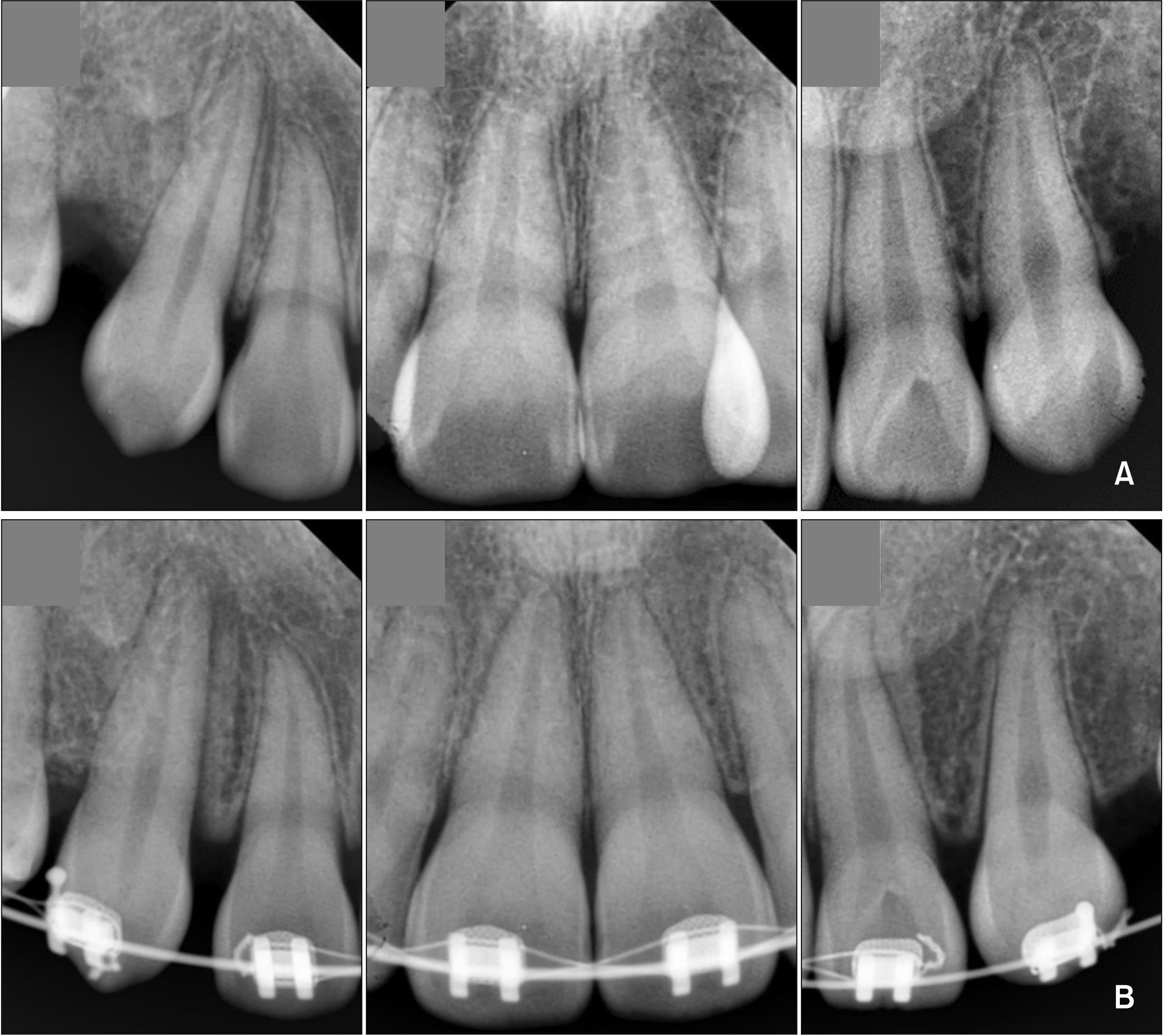
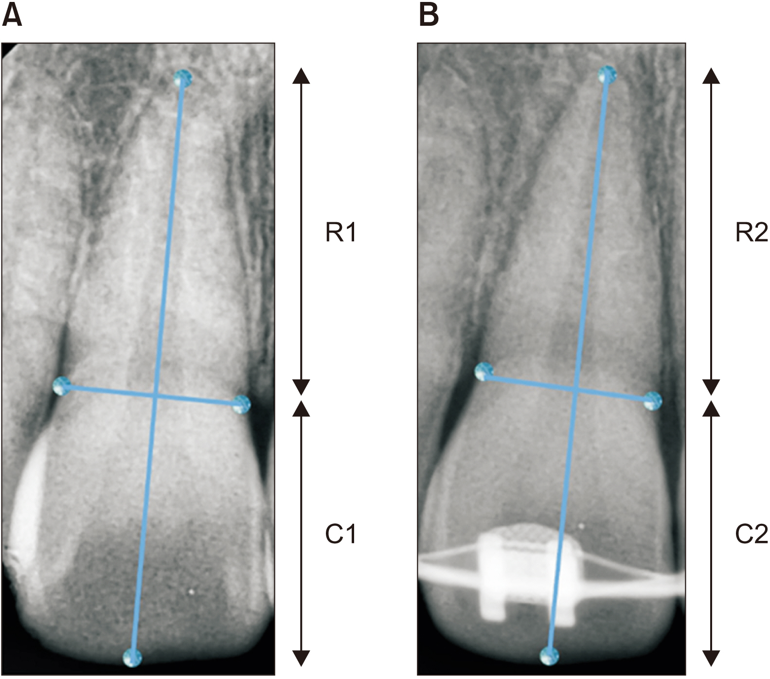
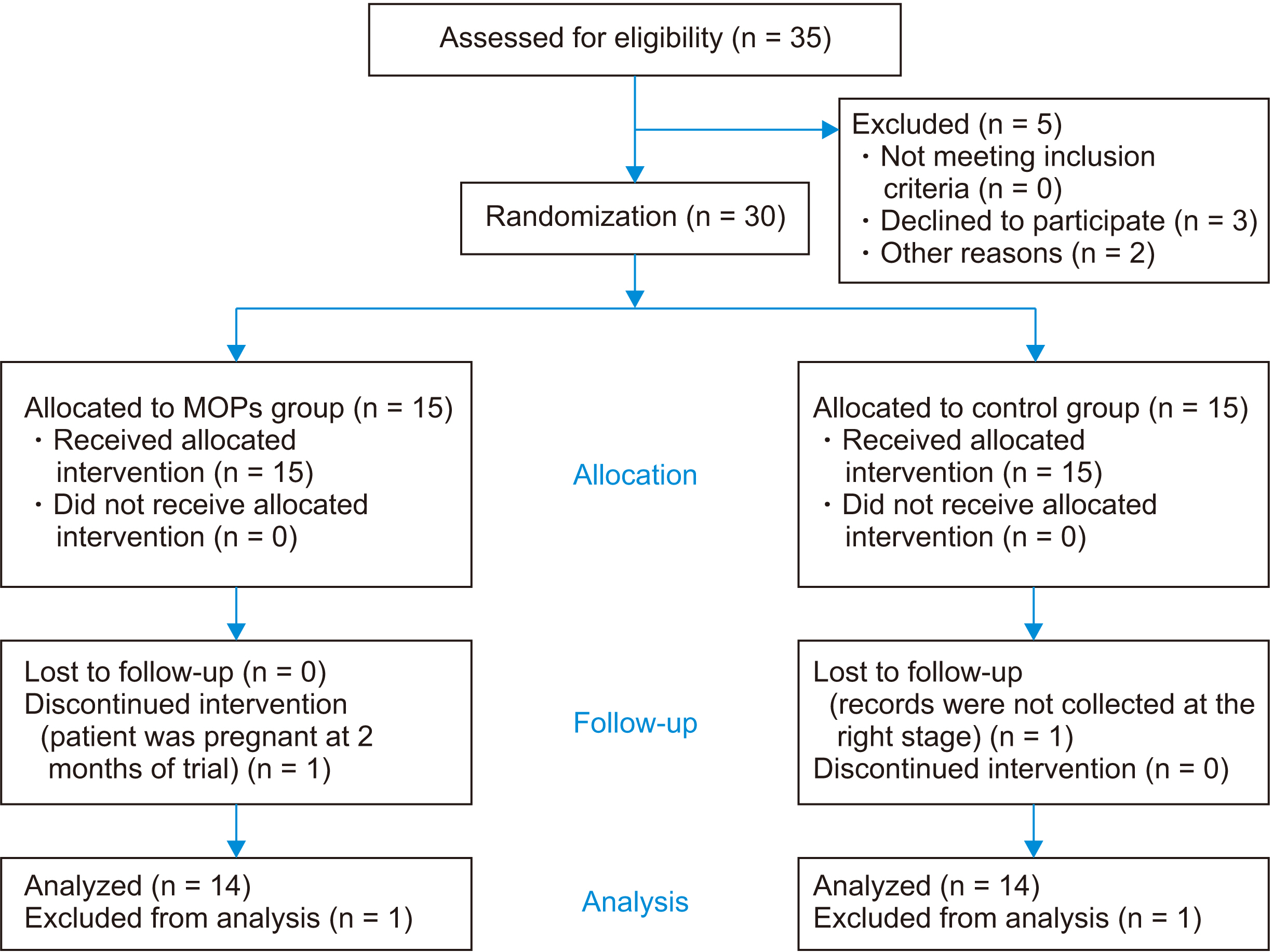
Thirty patients (25 females, 5 males; mean age, 22.66 ± 3.27 years) who presented with moderate crowding of the upper labial segment and underwent extraction-based fixed appliance treatment were recruited. They were randomly allocated to receive adjunctive therapy with MOPs (n = 15) or treatment with fixed appliances only (control group; n = 15). EARR was measured from long-cone periapical radiographs taken at the start and the sixth month of treatment. A correction factor for the enlargement difference was used to calculate EARR. Data were analyzed with descriptive statistics and repeated-measures analysis of variance.
Results
The mean root lengths of 168 teeth were measured and showed no statistically significant difference (p > 0.05) after six months of fixed appliance treatment in the MOP (mean difference [MD] = 0.13 mm; 95% confidence interval [CI] = −0.10–0.35) and control group (MD = 0.14 mm; 95% CI = −0.10–0.37). Most of the roots in the MOP and control groups (42.86% and 52.38%, respectively) showed only mild resorption. Less than 8% of the roots in both groups (7.14% in the MOP group and 4.76% in the control group) showed moderate resorption.
Conclusions
Acceleration of orthodontic tooth movement with adjunctive MOPs therapy during the alignment phase does not exacerbate EARR in patients with moderate crowding of the upper labial segment in comparison with controls.
Keyword
Figure
Reference
-
1. Iglesias-Linares A, Hartsfield JK Jr. 2017; Cellular and molecular pathways leading to external root resorption. J Dent Res. 96:145–52. DOI: 10.1177/0022034516677539. PMID: 27811065. PMCID: PMC5331617.
Article2. Fahey T, Stephenson P. 2016; Root resorption during fixed appliance therapy- mechanism, risk factors and prognosis. Orthod Update. 9:96–9. DOI: 10.12968/ortu.2016.9.3.96.3. Nowrin SA, Jaafar S, Ab Rahman N, Basri R, Alam MK, Shahid F. 2018; Association between genetic polymorphisms and external apical root resorption: a systematic review and meta-analysis. Korean J Orthod. 48:395–404. DOI: 10.4041/kjod.2018.48.6.395. PMID: 30450332. PMCID: PMC6234115.
Article4. Roscoe MG, Meira JB, Cattaneo PM. 2015; Association of orthodontic force system and root resorption: a systematic review. Am J Orthod Dentofacial Orthop. 147:610–26. DOI: 10.1016/j.ajodo.2014.12.026. PMID: 25919107.
Article5. Weiland F. 2003; Constant versus dissipating forces in orthodontics: the effect on initial tooth movement and root resorption. Eur J Orthod. 25:335–42. DOI: 10.1093/ejo/25.4.335. PMID: 12938838.
Article6. Kalemaj Z, DebernardI CL, Buti J. 2015; Efficacy of surgical and non-surgical interventions on accelerating orthodontic tooth movement: a systematic review. Eur J Oral Implantol. 8:9–24. PMID: 25738176.7. Al-Naoum F, Hajeer MY, Al-Jundi A. 2014; Does alveolar corticotomy accelerate orthodontic tooth movement when retracting upper canines? A split-mouth design randomized controlled trial. J Oral Maxillofac Surg. 72:1880–9. DOI: 10.1016/j.joms.2014.05.003. PMID: 25128922.
Article8. Gkantidis N, Mistakidis I, Kouskoura T, Pandis N. 2014; Effectiveness of non-conventional methods for accelerated orthodontic tooth movement: a systematic review and meta-analysis. J Dent. 42:1300–19. DOI: 10.1016/j.jdent.2014.07.013. PMID: 25072362.
Article9. Abbas NH, Sabet NE, Hassan IT. 2016; Evaluation of corticotomy-facilitated orthodontics and piezocision in rapid canine retraction. Am J Orthod Dentofacial Orthop. 149:473–80. DOI: 10.1016/j.ajodo.2015.09.029. PMID: 27021451.
Article10. Charavet C, Lecloux G, Jackers N, Maes N, Lambert F. 2019; Patient-reported outcomes measures (PROMs) following a piezocision-assisted versus conventional orthodontic treatments: a randomized controlled trial in adults. Clin Oral Investig. 23:4355–63. DOI: 10.1007/s00784-019-02887-z. PMID: 30955096.
Article11. Charavet C, Lecloux G, Bruwier A, Rompen E, Maes N, Limme M, et al. 2016; Localized piezoelectric alveolar decortication for orthodontic treatment in adults: a randomized controlled trial. J Dent Res. 95:1003–9. DOI: 10.1177/0022034516645066. PMID: 27129491.
Article12. Charavet C, Van Hede D, Anania S, Maes N, Lambert F. 2019; Multilevel biological responses following piezocision to accelerate orthodontic tooth movement: a study in rats. J World Fed Orthod. 8:100–6. DOI: 10.1016/j.ejwf.2019.07.002.
Article13. Charavet C, Lecloux G, Jackers N, Albert A, Lambert F. 2019; Piezocision-assisted orthodontic treatment using CAD/CAM customized orthodontic appliances: a randomized controlled trial in adults. Eur J Orthod. 41:495–501. DOI: 10.1093/ejo/cjy082. PMID: 30649257.
Article14. Sivarajan S, Ringgingon LP, Fayed MMS, Wey MC. 2020; The effect of micro-osteoperforations on the rate of orthodontic tooth movement: a systematic review and meta-analysis. Am J Orthod Dentofacial Orthop. 157:290–304. DOI: 10.1016/j.ajodo.2019.10.009. PMID: 32115107.
Article15. Alikhani M, Raptis M, Zoldan B, Sangsuwon C, Lee YB, Alyami B, et al. 2013; Effect of micro-osteoperforations on the rate of tooth movement. Am J Orthod Dentofacial Orthop. 144:639–48. DOI: 10.1016/j.ajodo.2013.06.017. PMID: 24182579.
Article16. Nanekrungsan K, Patanaporn V, Janhom A, Korwanich N. 2012; External apical root resorption in maxillary incisors in orthodontic patients: associated factors and radiographic evaluation. Imaging Sci Dent. 42:147–54. DOI: 10.5624/isd.2012.42.3.147. PMID: 23071964. PMCID: PMC3465756.
Article17. DiBiase AT, Woodhouse NR, Papageorgiou SN, Johnson N, Slipper C, Grant J, et al. 2016; Effect of supplemental vibrational force on orthodontically induced inflammatory root resorption: a multicenter randomized clinical trial. Am J Orthod Dentofacial Orthop. 150:918–27. DOI: 10.1016/j.ajodo.2016.06.025. PMID: 27894540.
Article18. Levander E, Malmgren O. 1988; Evaluation of the risk of root resorption during orthodontic treatment: a study of upper incisors. Eur J Orthod. 10:30–8. DOI: 10.1093/ejo/10.1.30. PMID: 3162877.
Article19. Kocadereli I, Yesil TN, Veske PS, Uysal S. 2011; Apical root resorption: a prospective radiographic study of maxillary incisors. Eur J Dent. 5:318–23. DOI: 10.1055/s-0039-1698898. PMID: 21769274. PMCID: PMC3137446.
Article20. Smale I, Artun J, Behbehani F, Doppel D, van't Hof M, Kuijpers-Jagtman AM. 2005; Apical root resorption 6 months after initiation of fixed orthodontic appliance therapy. Am J Orthod Dentofacial Orthop. 128:57–67. DOI: 10.1016/j.ajodo.2003.12.030. PMID: 16027626.
Article21. Alkebsi A, Al-Maaitah E, Al-Shorman H, Abu Alhaija E. 2018; Three-dimensional assessment of the effect of micro-osteoperforations on the rate of tooth movement during canine retraction in adults with Class II malocclusion: a randomized controlled clinical trial. Am J Orthod Dentofacial Orthop. 153:771–85. DOI: 10.1016/j.ajodo.2017.11.026. PMID: 29853235.
Article22. Aboalnaga AA, Salah Fayed MM, El-Ashmawi NA, Soliman SA. 2019; Effect of micro-osteoperforation on the rate of canine retraction: a split-mouth randomized controlled trial. Prog Orthod. 20:21. DOI: 10.1186/s40510-019-0274-0. PMID: 31155698. PMCID: PMC6545296.
Article23. Chan E, Dalci O, Petocz P, Papadopoulou AK, Darendeliler MA. 2018; Physical properties of root cementum: part 26. Effects of micro-osteoperforations on orthodontic root resorption: a microcomputed tomography study. Am J Orthod Dentofacial Orthop. 153:204–13. DOI: 10.1016/j.ajodo.2017.05.036. PMID: 29407497.
Article24. Motokawa M, Sasamoto T, Kaku M, Kawata T, Matsuda Y, Terao A, et al. 2012; Association between root resorption incident to orthodontic treatment and treatment factors. Eur J Orthod. 34:350–6. DOI: 10.1093/ejo/cjr018. PMID: 21811005.
Article25. Dudic A, Giannopoulou C, Martinez M, Montet X, Kiliaridis S. 2008; Diagnostic accuracy of digitized periapical radiographs validated against micro-computed tomography scanning in evaluating orthodontically induced apical root resorption. Eur J Oral Sci. 116:467–72. DOI: 10.1111/j.1600-0722.2008.00559.x. PMID: 18821990.
Article26. Signorelli L, Patcas R, Peltomäki T, Schätzle M. 2016; Radiation dose of cone-beam computed tomography compared to conventional radiographs in orthodontics. J Orofac Orthop. 77:9–15. DOI: 10.1007/s00056-015-0002-4. PMID: 26747662.
Article27. Janson GR, De Luca Canto G, Martins DR, Henriques JF, De Freitas MR. 2000; A radiographic comparison of apical root resorption after orthodontic treatment with 3 different fixed appliance techniques. Am J Orthod Dentofacial Orthop. 118:262–73. DOI: 10.1067/mod.2000.99136. PMID: 10982926.
Article28. Levander E, Malmgren O. 2000; Long-term follow-up of maxillary incisors with severe apical root resorption. Eur J Orthod. 22:85–92. DOI: 10.1093/ejo/22.1.85. PMID: 10721249.29. Sondeijker CFW, Lamberts AA, Beckmann SH, Kuitert RB, van Westing K, Persoon S, et al. 2020; Development of a clinical practice guideline for orthodontically induced external apical root resorption. Eur J Orthod. 42:115–24. DOI: 10.1093/ejo/cjz034. PMID: 31087032. PMCID: PMC7109605.
Article
- Full Text Links
- Actions
-
Cited
- CITED
-
- Close
- Share
- Similar articles
-
- A study on the affecting factors on root resorption
- External apical root resorption 6 months after initiation of orthodontic treatment: A randomized clinical trial comparing fixed appliances and orthodontic aligners
- A roentgenographic study on apical root resorption of human permanent teeth
- A study on the pattern of the root resorption following maxillary incisor retraction
- External apical root resorption in maxillary incisors in orthodontic patients: associated factors and radiographic evaluation


