Prompt CT Diagnosis of Epicardial Coronary Abscess after Percutaneous Coronary Intervention Caused by Klebsiella Pneumoniae
- Affiliations
-
- 1Military Service in Korean Army, Hongcheon, Korea
- 2Department of Cardiology, CHA University Bundang Medical Center, Seongnam, Korea
- 3Department of Radiology, CHA University Bundang Medical Center, Seongnam, Korea
- 4Smile Radiologic clinic, Seoul, Korea
- 5Department of Radiology, University of Maryland, Baltimore, MD, USA
- KMID: 2513527
- DOI: http://doi.org/10.4070/kcj.2020.0469
Figure
-
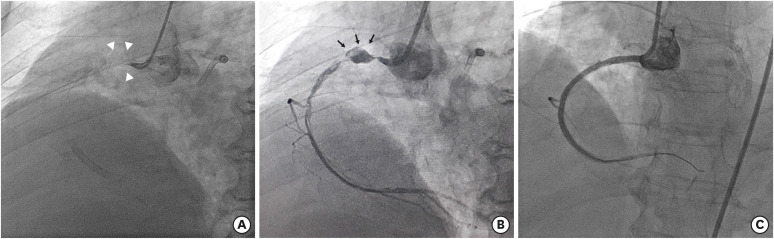
Figure 1 Percutaneous coronary angiography images. (A) Coronary angiographic image shows total occlusion at the origin of the right coronary artery (white arrowheads). Note multiple coronary stents in the RCA and left anterior descending coronary artery inserted in 16 months earlier. (B) Dissecting aneurysm or pseudoaneurysm (black arrows) is noted on a coronary angiographic image after balloon dilatation. (C) After completion of percutaneous coronary intervention by using Xience stent (3.5×33 mm), there is no evidence of residual pseudoaneurysm on coronary angiography.
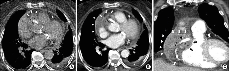
Figure 2 Chest CT images. (A) An air containing mass with high attenuation is noted on preconstrast CT around the proximal right coronary artery stent and along the right atrioventricular groove at the aortic sinus level (white arrows). Left pleural effusion is also seen (white asterisk). (B) On contrast enhanced axial CT image at the level of the left atrium, a low attenuation mass (arrows) with peripheral enhancement and multiple air-pockets is noted, consistent with air-forming epicardial coronary abscess (white and black arrows). Pericardial effusion (arrowheads) with linear pericardial thickening and enhancement is also demonstrated, indicating pericarditis (white arrowheads). (C) Coronal CT image at the level of the right ventricle shows similar findings.CT = computed tomography.
Reference
-
1. Elieson M, Mixon T, Carpenter J. Coronary stent infections: a case report and literature review. Tex Heart Inst J. 2012; 39:884–889. PMID: 23304047.2. Del Trigo M, Jimenez-Quevedo P, Fernandez-Golfin C, et al. Very late mycotic pseudoaneurysm associated with drug-eluting stent fracture. Circulation. 2012; 125:390–392. PMID: 22249529.3. Reddy K V C, Sanzgiri P, Thanki F, Suratkal V. Coronary stent infection: interesting cases with varied presentation. J Cardiol Cases. 2018; 19:5–8. PMID: 30693049.
- Full Text Links
-
- Actions
-
Cited
- CITED
-
- Close
- Share
-
- Similar articles
-
- Spondylodiscitis with Epidural Abscess Caused by Klebsiella pneumoniae
- Klebsiella pneumoniae Brain Abscess and Endophthalmitis after Acute Epiglottitis
- Ventriculitis Associated with Liver Abscess Caused by Klebsiella Pneumoniae
- A Case of Splenic Abscess with Multiple Fistulas Caused by Klebsiella pneumoniae
- Hematogenous Brain Abscess Resulting from Prostatic Abscess Caused by Klebsiella pneumoniae
