An accurate diagnosis of odontogenic cutaneous sinus tract by different computed tomography unit setting
- Affiliations
-
- 1Department of Oral and Maxillofacial Surgery, Dental Research Institute, School of Dentistry, Seoul National University, Seoul, Korea
- KMID: 2513430
- DOI: http://doi.org/10.5125/jkaoms.2021.47.1.51
Abstract
- Due to their rarity and the lack of associated dental symptoms, odontogenic cutaneous sinus tracts (OCSTs) are often misdiagnosed and confused with cutaneous lesions or non-odontogenic infections. It has been estimated that 50% of individuals affected by OCSTs are subjected to inappropriate treatments before the correct diagnosis is established. We describe the diagnosis and treatment of two cases of OCSTs. By using a computed tomography (CT) with soft tissue window setting, the extent of cortical bone destruction and the path of the sinus tract in the soft tissue was easily identified. Thus, we recommend the use of imaging techniques such as CT, which can confirm the odontogenic origin and the exact location of the OCST.
Figure
-
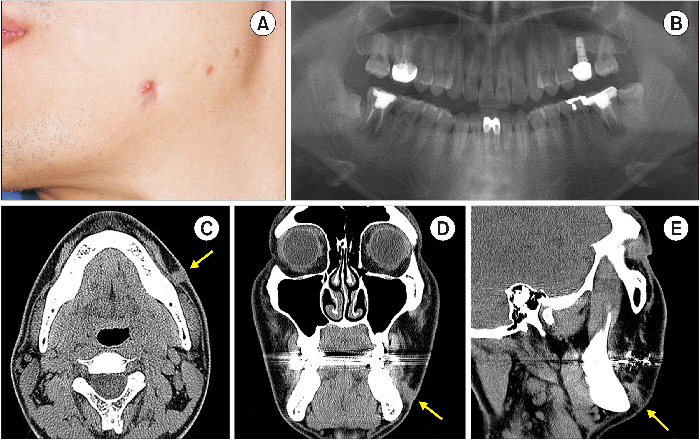
Fig. 1 A. Clinical examination revealed a non-tender, erythematous nodule measuring approximately 0.7 cm in diameter on the left submandibular region. B. Preoperative panoramic view revealed a periapical rarefaction of the #31, #37, #47 teeth and impacted #38, #48 third molar teeth. C-E. Axial, coronal, and sagittal computed tomography showing the exact path of the odontogenic sinus tract marked with arrows.
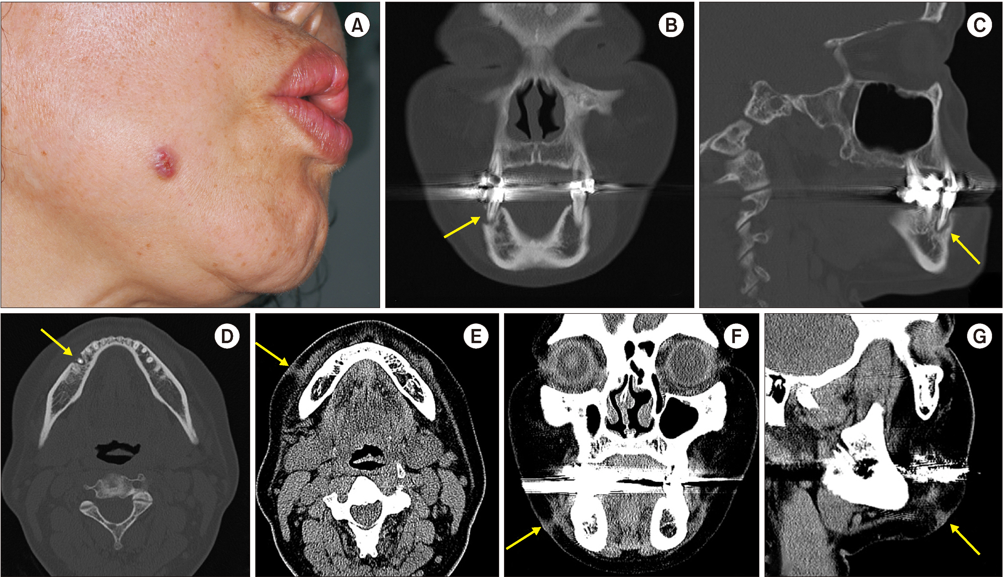
Fig. 2 A. Extraoral view of a patient with an orocutaneous fistula in the right mandible cheek. B-D. Preoperative axial, coronal, and sagittal computed tomography (CT) reveal lingual bone destruction of the #45 tooth and mild swelling of the adjoining buccal soft tissue marked with arrows. E-G. Axial, coronal, and sagittal CT with the soft tissue setting demonstrate the origin and the route of the fistula connected to the subcutaneous layer of the skin marked with arrows.
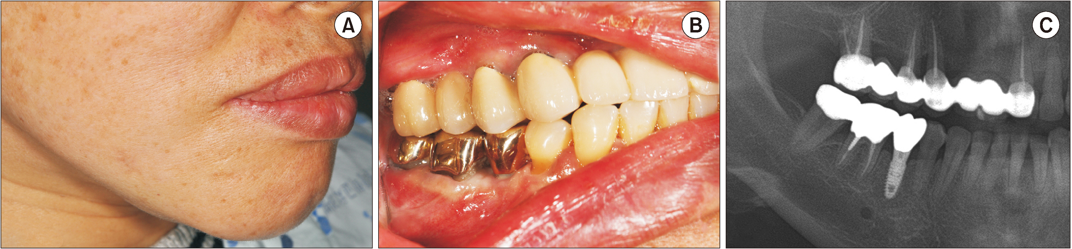
Fig. 3 A. Postoperative clinical photo of nearly imperceptible scarring after ten months. B. Clinical view of the final prosthesis delivery. C. Panoramic radiogram showing a well-osseointegrated implant along with prosthetic delivery at the 7-month follow-up after implant installation.
Reference
-
References
1. Guevara-Gutiérrez E, Riera-Leal L, Gómez-Martínez M, Amezcua-Rosas G, Chávez-Vaca CL, Tlacuilo-Parra A. 2015; Odontogenic cutaneous fistulas: clinical and epidemiologic characteristics of 75 cases. Int J Dermatol. 54:50–5. https://doi.org/10.1111/ijd.12262 . DOI: 10.1111/ijd.12262. PMID: 24134798.
Article2. Pasternak-Júnior B, Teixeira CS, Silva-Sousa YT, Sousa-Neto MD. 2009; Diagnosis and treatment of odontogenic cutaneous sinus tracts of endodontic origin: three case studies. Int Endod J. 42:271–6. https://doi.org/10.1111/j.1365-2591.2008.01519.x . DOI: 10.1111/j.1365-2591.2008.01519.x. PMID: 19228218.
Article3. Mittal N, Gupta P. 2004; Management of extra oral sinus cases: a clinical dilemma. J Endod. 30:541–7. https://doi.org/10.1097/00004770-200407000-00019 . DOI: 10.1097/00004770-200407000-00019. PMID: 15220655.
Article4. Mukerji R, Jones DC. 2002; Facial sinus of dental origin: a case report. Dent Update. 29:170–1. https://doi.org/10.12968/denu.2002.29.4.170 . DOI: 10.12968/denu.2002.29.4.170. PMID: 12050882.
Article5. Tian J, Liang G, Qi W, Jiang H. 2015; Odontogenic cutaneous sinus tract associated with a mandibular second molar having a rare distolingual root: a case report. Head Face Med. 11:13. https://doi.org/10.1186/s13005-015-0072-y . DOI: 10.1186/s13005-015-0072-y. PMID: 25885921. PMCID: PMC4414428.
Article6. Gupta M, Das D, Kapur R, Sibal N. 2011; A clinical predicament--diagnosis and differential diagnosis of cutaneous facial sinus tracts of dental origin: a series of case reports. Oral Surg Oral Med Oral Pathol Oral Radiol Endod. 112:e132–6. https://doi.org/10.1016/j.tripleo.2011.05.037 . DOI: 10.1016/j.tripleo.2011.05.037. PMID: 21873085.
Article7. Sammut S, Malden N, Lopes V. 2013; Facial cutaneous sinuses of dental origin - a diagnostic challenge. Br Dent J. 215:555–8. https://doi.org/10.1038/sj.bdj.2013.1141 . DOI: 10.1038/sj.bdj.2013.1141. PMID: 24309783.
Article8. Kishore Kumar RV, Devireddy SK, Gali RS, Chaithanyaa N, Chakravarthy C, Kumarvelu C. 2012; Cutaneous sinuses of cervicofacial region: a clinical study of 200 cases. J Maxillofac Oral Surg. 11:411–5. https://doi.org/10.1007/s12663-012-0353-y . DOI: 10.1007/s12663-012-0353-y. PMID: 24293932. PMCID: PMC3485468.
Article9. Chowdri NA, Sheikh S, Gagloo MA, Parray FQ, Sheikh MA, Khan FA. 2009; Clinicopathological profile and surgical results of nonhealing sinuses and fistulous tracts of the head and neck region. J Oral Maxillofac Surg. 67:2332–6. https://doi.org/10.1016/j.joms.2008.06.084 . DOI: 10.1016/j.joms.2008.06.084. PMID: 19837299.
Article10. Kim SM, Lee SK. 2019; Chronic non-bacterial osteomyelitis in the jaw. J Korean Assoc Oral Maxillofac Surg. 45:68–75. https://doi.org/10.5125/jkaoms.2019.45.2.68 . DOI: 10.5125/jkaoms.2019.45.2.68. PMID: 31106134. PMCID: PMC6502749.
Article11. Moon WK, Han MH, Kim IO, Sung MW, Chang KH, Choo SW, et al. 1995; Congenital fistula from ectopic accessory parotid gland: diagnosis with CT sialography and CT fistulography. AJNR Am J Neuroradiol. 16(4 Suppl):997–9. PMID: 7611095.12. Shemesh A, Hadad A, Azizi H, Lvovsky A, Ben Itzhak J, Solomonov M. 2019; Cone-beam computed tomography as a noninvasive assistance tool for oral cutaneous sinus tract diagnosis: a case series. J Endod. 45:950–6. https://doi.org/10.1016/j.joen.2019.03.017 . DOI: 10.1016/j.joen.2019.03.017. PMID: 31104817.
Article13. Angelopoulos C, Scarfe WC, Farman AG. 2012; A comparison of maxillofacial CBCT and medical CT. Atlas Oral Maxillofac Surg Clin North Am. 20:1–17. https://doi.org/10.1016/j.cxom.2011.12.008 . DOI: 10.1016/j.cxom.2011.12.008. PMID: 22365427.
Article14. Miracle AC, Mukherji SK. 2009; Conebeam CT of the head and neck, part 1: physical principles. AJNR Am J Neuroradiol. 30:1088–95. https://doi.org/10.3174/ajnr.A1653 . DOI: 10.3174/ajnr.A1653. PMID: 19439484. PMCID: PMC7051341.
Article15. Weiss R 2nd, Read-Fuller A. 2019; Cone beam computed tomography in oral and maxillofacial surgery: an evidence-based review. Dent J (Basel). 7:52. https://doi.org/10.3390/dj7020052 . DOI: 10.3390/dj7020052. PMID: 31052495. PMCID: PMC6631689.
Article16. MDC PACS viewer user guide R2.3 SP2 [Internet]. Amsterdam;Philips: Available from: https://home.itp.ac.ru/~alexd/DiagNET/Help/MDC_PACS_Viewer_UM_R2.3SP2_ENGLISH.pdf . cited 2020 Nov 1.17. 2020. Medlink PACS Plus viewer user manual [Internet]. Available from: https://vdocuments.mx/pacs-plus-viewer-user-manual.html . cited 2020 Nov 1.18. 2020. Acquisition and diagnostic software for X-ray images from DR flat panels or CR systems [Internet]. Available from: https://www.or-technology.com/media/downloads/Brochure%20dicomPACS%20DX-R_EN.pdf . cited 2020 Nov 1.19. 2020. RadiAnt DICOM Viewer [Internet]. Available from: https://www.radiantviewer.com/dicom-viewer-manual/change_brightness_contrast.html . cited 2020 Nov 1.
- Full Text Links
-
- Actions
-
Cited
- CITED
-
- Close
- Share
-
- Similar articles
-
- A Case Repot of Chronic Unhealing Wound Related to Odontogenic Cutaneous Sinus Tract
- A Case of Cutaneous Sinus Tract of Odontogenic Origin
- A Cutaneous Odontogenic Sinus
- A Case of Odontogenic Fistula Misdiagnosed as Cutaneous Ulcer on the Alar-Facial Angle
- Common pitfall of plastic surgeon for diagnosing cutaneous odontogenic sinus
