Neurointervention.
2021 Mar;16(1):70-77. 10.5469/neuroint.2020.00255.
Peri-Aneurysmal Brain Edema in Native and Treated Aneurysms: The Role of Thrombosis
- Affiliations
-
- 1Department of Neuroradiology, Neurocenter of Southern Switzerland, Ospedale Regionale di Lugano, Lugano, Switzerland
- 2Department of Radiology, Queen’s University at Kingston, Kingston, ON, Canada
- 3Department of Neuroradiology, Inselspital, Bern University Hospital, University of Bern, Bern, Switzerland
- KMID: 2513347
- DOI: http://doi.org/10.5469/neuroint.2020.00255
Abstract
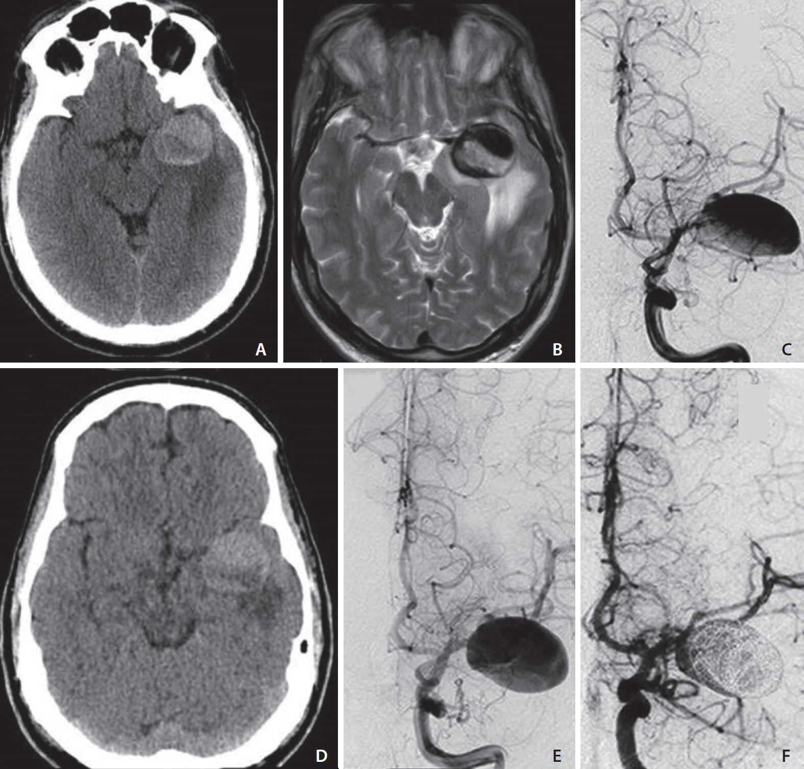
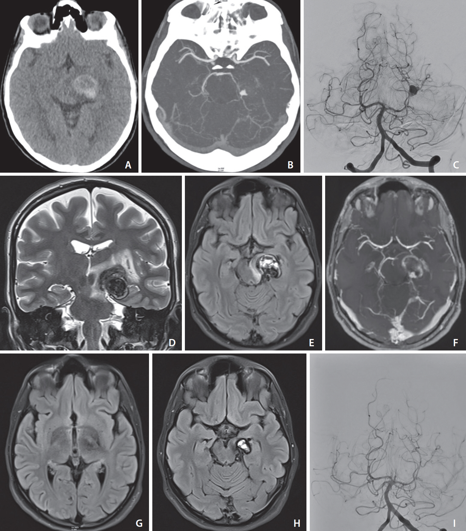
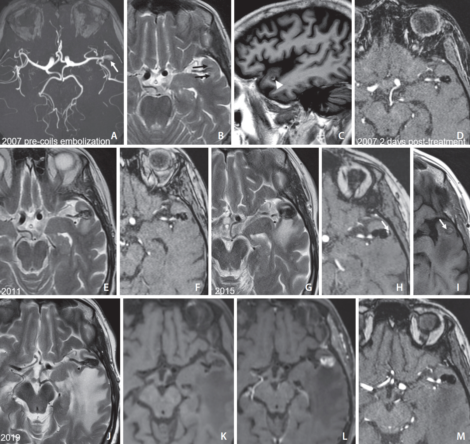
- Cerebral peri-aneurysmal edema (PE) is typically associated with giant partially-thrombosed aneurysms and less frequently with smaller aneurysms treated with endovascular embolization. An understanding of the pathophysiologic mechanism of PE is still limited. We report 3 cases of cerebral aneurysms associated with PE. We describe 2 cases of giant partially thrombosed aneurysms surrounded by vasogenic edema with apposition of an intramural and juxtamural thrombus. Our third case is a smaller aneurysm inciting vasogenic edema several years after coil embolization. Vessel-wall magnetic resonance imaging (MRI) showed avid wall enhancement and an enhancing thrombus embedded within the coils, reflecting inflammation of the aneurysm wall and proliferation of the vasa vasorum. Thrombosis within the aneurysmal sac and walls, both in native and treated aneurysms, may promote inflammatory changes and sustain the occurrence of PE. Vessel-wall MRI has a potential role in the evaluation process of this subgroup of aneurysms.
Keyword
Figure
Reference
-
1. Bose B, Northrup B, Osterholm J. Giant basilar artery aneurysm presenting as a third ventricular tumor. Neurosurgery. 1983; 13:699–702.
Article2. Heros RC, Kolluri S. Giant intracranial aneurysms presenting with massive cerebral edema. Neurosurgery. 1984; 15:572–577.
Article3. Craven I, Patel UJ, Gibson A, Coley SC. Symptomatic perianeurysmal edema following bare platinum embolization of a small unruptured cerebral aneurysm. AJNR Am J Neuroradiol. 2009; 30:1998–2000.
Article4. Lanzino G. Inflammation after embolization of intracranial aneurysms. J Neurosurg. 2008; 108:1071–1073.
Article5. Lehman VT, Brinjikji W, Kallmes DF, Huston J Rd, Lanzino G, Rabinstein AA, et al. Clinical interpretation of high-resolution vessel wall MRI of intracranial arterial diseases. Br J Radiol. 2016; 89:20160496.
Article6. Shimonaga K, Matsushige T, Ishii D, Sakamoto S, Hosogai M, Kawasumi T, et al. Clinicopathological insights from vessel wall imaging of unruptured intracranial aneurysms. Stroke. 2018; 49:2516–2519.
Article7. Horie N, Kitagawa N, Morikawa M, Tsutsumi K, Kaminogo M, Nagata I. Progressive perianeurysmal edema induced after endovascular coil embolization. Report of three cases and review of the literature. J Neurosurg. 2007; 106:916–920.8. Iihara K, Murao K, Sakai N, Soeda A, Ishibashi-Ueda H, Yutani C, et al. Continued growth of and increased symptoms from a thrombosed giant aneurysm of the vertebral artery after complete endovascular occlusion and trapping: the role of vasa vasorum. Case report. J Neurosurg. 2003; 98:407–413.9. Krings T, Alvarez H, Reinacher P, Ozanne A, Baccin CE, Gandolfo C, et al. Growth and rupture mechanism of partially thrombosed aneurysms. Interv Neuroradiol. 2007; 13:117–126.
Article10. Dengler J, Maldaner N, Bijlenga P, Burkhardt JK, Graewe A, Guhl S, Giant Intracranial Aneurysm Study Group, et al. Perianeurysmal edema in giant intracranial aneurysms in relation to aneurysm location, size, and partial thrombosis. J Neurosurg. 2015; 123:446–452.
Article11. Krings T, Mandell DM, Kiehl TR, Geibprasert S, Tymianski M, Alvarez H, et al. Intracranial aneurysms: from vessel wall pathology to therapeutic approach. Nat Rev Neurol. 2011; 7:547–559.
Article12. Im SH, Han MH, Kwon BJ, Jung C, Kim JE, Han DH. Aseptic meningitis after embolization of cerebral aneurysms using hydrogel-coated coils: report of three cases. AJNR Am J Neuroradiol. 2007; 28:511–512.13. Samaniego EA, Roa JA, Hasan D. Vessel wall imaging in intracranial aneurysms. J Neurointerv Surg. 2019; 11:1105–1112.
Article14. Sexton T, Smyth SS. Novel mediators and biomarkers of thrombosis. J Thromb Thrombolysis. 2014; 37:1–3.
Article15. Stenmark KR, Yeager ME, El Kasmi KC, Nozik-Grayck E, Gerasimovskaya EV, Li M, et al. The adventitia: essential regulator of vascular wall structure and function. Annu Rev Physiol. 2013; 75:23–47.
Article16. Larsen N, von der Brelie C, Trick D, Riedel CH, Lindner T, Madjidyar J, et al. Vessel wall enhancement in unruptured intracranial aneurysms: an indicator for higher risk of rupture? High-resolution MR imaging and correlated histologic findings. AJNR Am J Neuroradiol. 2018; 39:1617–1621.
Article17. Becske T, Kallmes DF, Saatci I, McDougall CG, Szikora I, Lanzino G, et al. Pipeline for uncoilable or failed aneurysms: results from a multicenter clinical trial. Radiology. 2013; 267:858–868.
Article18. Szikora I, Marosfoi M, Salomváry B, Berentei Z, Gubucz I. Resolution of mass effect and compression symptoms following endoluminal flow diversion for the treatment of intracranial aneurysms. AJNR Am J Neuroradiol. 2013; 34:935–939.
Article
- Full Text Links
- Actions
-
Cited
- CITED
-
- Close
- Share
- Similar articles
-
- Ethic Statement Correction: Peri-Aneurysmal Brain Edema in Native and Treated Aneurysms: The Role of Thrombosis
- Analysis of Initial Angiographic False Negative Aneurysmal Patients and False Positive Non-Aneurysmal Patients
- The Role of Neurosurgeons in the Era of Intra-Aneurysmal Treatment
- Unilateral Thrombosis of a Deep Cerebral Vein Associated with Transient Unilateral Thalamic Edema
- Kissing Aneurysms of Distal Anterior Cerebral Arteries


