Dual-Energy CT Angiography Improves Accuracy of Spot Sign for Predicting Hematoma Expansion in Intracerebral Hemorrhage
- Affiliations
-
- 1Department of Neurology and Cardiovascular Research Institute Maastricht, Maastricht University Medical Center, Maastricht, Netherlands
- 2Department of Radiology and Nuclear Medicine, MHeNS School for Mental Health and Neuroscience, Maastricht University Medical Center, Maastricht, Netherlands
- KMID: 2512357
- DOI: http://doi.org/10.5853/jos.2020.03531
Abstract
- Background and Purpose
Spot sign (SS) on computed tomography angiography (CTA) is associated with hematoma expansion (HE) and poor outcome after intracerebral hemorrhage (ICH). However, its predictive performance varies across studies, possibly because differentiating hyperdense hemorrhage from contrast media is difficult. We investigated whether dual-energy-CTA (DE-CTA), which can separate hemorrhage from iodinated contrast, improves the diagnostic accuracy of SS for predicting HE.
Methods
Primary ICH patients undergoing DE-CTA (both arterial as well as delayed venous phase) and follow-up computed tomography were prospectively included between 2014 and 2019. SS was assessed on both arterial and delayed phase images of the different DE-CTA datasets, i.e., conventional-like mixed images, iodine images, and fusion images. Diagnostic accuracy of SS for prediction of HE was determined on all datasets. The association between SS and HE, and between SS and poor outcome (modified Rankin Scale at 3 months ≥3) was assessed with multivariable logistic regression, using the dataset with highest diagnostic accuracy.
Results
Of 139 included patients, 47 showed HE (33.8%). Sensitivity of SS for HE was 32% (accuracy 0.72) on conventional-like mixed arterial images which increased to 76% (accuracy 0.80) on delayed fusion images. Presence of SS on delayed fusion images was independently associated with HE (odds ratio [OR], 17.5; 95% confidence interval [CI], 6.14 to 49.82) and poor outcome (OR, 3.84; 95% CI, 1.16 to 12.73).
Conclusions
Presence of SS on DE-CTA, in particular on delayed phase fusion images, demonstrates higher diagnostic performance in predicting HE compared to conventional-like mixed imaging, and it is associated with poor outcome.
Keyword
Figure
-
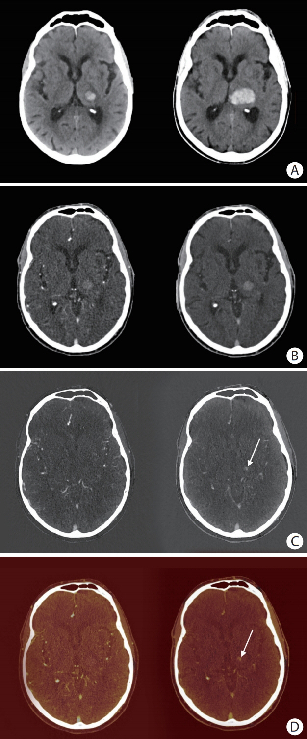
Figure 1. An 81-year old man with primary intracerebral hemorrhage (ICH) in the left thalamus. (A) Non-contrast baseline computed tomography (CT) (left) and non-contrast follow-up CT after 24 hours (right). (B) Mixed arterial image (left) and mixed delayed image (right). (C) Arterial iodine image (left) and delayed iodine image (right) with spot sign (SS) (arrow). (D) Arterial fusion image (left) and delayed fusion image (right) with SS (arrow). Arterial images did not show a SS. A SS can clearly be seen on delayed iodine (C, right panel) and fusion images (D, right panel), but could not be distinguished from hyperintense hemorrhage signal on conventional-like mixed delayed image (B, right panel). In this case, significant hematoma expansion took place (A).
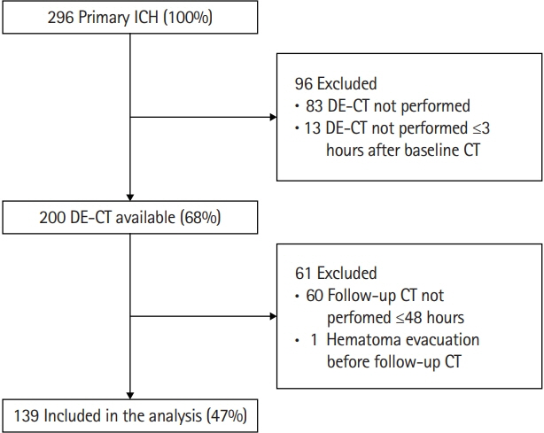
Figure 2. Flow-chart of selection study cohort. ICH, intracerebral hemorrhage; DE-CT, dual-energy computed tomography; CT, computed tomography.
Reference
-
References
1. Van Asch CJ, Luitse MJ, Rinkel GJ, van der Tweel I, Algra A, Klijn CJ. Incidence, case fatality, and functional outcome of intracerebral haemorrhage over time, according to age, sex, and ethnic origin: a systematic review and meta-analysis. Lancet Neurol. 2010; 9:167–176.
Article2. Xu X, Zhang J, Yang K, Wang Q, Xu B, Chen X. Accuracy of spot sign in predicting hematoma expansion and clinical outcome: a meta-analysis. Medicine (Baltimore). 2018; 97:e11945.3. Brouwers HB, Greenberg SM. Hematoma expansion following acute intracerebral hemorrhage. Cerebrovasc Dis. 2013; 35:195–201.
Article4. Davis SM, Broderick J, Hennerici M, Brun NC, Diringer MN, Mayer SA, et al. Hematoma growth is a determinant of mortality and poor outcome after intracerebral hemorrhage. Neurology. 2006; 66:1175–1181.
Article5. Brott T, Broderick J, Kothari R, Barsan W, Tomsick T, Sauerbeck L, et al. Early hemorrhage growth in patients with intracerebral hemorrhage. Stroke. 1997; 28:1–5.
Article6. Demchuk AM, Dowlatshahi D, Rodriguez-Luna D, Molina CA, Blas YS, Dzialowski I, et al. Prediction of haematoma growth and outcome in patients with intracerebral haemorrhage using the CT-angiography spot sign (PREDICT): a prospective observational study. Lancet Neurol. 2012; 11:307–314.
Article7. Phan TG, Krishnadas N, Lai VW, Batt M, Slater LA, Chandra RV, et al. Meta-analysis of accuracy of the spot sign for predicting hematoma growth and clinical outcomes. Stroke. 2019; 50:2030–2036.
Article8. Postma AA, Das M, Stadler AA, Wildberger JE. Dual-energy CT: what the neuroradiologist should know. Curr Radiol Rep. 2015; 3:16.
Article9. Watanabe Y, Tsukabe A, Kunitomi Y, Nishizawa M, Arisawa A, Tanaka H, et al. Dual-energy CT for detection of contrast enhancement or leakage within high-density haematomas in patients with intracranial haemorrhage. Neuroradiology. 2014; 56:291–295.
Article10. Tijssen MP, Hofman PA, Stadler AA, van Zwam W, de Graaf R, van Oostenbrugge RJ, et al. The role of dual energy CT in differentiating between brain haemorrhage and contrast medium after mechanical revascularisation in acute ischaemic stroke. Eur Radiol. 2014; 24:834–840.
Article11. Tawfik AM, Kerl JM, Razek AA, Bauer RW, Nour-Eldin NE, Vogl TJ, et al. Image quality and radiation dose of dual-energy CT of the head and neck compared with a standard 120- kVp acquisition. AJNR Am J Neuroradiol. 2011; 32:1994–1999.12. Postma AA, Hofman PA, Stadler AA, van Oostenbrugge RJ, Tijssen MP, Wildberger JE. Dual-energy CT of the brain and intracranial vessels. AJR Am J Roentgenol. 2012; 199(5 Suppl):S26–S33.
Article13. Potter CA, Sodickson AD. Dual-energy CT in emergency neuroimaging: added value and novel applications. Radiographics. 2016; 36:2186–2198.
Article14. Huynh TJ, Flaherty ML, Gladstone DJ, Broderick JP, Demchuk AM, Dowlatshahi D, et al. Multicenter accuracy and interobserver agreement of spot sign identification in acute intracerebral hemorrhage. Stroke. 2014; 45:107–112.
Article15. Kothari RU, Brott T, Broderick JP, Barsan WG, Sauerbeck LR, Zuccarello M, et al. The ABCs of measuring intracerebral hemorrhage volumes. Stroke. 1996; 27:1304–1305.
Article16. Tan CO, Lam S, Kuppens D, Bergmans RH, Parameswaran BK, Forghani R, et al. Spot and diffuse signs: quantitative markers of intracranial hematoma expansion at dual-energy CT. Radiology. 2019; 290:179–186.17. Brouwers HB, Chang Y, Falcone GJ, Cai X, Ayres AM, Battey TW, et al. Predicting hematoma expansion after primary intracerebral hemorrhage. JAMA Neurol. 2014; 71:158–164.
Article18. Moon BH, Jang DK, Han YM, Jang KS, Huh R, Park YS. Association factors for CT angiography spot sign and hematoma growth in Korean patients with acute spontaneous intracerebral hemorrhage: a single-center cohort study. J Korean Neurosurg Soc. 2014; 56:295–302.19. Rodriguez-Luna D, Rubiera M, Ribo M, Coscojuela P, Piñeiro S, Pagola J, et al. Ultraearly hematoma growth predicts poor outcome after acute intracerebral hemorrhage. Neurology. 2011; 77:1599–1604.
Article20. Kim SH, Jung HH, Whang K, Kim JY, Pyen JS, Oh JW. Which emphasizing factors are most predictive of hematoma expansion in spot sign positive intracerebral hemorrhage? J Korean Neurosurg Soc. 2014; 56:86–90.
Article21. Ciura VA, Brouwers HB, Pizzolato R, Ortiz CJ, Rosand J, Goldstein JN, et al. Spot sign on 90-second delayed computed tomography angiography improves sensitivity for hematoma expansion and mortality: prospective study. Stroke. 2014; 45:3293–3297.22. Ederies A, Demchuk A, Chia T, Gladstone DJ, Dowlatshahi D, Bendavit G, et al. Postcontrast CT extravasation is associated with hematoma expansion in CTA spot negative patients. Stroke. 2009; 40:1672–1676.
Article23. Wada R, Aviv RI, Fox AJ, Sahlas DJ, Gladstone DJ, Tomlinson G, et al. CT angiography “spot sign” predicts hematoma expansion in acute intracerebral hemorrhage. Stroke. 2007; 38:1257–1262.
Article24. Goldstein JN, Fazen LE, Snider R, Schwab K, Greenberg SM, Smith EE, et al. Contrast extravasation on CT angiography predicts hematoma expansion in intracerebral hemorrhage. Neurology. 2007; 68:889–894.
Article25. Han JH, Lee JM, Koh EJ, Choi HY. The spot sign predicts hematoma expansion, outcome, and mortality in patients with primary intracerebral hemorrhage. J Korean Neurosurg Soc. 2014; 56:303–309.
Article26. Dowlatshahi D, Brouwers HB, Demchuk AM, Hill MD, Aviv RI, Ufholz LA, et al. Predicting intracerebral hemorrhage growth with the spot sign: the effect of onset-to-scan time. Stroke. 2016; 47:695–700.27. Rodriguez-Luna D, Dowlatshahi D, Aviv RI, Molina CA, Silva Y, Dzialowski I, et al. Venous phase of computed tomography angiography increases spot sign detection, but intracerebral hemorrhage expansion is greater in spot signs detected in arterial phase. Stroke. 2014; 45:734–739.
Article28. Wu TC, Chen TY, Shiue YL, Chen JH, Hsieh TJ, Ko CC, et al. Added value of delayed computed tomography angiography in primary intracranial hemorrhage and hematoma size for predicting spot sign. Acta Radiol. 2018; 59:485–490.
Article29. Rodriguez-Luna D, Coscojuela P, Rodriguez-Villatoro N, Juega JM, Boned S, Muchada M, et al. Multiphase CT angiography improves prediction of intracerebral hemorrhage expansion. Radiology. 2017; 285:932–940.
Article30. Kim J, Smith A, Hemphill JC 3rd, Smith WS, Lu Y, Dillon WP, et al. Contrast extravasation on CT predicts mortality in primary intracerebral hemorrhage. AJNR Am J Neuroradiol. 2008; 29:520–525.
Article31. Roele ED, Timmer VC, Vaassen LA, van Kroonenburgh AM, Postma AA. Dual-energy CT in head and neck imaging. Curr Radiol Rep. 2017; 5:19.
Article32. Morotti A, Romero JM, Jessel MJ, Brouwers HB, Gupta R, Schwab K, et al. Effect of CTA tube current on spot sign detection and accuracy for prediction of intracerebral hemorrhage expansion. AJNR Am J Neuroradiol. 2016; 37:1781–1786.
Article
- Full Text Links
-
- Actions
-
Cited
- CITED
-
- Close
- Share
-
- Similar articles
-
- Role of 'Spot Sign' on CT Angiography to Predict Hematoma Expansion in Spontaneous Intracerebral Hemorrhage
- Which Emphasizing Factors Are Most Predictive of Hematoma Expansion in Spot Sign Positive Intracerebral Hemorrhage?
- The Spot Sign Predicts Hematoma Expansion, Outcome, and Mortality in Patients with Primary Intracerebral Hemorrhage
- Leak Sign on Dynamic-SusceptibilityContrast Magnetic Resonance Imaging in Acute Intracerebral Hemorrhage
- Association Factors for CT Angiography Spot Sign and Hematoma Growth in Korean Patients with Acute Spontaneous Intracerebral Hemorrhage : A Single-Center Cohort Study
