Linked Color Imaging Demonstrates Characteristic Findings in Semi-Pedunculated Gastric Adenocarcinoma in Helicobacter Pylori-Negative Normal Mucosa
- Affiliations
-
- 1Division of Gastroenterology, Department of Medicine, Jichi Medical University, Tochigi, Japan
- 2Department of Pathology, Jichi Medical University, Tochigi, Japan
- 3Department of Surgery, Jichi Medical University, Tochigi, Japan
- KMID: 2512320
- DOI: http://doi.org/10.5946/ce.2020.059
Figure
-

Fig. 1. (A) White light imaging shows a 7-mm red semi-pedunculated polyp along the greater curvature in the proximal gastric body. (B) The polyp appears purple–red in an area of light orange normal mucosa using linked color imaging. (C) The near-view using linked color imaging shows an irregular polygonal shaped white zone, suggestive of a malignant lesion. (D) Magnifying blue laser imaging shows an irregular surface pattern with a varied size white zone. (E) The near-view of a hyperplastic polyp using linked color imaging shows elongated white zones distinct from an irregular polygonal shaped white zone, suggestive of a benign lesion. Purple intestinal metaplasia is seen in the background mucosa. (F) Magnifying blue laser imaging of a hyperplastic polyp shows elongated white zones.
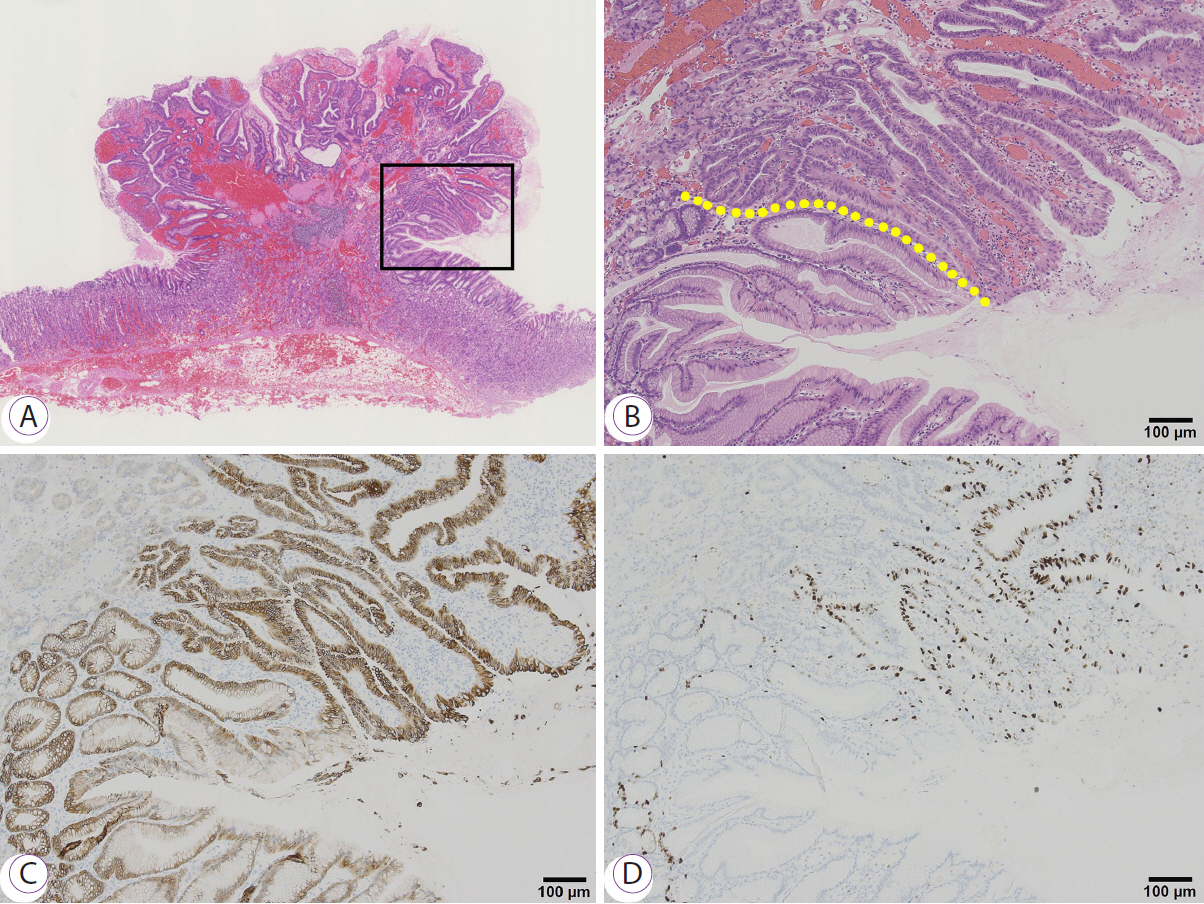
Fig. 2. The resected specimen (A) (loupe, hematoxylin and eosin [H&E] stain) and (B) (H&E stain, ×10). The area in the black box shows papillary proliferation of atypical cells, suggesting well-differentiated adenocarcinoma and hyperplastic foveolar epithelium in the deeper layer. The yellow line shows the border between the malignant portion and the foveolar epithelium. Cancer is found on most of the polyp’s surface and is limited to the mucosa and broad interstitial areas are seen. Immunostaining for mucin 5AC (C) in the malignant area stains in a manner similar to that observed in the background foveolar epithelium and MIB-1 (D) is positive in the entire area of adenocarcinoma.
Reference
-
1. Osawa H, Miura Y, Takezawa T, et al. Linked color imaging and blue laser imaging for upper gastrointestinal screening. Clin Endosc. 2018; 51:513–526.
Article2. Kanzaki H, Takenaka R, Kawahara Y, et al. Linked color imaging (LCI), a novel image-enhanced endoscopy technology, emphasizes the color of early gastric cancer. Endosc Int Open. 2017; 5:E1005–E1013.
Article3. Fukuda H, Miura Y, Osawa H, et al. Linked color imaging can enhance recognition of early gastric cancer by high color contrast to surrounding gastric intestinal metaplasia. J Gastroenterol. 2019; 54:396–406.
Article4. Shinozaki S, Osawa H, Hayashi Y, Lefor AK, Yamamoto H. Linked color imaging for the detection of early gastrointestinal neoplasms. Therap Adv Gastroenterol. 2019; 12:1756284819885246.
Article5. Dohi O, Yagi N, Onozawa Y, et al. Linked color imaging improves endoscopic diagnosis of active Helicobacter pylori infection. Endosc Int Open. 2016; 4:E800–E805.
Article6. Yasuda T, Hiroyasu T, Hiwa S, et al. Potential of automatic diagnosis system with linked color imaging for diagnosis of Helicobacter pylori infection. Dig Endosc. 2020; 32:373–381.7. Jiang ZX, Nong B, Liang LX, Yan YD, Zhang G. Differential diagnosis of Helicobacter pylori-associated gastritis with the linked-color imaging score. Dig Liver Dis. 2019; 51:1665–1670.
Article8. Yamamoto Y, Fujisaki J, Omae M, Hirasawa T, Igarashi M. Helicobacter pylori-negative gastric cancer: characteristics and endoscopic findings. Dig Endosc. 2015; 27:551–561.
Article9. Isono Y, Baba Y, Mukai K, et al. Gastric adenocarcinoma coexisting with a reddish semipedunculated polyp arising from Helicobacter pylori-negative normal gastric mucosa: a report of two cases. Clin J Gastroenterol. 2018; 11:481–486.
Article10. Togo K, Ueo T, Yonemasu H, et al. Two cases of adenocarcinoma occurring in sporadic fundic gland polyps observed by magnifying endoscopy with narrow band imaging. World J Gastroenterol. 2016; 22:9028–9034.
Article
- Full Text Links
-
- Actions
-
Cited
- CITED
-
- Close
- Share
-
- Similar articles
-
- Vaccine Development and Future for Helicobacter pylori
- Helicobacter pylori-negative Gastric Mucosa-associated Lymphoid Tissue Lymphoma
- Linked Color Imaging and Blue Laser Imaging for Upper Gastrointestinal Screening
- Helicobacter pylori-negative Gastric Cancer
- Histochemical and Immunohistochemical Stain of Helicobacter pylori from the Gastric Mucosa
