Missing teeth after maxillofacial trauma:a case report and management protocol
- Affiliations
-
- 1Department of Oral and Maxillofacial Surgery, K.V.G. Dental College and Hospital, Sullia, India
- KMID: 2510017
- DOI: http://doi.org/10.5125/jkaoms.2020.46.6.422
Abstract
- Management of maxillofacial trauma includes primary care, in which diagnosis and management of dentoalveolar injury play a vital role. Due to the impact sustained during a maxillofacial injury (whether direct or indirect), dentoalveolar injuries can occur, leading to fracture and displacement of teeth and associated alveolar bone into the surrounding soft tissues and associated structures, such as the maxillary sinus, nasal cavity, upper respiratory tract, tracheobronchial tree, or gastrointestinal tract. Undiagnosed displaced teeth may cause complications such as airway obstruction. This paper reports a case of displaced teeth in the nasal cavity and gastrointestinal tract and highlights the management protocol for displaced teeth secondary to maxillofacial trauma.
Figure
-
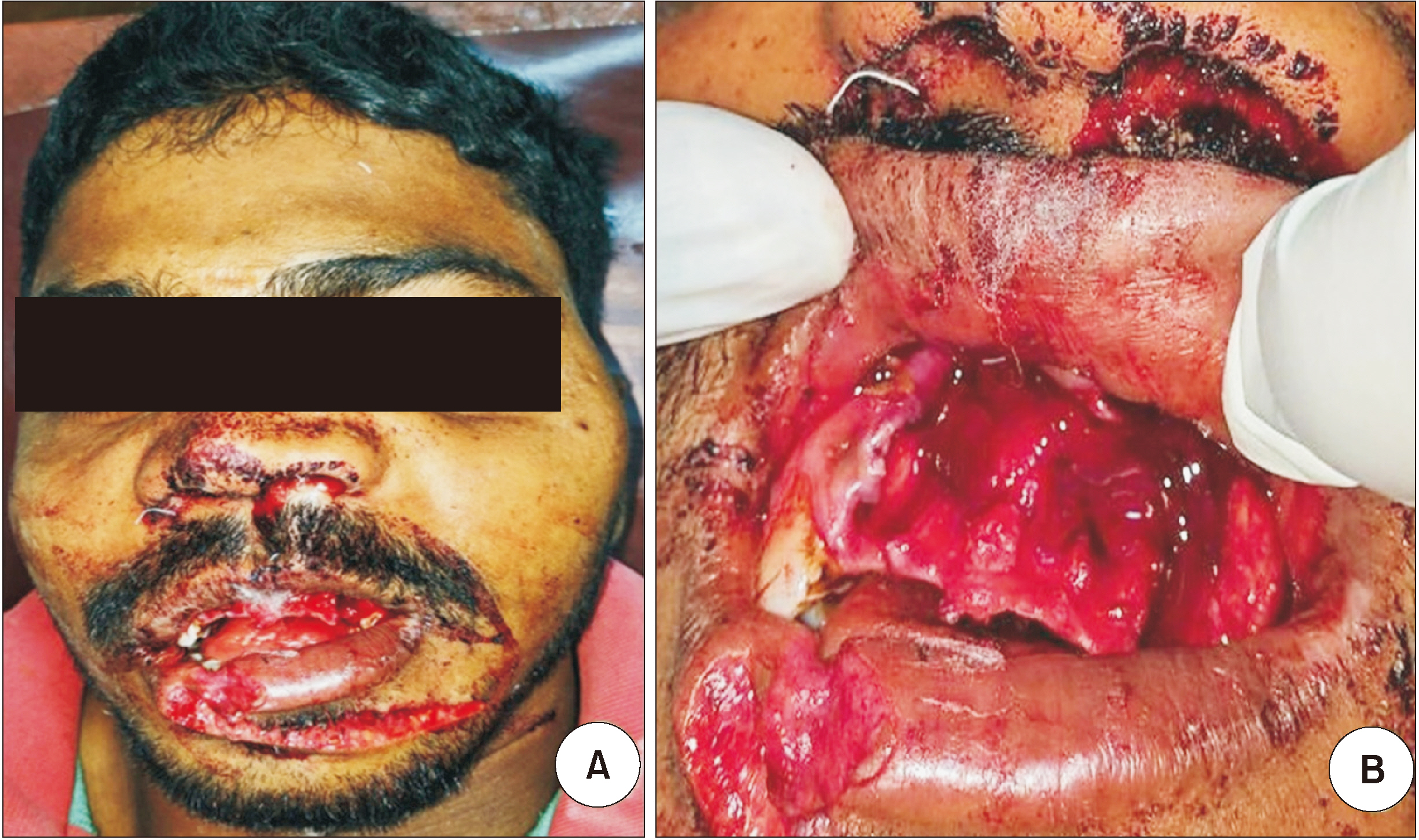
Fig. 1 Preoperative photographs of the patient. A. Frontal view. B. Dentoalveolar fracture with respect to maxillary anterior segments.

Fig. 2 Computed tomography of the head. A. Axial view: Note the arrows indicating teeth in the nasal cavity and in the soft tissue near the left maxillary sinus. B. Coronal view: Note the arrows showing teeth in the floor of the nasal cavity.
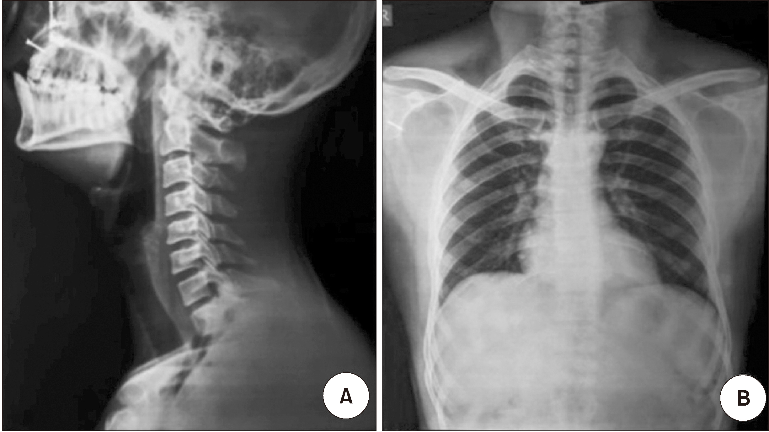
Fig. 3 Radiographs of the tracheobronchial tree and lungs. A. Lateral neck radiograph. B. Anteroposterior chest radiograph.

Fig. 4 Nasal endoscopic view. A. The left naris revealed perforation, but no teeth were located. B. The right naris showed no teeth.
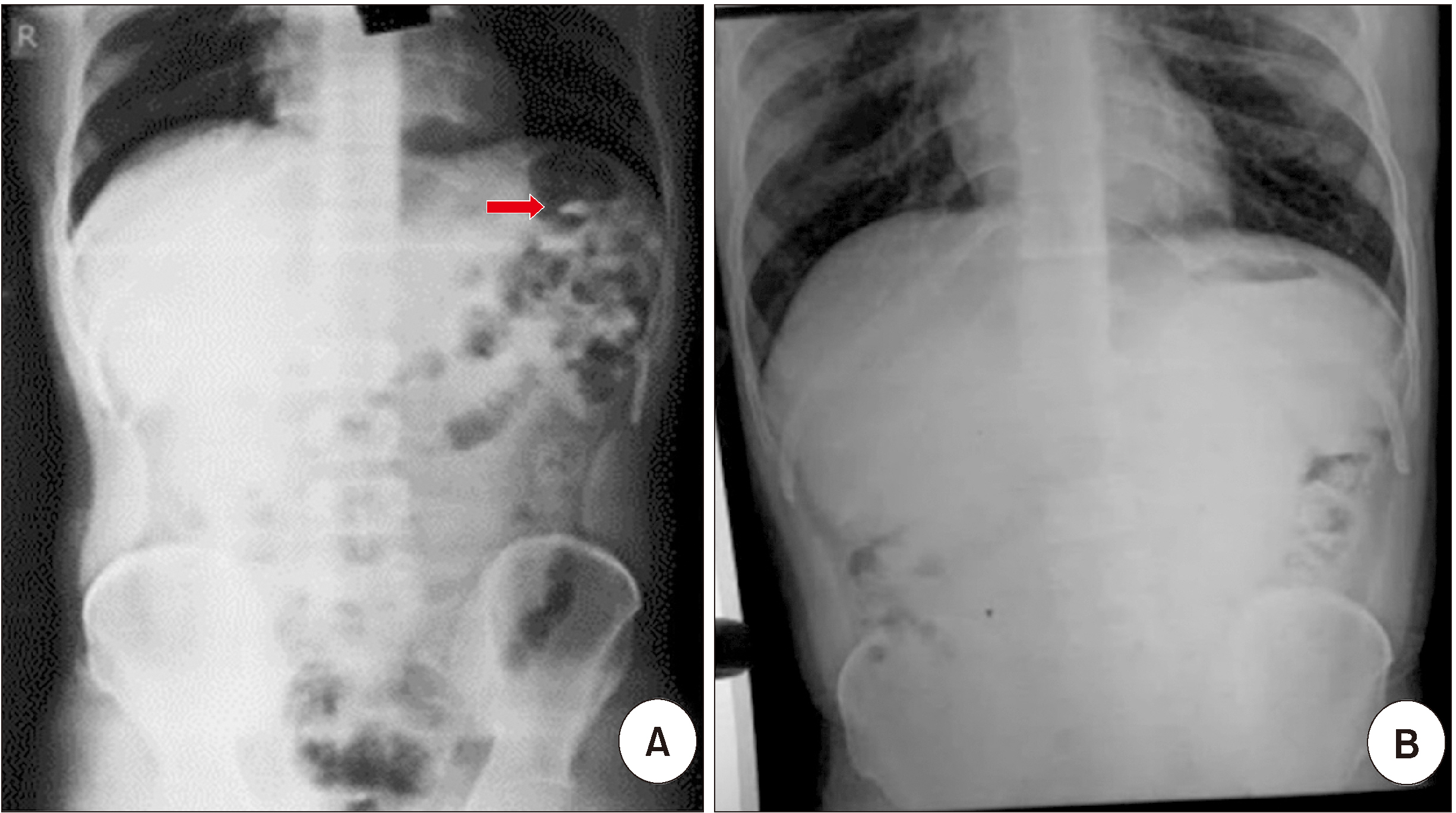
Fig. 5 Radiograph of the gastrointestinal tract. A. Erect abdominal radiograph: Note the arrow showing the hyperdense structure, i.e., a tooth, in the bowel. B. Erect abdominal radiograph: Follow-up radiograph after 7 days of laxative use.
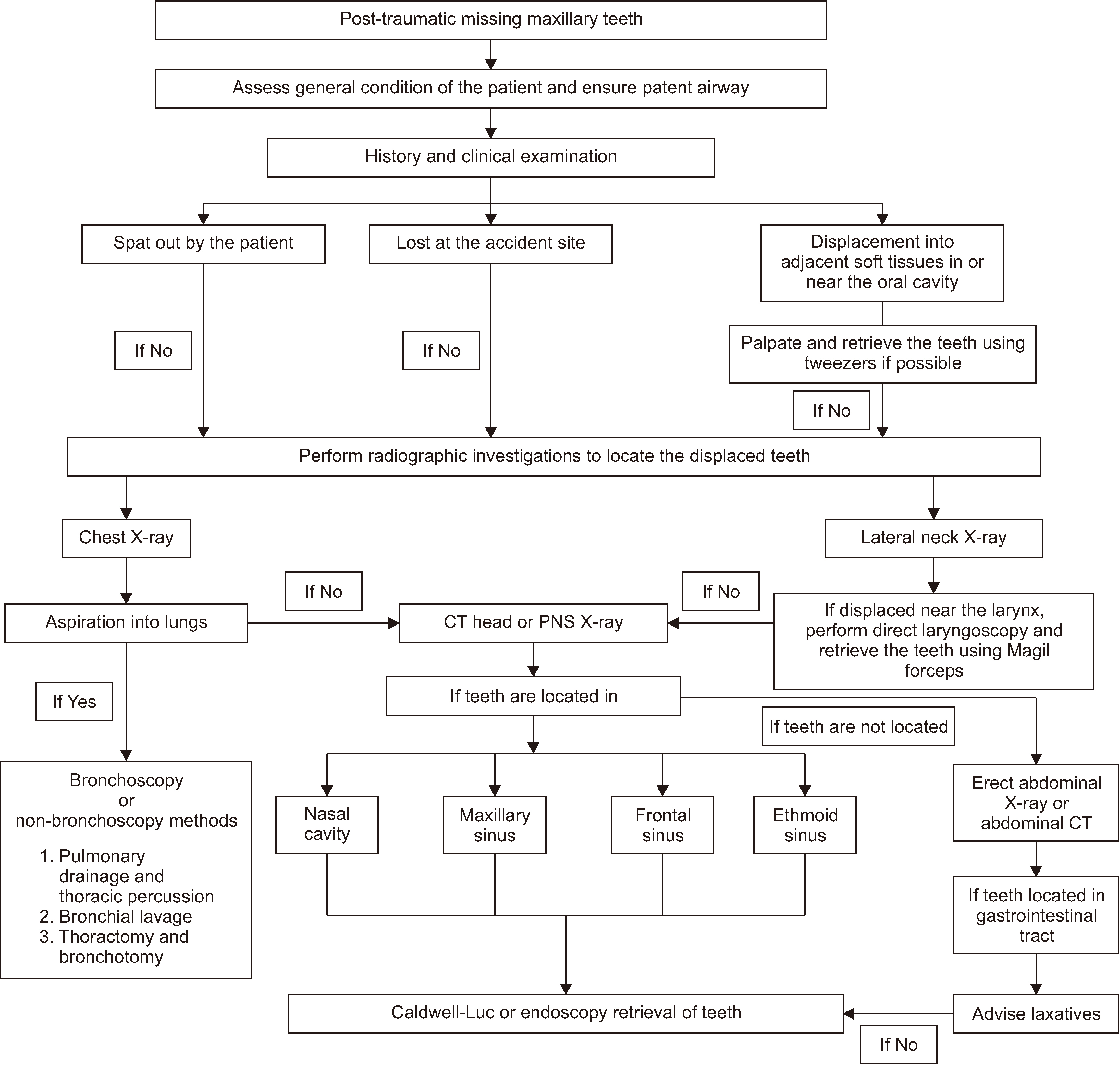
Fig. 6 Protocol for management of displaced missing teeth after maxillofacial trauma. (CT: computed tomography, PNS: paranasal sinus view)
Reference
-
References
1. Glendor U. 2008; Epidemiology of traumatic dental injuries--a 12 year review of the literature. Dent Traumatol. 24:603–11. https://doi.org/10.1111/j.1600-9657.2008.00696.x . DOI: 10.1111/j.1600-9657.2008.00696.x. PMID: 19021651.
Article2. Gassner R, Bösch R, Tuli T, Emshoff R. 1999; Prevalence of dental trauma in 6000 patients with facial injuries: implications for prevention. Oral Surg Oral Med Oral Pathol Oral Radiol Endod. 87:27–33. https://doi.org/10.1016/s1079-2104(99)70290-8 . DOI: 10.1016/s1079-2104(99)70290-8. PMID: 9927076.
Article3. Ceallaigh PO, Ekanaykaee K, Beirne CJ, Patton DW. 2007; Diagnosis and management of common maxillofacial injuries in the emergency department. Part 5: dentoalveolar injuries. Emerg Med J. 24:429–30. https://doi.org/10.1136/emj.2006.035949 . DOI: 10.1136/emj.2006.035949. PMID: 17513544. PMCID: PMC2658285.
Article4. Arunprasad G, Ramasamy A, Madhan B, Krishnan B. 2015; Traumatic dislocation of the mandibular lateral incisor into the nasal floor. BMJ Case Rep. 2015:bcr2015213553. https://doi.org/10.1136/bcr-2015-213553 . DOI: 10.1136/bcr-2015-213553. PMID: 26678697. PMCID: PMC4691925.
Article5. Ilyas N, Rajendran VR, Thottath J, Saanida MP, Menon I. Sherinas. 2017; Curious odyssey of teeth through body spaces: report of a rare case. Int J Med Res Prof. 3:268–70.6. Jain V, Dubey A, Kumar J, Srivastava S, Tripathy M. 2015; Accidental ingestion of a hypodermic needle during root canal treatment: a case report. Gen Dent. 63:30–2. PMID: 26325638.7. Fields RT Jr, Schow SR. 1998; Aspiration and ingestion of foreign bodies in oral and maxillofacial surgery: a review of the literature and report of five cases. J Oral Maxillofac Surg. 56:1091–8. https://doi.org/10.1016/s0278-2391(98)90263-4 . DOI: 10.1016/s0278-2391(98)90263-4. PMID: 9734773.
Article8. Zerella JT, Dimler M, McGill LC, Pippus KJ. 1998; Foreign body aspiration in children: value of radiography and complications of bronchoscopy. J Pediatr Surg. 33:1651–4. https://doi.org/10.1016/s0022-3468(98)90601-7 . DOI: 10.1016/s0022-3468(98)90601-7. PMID: 9856887.
Article9. Tung TC, Chen YR, Chen CT, Lin CJ. 1997; Full intrusion of a tooth after facial trauma. J Trauma. 43:357–9. https://doi.org/10.1097/00005373-199708000-00026 . DOI: 10.1097/00005373-199708000-00026. PMID: 9291387.
Article10. Zitzmann NU, Elsasser S, Fried R, Marinello CP. 1999; Foreign body ingestion and aspiration. Oral Surg Oral Med Oral Pathol Oral Radiol Endod. 88:657–60. https://doi.org/10.1016/s1079-2104(99)70004-1 . DOI: 10.1016/s1079-2104(99)70004-1. PMID: 10625844.
Article11. Kumar N, Goyal H, Bindra A, Goyal K. 2014; Management of aspirated tooth in an adult head injury patient: Report of two cases. Saudi J Anaesth. 8:276–8. https://doi.org/10.4103/1658-354X.130747 . DOI: 10.4103/1658-354X.130747. PMID: 24843346. PMCID: PMC4024690.
Article12. Webb WA. 1988; Management of foreign bodies of the upper gastrointestinal tract. Gastroenterology. 94:204–16. https://doi.org/10.1016/0016-5085(88)90632-4 . DOI: 10.1016/0016-5085(88)90632-4. PMID: 3275566.
Article13. Kuo SC, Chen YL. 2008; Accidental swallowing of an endodontic file. Int Endod J. 41:617–22. https://doi.org/10.1111/j.1365-2591.2008.01392.x . DOI: 10.1111/j.1365-2591.2008.01392.x. PMID: 18479380.
Article
- Full Text Links
-
- Actions
-
Cited
- CITED
-
- Close
- Share
-
- Similar articles
-
- The case report of the skeletal Angle's Class II malocclusion with the upper central incisor missing
- The study on the prognosis of dental implants which has been installed after maxillofacial trauma
- Orthodontic protraction of the third molars to the posterior teeth missing area
- A Study of Congenitally Missing Permanent Teeth in Wonju Severance Christian Hospital
- The relationship between the congenitally missing third molar and variation of number of the other teeth
