Duplication of the odontoid process with other congenital defects of the craniocervical Junction: case report and review of the literature
- Affiliations
-
- 1Department of Neurosurgery, Tulane Center for Clinical Neurosciences, Tulane University School of Medicine, New Orleans, LA, USA
- 2Department of Structural & Cellular Biology, Tulane University School of Medicine, New Orleans, LA, USA
- 3Department of Neurosurgery and Ochsner Neuroscience Institute, Ochsner Health System, New Orleans, LA, USA
- 4Department of Anatomical Sciences, St. George’s University, St. George’s, Grenada, West Indies
- 5Department of Neurology, Tulane Center for Clinical Neurosciences, Tulane University School of Medicine, New Orleans, LA, USA
- KMID: 2509702
- DOI: http://doi.org/10.5115/acb.20.189
Abstract
- Duplication of the odontoid process remains a rare developmental pathology that is underrepresented in the current literature. As the pivot point for the craniovertebral junction, the odontoid process is vital for the integrity of the atlanto-axial joint and the ability of the head and cervical spine to rotate correctly. The pathogenesis being incompletely understood, it has been proposed that odontoid process duplication involves faulty sclerotome migration and disruption of the axis ossification center. Patients presenting with this pathology usually have associated structural abnormalities. A detailed anatomical and embryological understanding of the odontoid process is necessary for successful management and treatment of patients presenting with odontoid process duplication. We present a rare case of a patient with a duplicated odontoid process in association with C2–C3 fusion, incomplete anterior arch of C1, variant inferior bony process of the transverse process of C1, and enlarged right jugular foramen.
Figure
-
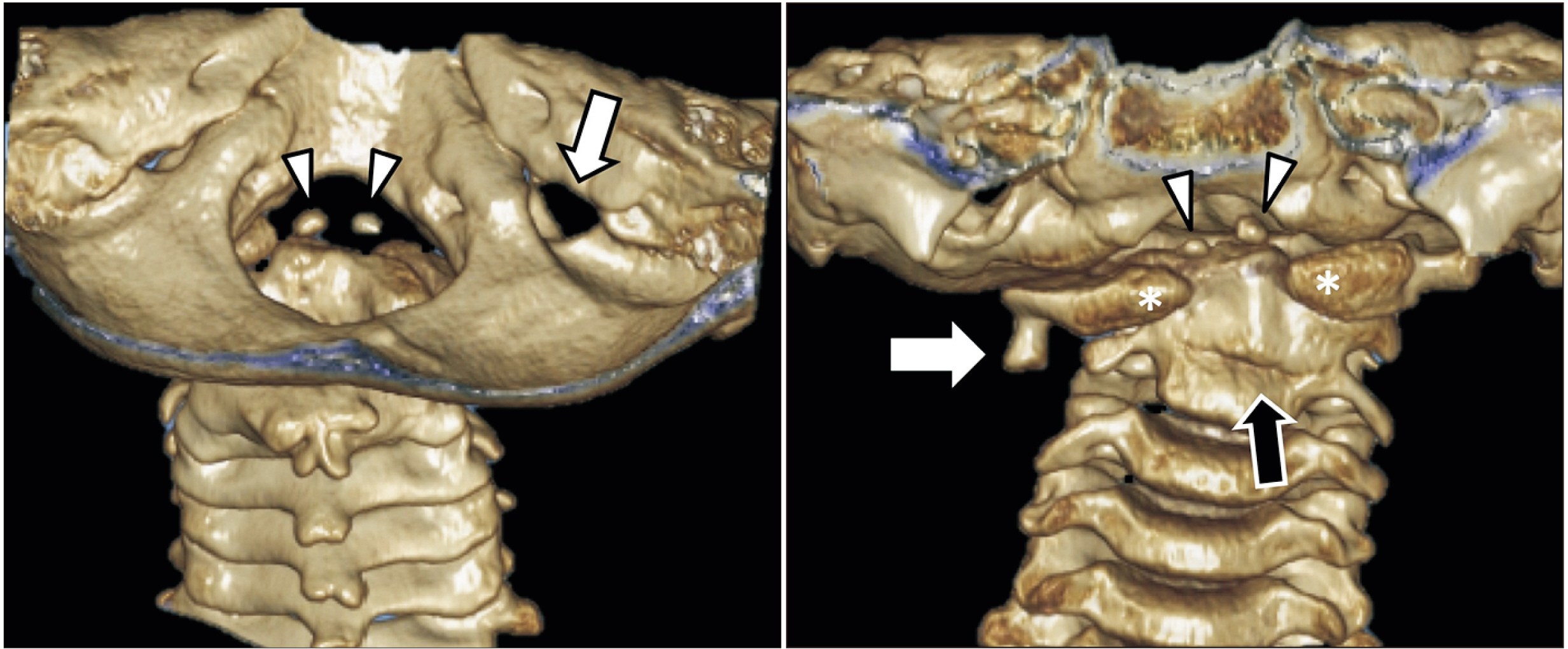
Fig. 1 Three-dimensional reconstructed computed tomography of the craniocervical junction in the patient presented herein. Note the two ossification centers (arrow heads) of the duplicated odontoid process via a posterior view through the foramen magnum and from a posterior view (left). On this same image, the right jugular foramen (arrow) was significantly enlarged compared to the left. From an anterior view (right), note again the two ossification centers for the apical parts of the odontoid processes (arrow heads), the split anterior arch of C1 (asterisks), and the fused C2 and C3 vertebrae (black arrow). Also, note the right-sided inferior bony extension from the transverse process of C1 (white arrow).
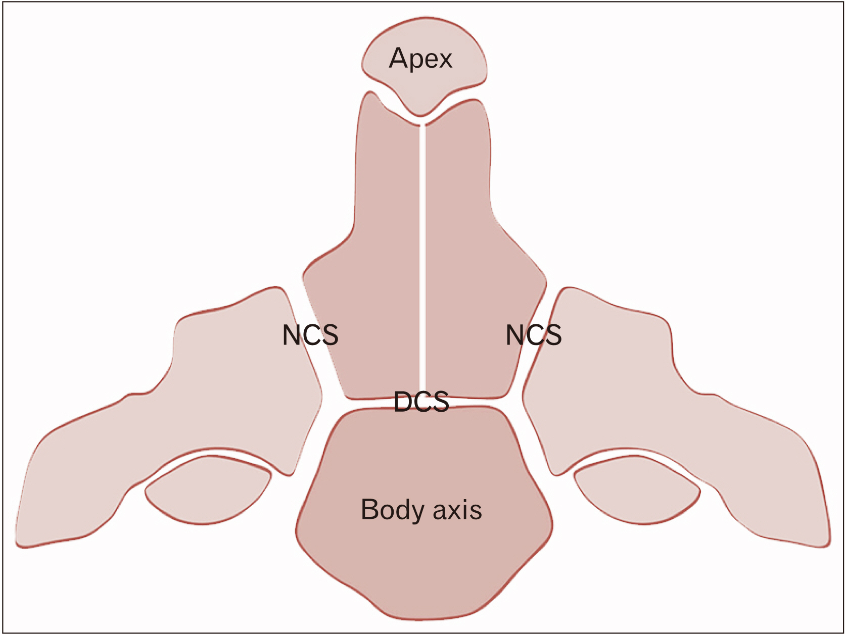
Fig. 2 Schematic drawing of the various ossification centers of the axis. Note the DCS between the body and odontoid process and the NCS separating the odontoid process and body of the axis from the remaining parts of C2. Smaller sits of fusion include the synchondrosis between the apex of the odontoid process and its lower parts and the midline fusion of left and right sides of the odontoid process. DCS, dentocentral synchondrosis; NCS, neurocentral synchondrosis.

Fig. 3 Schematic drawing of the embryological derivations of the craniocervical junction. Note the early sclerotome contributions to this region. The first two occipital somites (yellow) give rise to the regions of the clivus at the skullbase and the third occipital somite (green) gives rise to parts of the occipital bone and region of jugular foramen. The proatlas (fourth occipital somite) gives rise to the apical region of the odontoid process, occipital condyle, and lateral mass of the atlas. The first cervical sclerotome gives rise to the anterior and posterior arches of the atlas, and the body of the odontoid process.
Reference
-
References
1. Garant M, Oudjhane K, Sinsky A, O'Gorman AM. 1997; Duplicated odontoid process: plain radiographic and CT appearance of a rare congenital anomaly of the cervical spine. AJNR Am J Neuroradiol. 18:1719–20. PMID: 9367321.2. Dilettoso S, Uccello M, Dilettoso A, Gelardi S, Dilettoso B. 2012; Duplicated odontoid process and atlas clefts associated to Klippel-Feil syndrome. Spine J. 12:449–50. DOI: 10.1016/j.spinee.2012.03.031. PMID: 22513073.
Article3. Manjila S, Miller EA, Vadera S, Goel RK, Khan FR, Crowe C, Geertman RT. 2012; Duplication of the pituitary gland associated with multiple blastogenesis defects: duplication of the pituitary gland (DPG)-plus syndrome. Case report and review of literature. Surg Neurol Int. 3:23. DOI: 10.4103/2152-7806.92939. PMID: 22439114. PMCID: PMC3307243.
Article4. Usta Y, Sakha F, White WL, Little AS, Knecht L. 2012; Duplicated pituitary gland and odontoid process. A case report. Neuroradiol J. 25:360–3. DOI: 10.1177/197140091202500312. PMID: 24028990.5. Akobo S, Rizk E, Loukas M, Chapman JR, Oskouian RJ, Tubbs RS. 2015; The odontoid process: a comprehensive review of its anatomy, embryology, and variations. Childs Nerv Syst. 31:2025–34. DOI: 10.1007/s00381-015-2866-4. PMID: 26254085.
Article6. Kaplan KM, Spivak JM, Bendo JA. 2005; Embryology of the spine and associated congenital abnormalities. Spine J. 5:564–76. DOI: 10.1016/j.spinee.2004.10.044. PMID: 16153587.
Article7. Offiah CE, Day E. 2017; The craniocervical junction: embryology, anatomy, biomechanics and imaging in blunt trauma. Insights Imaging. 8:29–47. DOI: 10.1007/s13244-016-0530-5. PMID: 27815845. PMCID: PMC5265194.
Article8. Bambakidis NC, Dickman CA, Spetzler RF, Sonntag VKH. 2012. Surgery of the craniovertebral junction. 2nd ed. Thieme;New York: DOI: 10.1055/b-0034-84439.9. Muhleman M, Charran O, Matusz P, Shoja MM, Tubbs RS, Loukas M. 2012; The proatlas: a comprehensive review with clinical implications. Childs Nerv Syst. 28:349–56. DOI: 10.1007/s00381-012-1698-8. PMID: 22282080.
Article10. Sureisen M, Achannan R, Chong KC, Wong CC. 2015; What the mind does not know, the eyes do not see: a rare congenital fusion of the odontoid process to the atlantal hemiarch. BMJ Case Rep. 2015:bcr2015212748. DOI: 10.1136/bcr-2015-212748. PMID: 26508120. PMCID: PMC4636696.
Article11. Britton E, Barry M. Aresti M, Barry M, Paterson M, Ramachandran M, editors. 2015. Fractures of the spine in children. Paediatric orthopaedic trauma in clinical practice. Springer;London: p. 57–70. DOI: 10.1007/978-1-4471-6756-3_4.
Article12. Bonneville F, Jacamon M, Runge M, Jacquet G, Bonneville JF. 2004; Split atlas in a patient with odontoid fracture. Neuroradiology. 46:450–2. DOI: 10.1007/s00234-004-1189-z. PMID: 15105979.
Article13. O'Brien WT Sr, Shen P, Lee P. 2015; The dens: normal development, developmental variants and anomalies, and traumatic injuries. J Clin Imaging Sci. 5:38. DOI: 10.4103/2156-7514.159565. PMID: 26199787. PMCID: PMC4498315.14. Jain N, Verma R, Garga UC, Baruah BP, Jain SK, Bhaskar SN. 2016; CT and MR imaging of odontoid abnormalities: a pictorial review. Indian J Radiol Imaging. 26:108–19. DOI: 10.4103/0971-3026.178358. PMID: 27081234. PMCID: PMC4813060.
Article
- Full Text Links
-
- Actions
-
Cited
- CITED
-
- Close
- Share
-
- Similar articles
-
- Congenital Agenesis of Odonteid Process: A Case Report
- Congenital Hypoplasia of the Posterior Arch of the Atlas Associated with a Fracture of the Odontoid Process: A Case Report
- Anterior Craniocervical Junctional Neurenteric Cyst
- Atlanto-axial Dislocation with the Fracture of the Odontoid Process: A Case Report
- Unilateral Fusion of the Odontoid Process with the Atlas in Klippel-Feil syndrome: A Case Report
