Anterior Craniocervical Junctional Neurenteric Cyst
- Affiliations
-
- 1Department of Neurosurgery, CHA Bundang Medical Center, CHA University College of Medicine, Seongnam, Korea
- KMID: 2522228
- DOI: http://doi.org/10.14791/btrt.2021.9.e22
Abstract
- Intracranial neurenteric cyst at the anterior craniocervical junction is very rare, and its treatment and prognosis have not been established. We report a case of neurenteric cyst at the anterior craniocervical junction and review the relevant literature. A 16-year-old girl presented with a 2-month history of slowly progressive headache. MRI revealed a well-defined intradural extramedullary cyst in the anterior medulla and brain stem with C1 cord compression. We performed gross total resection of the cyst using a far-lateral transcondylar approach. Surgical resection is the treatment of choice for neurenteric cysts at anterior craniocervical junction, the far-lateral transcondylar approach might be the optimal surgical approach.
Keyword
Figure
-
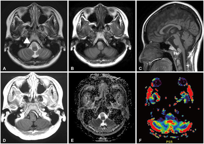
Fig. 1 Preoperative MR images of the craniocervical junction. A: The T2-weighted axial image shows a homogeneously hyperintense cyst (arrowhead) anterior to the medulla. B and C: Pre-gadolinium T1-weighted axial and sagittal images show a homogeneously hyperintense cyst that compresses the brain stem at the anterior craniocervical junction. D: The post-gadolinium T1-weighted axial image shows no enhancement of the cystic lesion. E: Diffusion MR image shows a homogeneously hyperintense cyst without diffusion restriction. F: Perfusion MR images reveal partial uptake of the contrast medium in the anterior part of the cystic lesion.
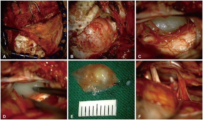
Fig. 2 Intraoperative photographs of neurenteric cyst resection using the far-lateral transcondylar approach. A: The right semispinalis captitis muscle was seen, and the dissection of the superior and inferior oblique muscles was performed. B: To secure the vertebral artery, the groove of the vertebral artery was located and lateral partial suboccipital craniotomy was performed. Condylectomy was performed to optimize the view of the anterior brain stem. C: A well-defined capsulated cyst that adheres to the lower cranial nerve and vertebral artery was seen. Posterior bulging of the medulla and cervical cord were seen. D: The lesion was punctured and filled with yellowish viscous fluid. E: The decompressed and lax cyst was completely resected after dissection of the adhesion, which was approximately 10 mm long. F: The premedullary cistern was subsequently secured. The medulla, cervical cord, low cranial nerve, C1 nerve root, and vertebral artery were intact.
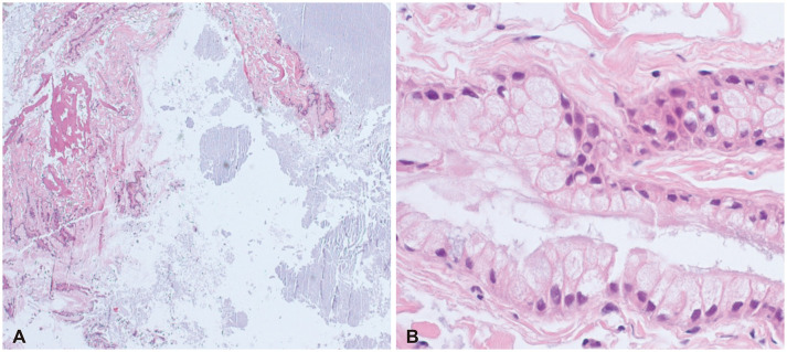
Fig. 3 Histopathologic findings. A: The low-magnification image shows mucin (hematoxylin and eosin stain, original magnification ×40). B: The high-magnification image shows non-ciliated, mucin-producing columnar epithelium lining the cyst wall and basal nuclei (hematoxylin and eosin stain, original magnification ×200).
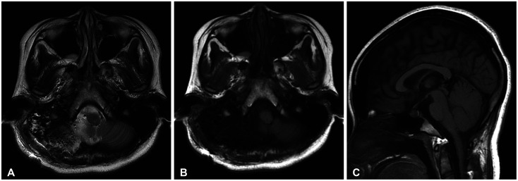
Fig. 4 Postoperative MR images of the craniocervical junction. T2, T1-weighted (A and B) and post-gadolinium T1-weight (C) images demonstrate grossly total resection of the cyst and decompression of the anterior part of the premedullary cistern.
Reference
-
1. Sundaram C, Paul TR, Raju BV, et al. Cysts of the central nervous system: a clinicopathologic study of 145 cases. Neurol India. 2001; 49:237–242. PMID: 11593239.2. Guilburd JN, Arieh YB, Peyser E. Spinal intradural enterogenous cyst: report of a case. Surg Neurol. 1980; 14:359–362. PMID: 7444743.3. Al Qadhi S, Laiq S, Salhotra N, et al. Neurenteric cyst at craniovertebral junction: an uncommon presentation. Am J Med Case Rep. 2021; 9:407–410.4. Prasad GL, Sharma BS, Mahapatra AK. Ventral foramen magnum neurenteric cysts: a case series and review of literature. Neurosurg Rev. 2016; 39:535–544. PMID: 26662045.5. Haque M, Rahman A, Ahmed N, Alam S. Huge ventral cervicomedullary neurenteric cyst: a rare entity with good surgical outcome and appraisal. Asian J Neurosurg. 2020; 15:1016–1019. PMID: 33708680.6. Wang L, Zhang J, Wu Z, et al. Diagnosis and management of adult intracranial neurenteric cysts. Neurosurgery. 2011; 68:44–52. PMID: 21150754.7. Preece MT, Osborn AG, Chin SS, Smirniotopoulos JG. Intracranial neurenteric cysts: imaging and pathology spectrum. AJNR Am J Neuroradiol. 2006; 27:1211–1216. PMID: 16775266.8. Anderson T, Kaufman T, Murtagh R. Intracranial neurenteric cyst: a case report and differential diagnosis of intracranial cystic lesions. Radiol Case Rep. 2020; 15:2649–2654. PMID: 33093931.9. Perry A, Scheithauer BW, Zaias BW, Minassian HV. Aggressive enterogenous cyst with extensive craniospinal spread: case report. Neurosurgery. 1999; 44:401–404. PMID: 9932896.10. Gavrjushin AV, Chelushkin DM. Intra-axial neurenteric cyst of medulla: case report and literature review. Cureus. 2021; 13:e15361. PMID: 34239793.11. Nelson SM, Mathis DA, Hobbs JK, Timpone VM. Intracranial neurenteric cyst mimicking an ependymoma: imaging features, pathologic correlation and review of literature. Clin Imaging. 2017; 44:117–120. PMID: 28505503.12. Chen CT, Lai HY, Jung SM, Lee CY, Wu CT, Lee ST. Neurenteric cyst or neuroendodermal cyst? Immunohistochemical study and pathogenesis. World Neurosurg. 2016; 96:85–90. PMID: 27586176.13. Warf BC. Comparison of endoscopic third ventriculostomy alone and combined with choroid plexus cauterization in infants younger than 1 year of age: a prospective study in 550 African children. J Neurosurg. 2005; 103:475–481. PMID: 16383244.14. Savage JJ, Casey JN, McNeill IT, Sherman JH. Neurenteric cysts of the spine. J Craniovertebr Junction Spine. 2010; 1:58–63. PMID: 20890417.15. Breeze RE, Nichols P, Segal H, Apuzzo ML. Intradural epithelial cyst at the craniovertebral junction. Case report. J Neurosurg. 1990; 73:788–791. PMID: 2213172.16. Ergun R, Akdemir G, Gezici AR, Kara C, Ergungor F. Craniocervical neurenteric cyst without associated abnormalities. Pediatr Neurosurg. 2000; 32:95–99. PMID: 10838509.17. Koksel T, Revesz T, Crockard HA. Craniospinal neurenteric cyst. Br J Neurosurg. 1990; 4:425–428. PMID: 2261106.18. Menezes AH, Ryken TC. Craniocervical intradural neurenteric cysts. Pediatr Neurosurg. 1995; 22:88–95. PMID: 7710978.19. Mehdi W, Niaz A, Irfan M, Tasdique S, Majeed S. Far lateral transcondylar approach for anterior foramen magnum lesions. Pak J Neurol Surg. 2020; 24:149–155.20. Liu JK, Couldwell WT. Far-lateral transcondylar approach: surgical technique and its application in neurenteric cysts of the cervicomedullary junction: report of two cases. Neurosurg Focus. 2005; 19:1–7.21. Menezes AH, Traynelis VC. Spinal neurenteric cysts in the magnetic resonance imaging era. Neurosurgery. 2006; 58:97–105. PMID: 16385333.22. Menezes AH, Dlouhy BJ. Neurenteric cysts at foramen magnum in children: presentation, imaging characteristics, and surgical management—case series and literature review. Childs Nerv Syst. 2020; 36:1379–1384. PMID: 32322975.23. Mazur MD, Couldwell WT, Cutler A, et al. Occipitocervical instability after far-lateral transcondylar surgery: a biomechanical analysis. Neurosurgery. 2017; 80:140–145. PMID: 28362894.24. Gauden AJ, Khurana VG, Tsui AE, Kaye AH. Intracranial neuroenteric cysts: a concise review including an illustrative patient. J Clin Neurosci. 2012; 19:352–359. PMID: 22260959.
