Anat Cell Biol.
2020 Dec;53(4):502-504. 10.5115/acb.20.062.
The terminal ventricle of Saguinus leucopus (Primate)
- Affiliations
-
- 1Department of Basic Sciences, Faculty of Health Sciences, Universidad de Caldas, Manizales, Colombia
- 2Medicine Program, Department of Basic Sciences, Universidad de Manizales, Manizales, Colombia
- 3Department of Animal Health, Faculty of Agricultural Sciences, Universidad de Caldas, Manizales, Colombia
- 4Department of Animal Health, Faculty of Veterinary Medicine and Zootechnics, Universidad del Tolima, Ibagué, Colombia
- KMID: 2509696
- DOI: http://doi.org/10.5115/acb.20.062
Abstract
- The terminal ventricle is a dilation of the ventricular system located within the spinal cord, which is enveloped in ependymal cells that are involved in the dynamic of the cerebrospinal liquid. In the present study, four Saguinus leucopus specimens were dissected, two males and two females, whose spinal cords were extracted and histologically processed via hematoxylin and eosin stains of cuts at the conus medullaris. The S. leucopus’ terminal ventricle was observed at the conus medullaris, and had an average diameter of 241.38 μm. Thus, the presence of the terminal ventricle in the S. leucopus at the level of the conus medullaris was established.
Keyword
Figure
-
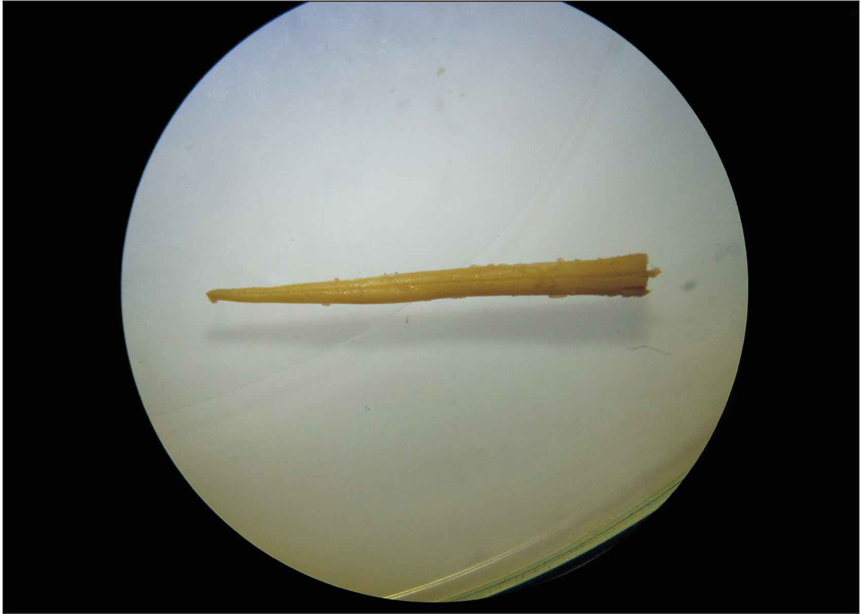
Fig. 1 Image taken with a stereoscope, in which the narrow form of the conus medullaris of Saguinus leucopus can be observed.
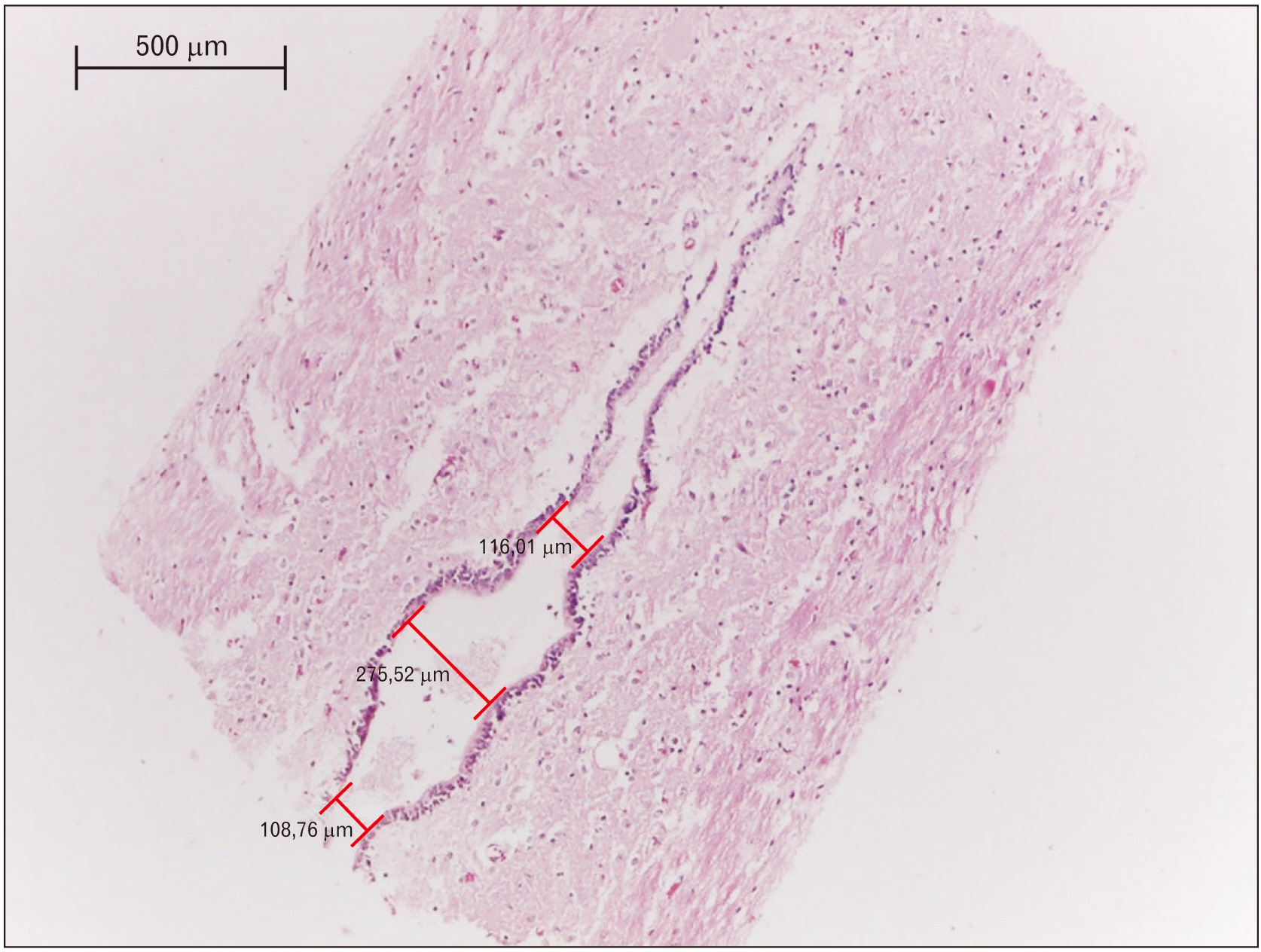
Fig. 2 Histological sagittal section of the conus medullaris of Saguinus leucopus. The terminal ventricle and the central canal are covered with ependymal cell.
Reference
-
References
1. Duque-Parra JE, Barco-Ríos J, García-Aguirre JF. 2017; A historical approach to the ventricular system of the brain. Rev Fac Med. 65:473–7. DOI: 10.15446/revfacmed.v65n3.57884.
Article2. Woodley-Cook J, Konieczny M, Spears J. 2016; The slowly enlarging ventriculus terminalis. Pol J Radiol. 81:529–31. DOI: 10.12659/PJR.895669. PMID: 27867442. PMCID: PMC5102252.
Article3. Liccardo G, Ruggeri F, De Cerchio L, Floris R, Lunardi P. 2005; Fifth ventricle: an unusual cystic lesion of the conus medullaris. Spinal Cord. 43:381–4. DOI: 10.1038/sj.sc.3101712. PMID: 15655569.
Article4. Vigh B, Vigh-Teichmann I, Manzano e Silva MJ, van den Pol AN. 1983; Cerebrospinal fluid-contacting neurons of the central canal and terminal ventricle in various vertebrates. Cell Tissue Res. 231:615–21. DOI: 10.1007/BF00218119. PMID: 6871973.
Article5. Uehara M, Ueshima T. 1985; Light and electron microscopy of the chicken coccygeal cord. Nihon Juigaku Zasshi. 47:963–70. DOI: 10.1292/jvms1939.47.963. PMID: 4094279.
Article6. Storer KP, Toh J, Stoodley MA, Jones NR. 1998; The central canal of the human spinal cord: a computerised 3-D study. J Anat. 192(Pt 4):565–72. DOI: 10.1046/j.1469-7580.1998.19240565.x. PMID: 9723983. PMCID: PMC1467810.
Article7. Fitzgerald P. 1967; The fifth ventricle. Ir J Med Sci. 6:133–6. DOI: 10.1007/BF02954269. PMID: 6042012.
Article8. Marín-García P, González-Soriano J, Martinez-Sainz P, Contreras-Rodríguez J, Del Corral-Gros C, Rodríguez-Veiga E. 1995; Spinal cord central canal of the German shepherd dog: morphological, histological, and ultrastructural considerations. J Morphol. 224:205–12. DOI: 10.1002/jmor.1052240209. PMID: 7745605.
Article9. Fletcher TF. Evans H, de Lahunta A, editors. 2013. Spinal cord and meninges. Miller's anatomy of the dog. 4th ed. Elsevier;Louis: p. 589–610.10. Sakata M, Yashika K, Hashimoto PH. 1993; Caudal aperture of the central canal at the filum terminale in primates. Kaibogaku Zasshi. 68:213–9. PMID: 8337935.11. Coleman LT, Zimmerman RA, Rorke LB. 1995; Ventriculus terminalis of the conus medullaris: MR findings in children. AJNR Am J Neuroradiol. 16:1421–6. PMID: 7484626.12. Lotfinia I, Mahdkhah A. 2018; The cystic dilation of ventriculus terminalis with neurological symptoms: three case reports and a literature review. J Spinal Cord Med. 41:741–7. DOI: 10.1080/10790268.2018.1474680. PMID: 29791269. PMCID: PMC6217512.
Article13. Suh SH, Chung TS, Lee SK, Cho YE, Kim KS. 2012; Ventriculus terminalis in adults: unusual magnetic resonance imaging features and review of the literature. Korean J Radiol. 13:557–63. DOI: 10.3348/kjr.2012.13.5.557. PMID: 22977322. PMCID: PMC3435852.
Article14. Rossi A, Biancheri R, Cama A, Piatelli G, Ravegnani M, Tortori-Donati P. 2004; Imaging in spine and spinal cord malformations. Eur J Radiol. 50:177–200. DOI: 10.1016/j.ejrad.2003.10.015. PMID: 15081131.
Article15. Nassar SI, Correll JW, Housepian EM. 1968; Intramedullary cystic lesions of the conus medullaris. J Neurol Neurosurg Psychiatry. 31:106–9. DOI: 10.1136/jnnp.31.2.106. PMID: 5684018. PMCID: PMC496312.
Article16. Nayak SB, Shetty SD. 2019; Partial duplication of tentorium cerebelli and complete duplication of falx cerebelli. Anat Cell Biol. 52:337–9. DOI: 10.5115/acb.19.017. PMID: 31598364. PMCID: PMC6773897.
Article17. Sigal R, Denys A, Halimi P, Shapeero L, Doyon D, Boudghène F. 1991; Ventriculus terminalis of the conus medullaris: MR imaging in four patients with congenital dilatation. AJNR Am J Neuroradiol. 12:733–7. PMID: 1882755.18. Bogin B, Varela-Silva MI. 2010; Leg length, body proportion, and health: a review with a note on beauty. Int J Environ Res Public Health. 7:1047–75. DOI: 10.3390/ijerph7031047. PMID: 20617018. PMCID: PMC2872302.
Article19. Castañeda FE, Buritica EF, Barbosa IX. 2010; White-footed tamarin Saguinus leucopus GUNTHER 1876: some biological aspects and issues of veterinary interest about the species. Rev Colombiana Cienc Anim. 3:81–9.
- Full Text Links
-
- Actions
-
Cited
- CITED
-
- Close
- Share
-
- Similar articles
-
- PrimateDB: Development of Primate Genome DB and Web Service
- Periodic Explosive Expansion of Human Retroelements Associated with the Evolution of the Hominoid Primate
- Establishment of a Research and Assessment System Using High Quality Non-Human Primates
- Genomic Features of Retroelements and Implications for Human Disease
- Glioblastoma Multiforme in the Trigone of the Lateral Ventricle: A Case Report
