Anat Cell Biol.
2020 Dec;53(4):493-501. 10.5115/acb.20.173.
Comparative evaluation of the ultrastructural morphology and distribution of filiform and fungiform tongue papillae in Egyptian mice, fruit bats and long-eared hedgehogs
- Affiliations
-
- 1Department of Oral Biology, Faculty of Dentistry, Cairo University, Cairo, Egypt
- 2Department of Oral Biology, Faculty of Oral and Dental Medicine, Suez Canal University, Ismaelia, Egypt
- 3Department of Oral Biology, Faculty of Oral and Dental Medicine, Ahram Canadian University, Cairo, Egypt
- KMID: 2509695
- DOI: http://doi.org/10.5115/acb.20.173
Abstract
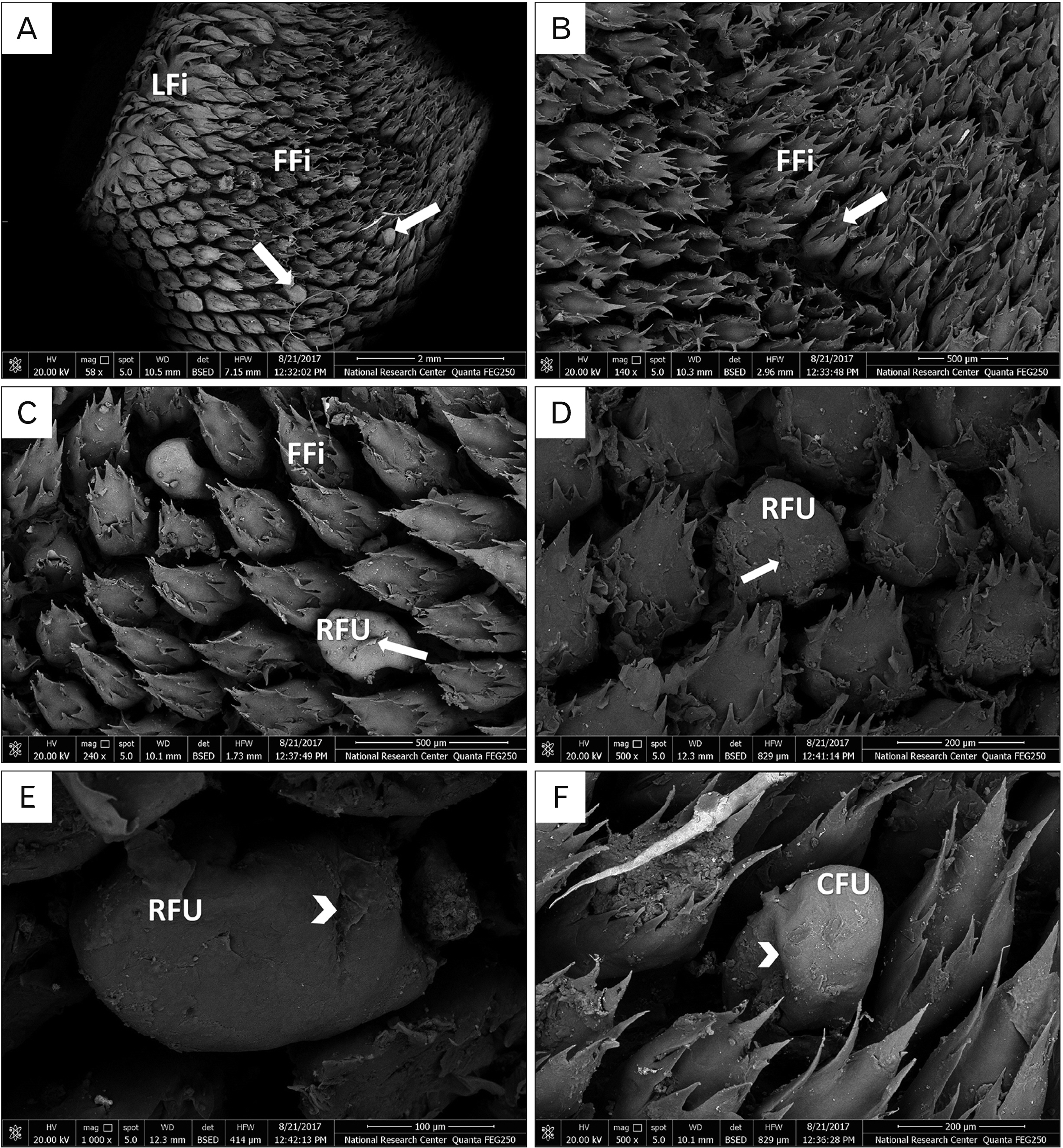
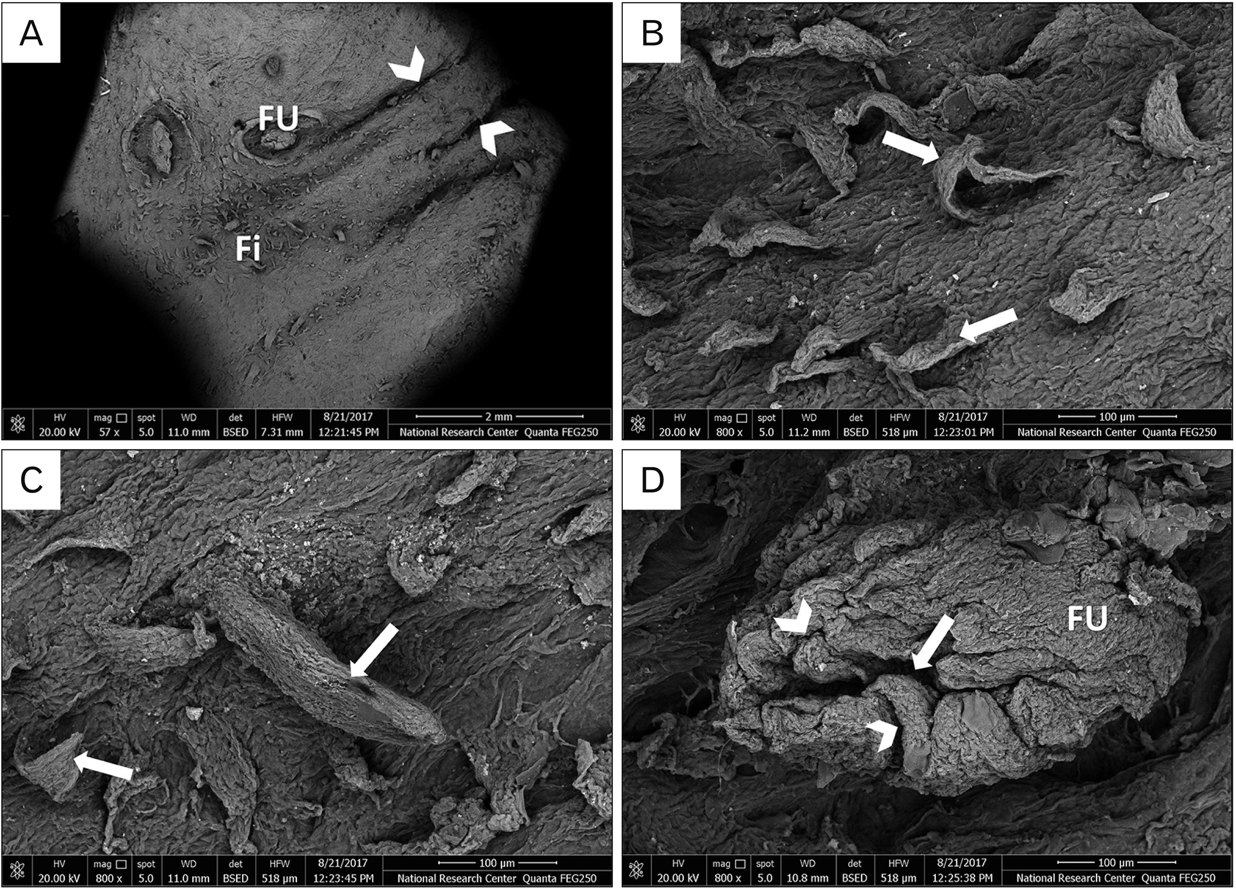
- The tongue is a specialized vital organ. It aids in mastication, deglutition and food digestion. It also shares in the perception of taste sensation as it possesses various gustatory papillae. It is being subjected to numerous anatomical and histological examinations aiming at exploring the correlation between its morphological features and animal adaptations to various types of nutrition and environmental conditions. The goal of the present work was to compare the ultrastructural features of the filiform and fungiform papillae of three various mammals possessing different feeding habits; Egyptian mice, fruit bats and long-eared hedgehogs. Specimens were obtained from the tongues of four healthy adult animals from each mammalian type. Tongues were fixed and all the appropriate procedures were done to perform scanning electron microscopic investigation. Scanning electron microscopic examination demonstrated that in mice, there were four different sub-types of filiform papillae (spike, leaf, conical and tongue-shaped). In bats, there were two sub-types (flower and leaf-like) and in hedgehogs, there was only one type (tongue-like). These filiform papillae showed different distribution and orientation. As for the fungiform papillae, they were cylindrical in mice, rounded or conical in bats and dome-shaped in hedgehogs. Fungiform papillae possessed taste pores containing taste buds. Ultrastructural variations of the filiform and fungiform papillae were suggested to be probably due to adaptation to various feeding habits and different environmental conditions of these animals.
Keyword
Figure
Reference
-
References
1. Hussein AJ, Al-Asadi FS. 2010; Histological, anatomical and embryonical study of fungiform papillae in tongue of Iraqi sheep. Basrah J Vet Res. 9:78–89. DOI: 10.33762/bvetr.2010.55080.2. Kulawik M, Nienartowicz-Zdrojewska A. 2006; The mucous membrane on the ventral surface of the apex and on the lateral surfaces of the body of the tongue in the raccoon dog (Nyctereutes procyonoides). Acta Sci Pol Med Vet. 5:67–73.3. Darwish ST. 2012; Comparative histological and ultrastructural study of the tongue in Ptyodactylus guttatus and Stenodactylus petrii (Lacertilia, Gekkonidae). J Am Sci. 8:603–12.4. Herrel A, Canbek M, Ozelmas U, Uyanoğlu M, Karakaya M. 2005; Comparative functional analysis of the hyolingual anatomy in lacertid lizards. Anat Rec A Discov Mol Cell Evol Biol. 284:561–73. DOI: 10.1002/ar.a.20195. PMID: 15880434.
Article5. Iwasaki S. 2002; Evolution of the structure and function of the vertebrate tongue. J Anat. 201:1–13. DOI: 10.1046/j.1469-7580.2002.00073.x. PMID: 12171472. PMCID: PMC1570891.
Article6. Carlesso Santos T, Yuri Fukuda K, Plácido Guimarães J, Franco Oliveira M, Angelica Miglino M, Watanabe LS. 2011; Light and scanning electron microcopy study of the tongue in Rhea americana. Zoolog Sci. 28:41–6. DOI: 10.2108/zsj.28.41. PMID: 21186946.7. Abd Al-Rhman SA, Al-Fartwsy AR, Al-Shuaily EH. 2016; Morphohistological study of the tongue in local mice species by using special stain. J Am Sci. 12:13–20.8. Jung HS, Akita K, Kim JY. 2004; Spacing patterns on tongue surface-gustatory papilla. Int J Dev Biol. 48:157–61. DOI: 10.1387/ijdb.15272380. PMID: 15272380.
Article9. Okada S, Schraufnagel DE. 2005; Scanning electron microscopic structure of the lingual papillae of the common opossum (Didelphis marsupialis). Microsc Microanal. 11:319–32. DOI: 10.1017/S1431927605050257. PMID: 16079016.
Article10. Kobayashi K, Miyata K, Takahashi K, Iwasaki S. 1989; [Three-dimensional architecture of the connective tissue papillae of the mouse tongue as viewed by scanning electron microscopy]. Kaibogaku Zasshi. 64:523–38. Japanese. PMID: 2634898.11. Shiels AB, Flores CA, Khamsing A, Krushelnycky PD, Mosher SM, Drake DR. 2013; Dietary niche differentiation among three species of invasive rodents (Rattus rattus, R. exulans, Mus musculus). Biol Invasions. 15:1037–48. DOI: 10.1007/s10530-012-0348-0.12. Marshall AG. 1983; Bats, flowers and fruit: evolutionary relationships in the Old World. Biol J Linn Soc Lond. 20:115–35. DOI: 10.1111/j.1095-8312.1983.tb01593.x.
Article13. Adams RA, Carter RT. 2017; Megachiropteran bats profoundly unique from microchiropterans in climbing and walking locomotion: evolutionary implications. PLoS One. 12:e0185634. DOI: 10.1371/journal.pone.0185634. PMID: 28957404. PMCID: PMC5619802.
Article14. Abumandour MM, El-Bakary RM. 2013; Morphological and scanning electron microscopic studies of the tongue of the Egyptian fruit bat (Rousettus aegyptiacus) and their lingual adaptation for its feeding habits. Vet Res Commun. 37:229–38. DOI: 10.1007/s11259-013-9567-9. PMID: 23709139.
Article15. Jabbar AI. 2014; Anatomical and histological study of tongue in the hedgehog (Hemiechinus auritus). Int J Recent Sci Res. 5:760–3.16. Massoud D, Lao-Pérez M, Hurtado A, Abdo W, Palomino-Morales R, Carmona FD, Burgos M, Jiménez R, Barrionuevo FJ. 2018; Germ cell desquamation-based testis regression in a seasonal breeder, the Egyptian long-eared hedgehog, Hemiechinus auritus. PLoS One. 13:e0204851. DOI: 10.1371/journal.pone.0204851. PMID: 30286149. PMCID: PMC6171879.
Article17. Kingdon J, Happold D, Hoffmann M, Butynski T, Happold M, Kalina J. 2013. Mammals of Africa. Bloomsbury;London:18. Qumsiyeh MB. 1996. Mammals of the holy land. Texas Tech University Press;Lubbock: p. 64.19. Nijman V, Bergin D. 2015; Trade in hedgehogs (Mammalia: Erinaceidae) in Morocco, with an overview of their trade for medicinal purposes throughout Africa and Eurasia. J Threat Taxa. 7:7131–7. DOI: 10.11609/JoTT.o4271.7131-7.
Article20. Pastor JF, Barbosa M, De Paz FJ. 2008; Morphological study of the lingual papillae of the giant panda (Ailuropoda melanoleuca) by scanning electron microscopy. J Anat. 212:99–105. DOI: 10.1111/j.1469-7580.2008.00850.x. PMID: 18254792. PMCID: PMC2408975.
Article21. Jackowiak H, Godynicki S. 2005; The distribution and structure of the lingual papillae on the tongue of the bank vole Clethrinomys glareolus. Folia Morphol (Warsz). 64:326–33. PMID: 16425161.22. Abumandour MMA, El-Bakary RMA. 2013; Anatomic reference for morphological and scanning electron microscopic studies of the New Zealand white rabbits tongue (Orycotolagus cuniculus) and their lingual adaptation for feeding habits. J Morphol Sci. 30:254–65.23. Shindo J, Yoshimura K, Kobayashi K. 2006; Comparative morphological study on the stereo-structure of the lingual papillae and their connective tissue cores of the American beaver (Castor canadensis). Okajimas Folia Anat Jpn. 82:127–37. DOI: 10.2535/ofaj.82.127. PMID: 16526571.
Article24. Iwasaki S, Yoshizawa H, Kawahara I. 1997; Study by scanning electron microscopy of the morphogenesis of three types of lingual papilla in the rat. Anat Rec. 247:528–41. DOI: 10.1002/(SICI)1097-0185(199704)247:4<528::AID-AR12>3.0.CO;2-R. PMID: 9096793.
Article25. El-Nahass EEM. 2019; The morphological development of lingual papillae at prenatal, postnatal, young and adult stages of white albino mouse. J Histol Histopathol. 6:6. DOI: 10.7243/2055-091X-6-6.
Article26. Jackowiak H, Trzcielińska-Lorych J, Godynicki S. 2009; The microstructure of lingual papillae in the Egyptian fruit bat (Rousettus aegyptiacus) as observed by light microscopy and scanning electron microscopy. Arch Histol Cytol. 72:13–21. DOI: 10.1679/aohc.72.13. PMID: 19789409.
Article27. Gregorin R. 2003; Comparative morphology of the tongue in free-tailed bats (Chiroptera, Molossidae). Iheringia Sér Zool. 93:213–21. DOI: 10.1590/S0073-47212003000200014.
Article28. Mqokeli BR, Downs CT. 2013; Palatal and lingual adaptations for frugivory and nectarivory in the Wahlberg's epauletted fruit bat (Epomophorus wahlbergi). Zoomorphology. 132:111–9. DOI: 10.1007/s00435-012-0170-3.29. Massoud D, Abumandour MMA. 2020; Anatomical features of the tongue of two chiropterans endemic in the Egyptian fauna; the Egyptian fruit bat (Rousettus aegyptiacus) and insectivorous bat (Pipistrellus kuhlii). Acta Histochem. 122:151503. DOI: 10.1016/j.acthis.2020.151503. PMID: 31955907.
Article30. Goodarzi N, Azarhoosh M. 2016; Morpholoical study of the Brandt's hedgehog, Paraechinus hypomelas (Eulipotyphla, Erinaceidae), tongue. Vestn Zool. 50:457–66. DOI: 10.1515/vzoo-2016-0052.
Article31. Massoud D, Abumandour MMA. 2019; Descriptive studies on the tongue of two micro-mammals inhabiting the Egyptian fauna; the Nile grass rat (Arvicanthis niloticus) and the Egyptian long-eared hedgehog (Hemiechinus auritus). Microsc Res Tech. 82:1584–92. DOI: 10.1002/jemt.23324. PMID: 31225934.32. Iwasaki S, Miyata K, Kobayashi K. 1987; Comparative studies of the dorsal surface of the tongue in three mammalian species by scanning electron microscopy. Acta Anat (Basel). 128:140–6. DOI: 10.1159/000146330. PMID: 3564886.
Article33. Iwasaki S, Miyata K, Kobayashi K. 1987; The surface structure of the dorsal epithelium of tongue in the mouse. Kaibogaku Zasshi. 62:69–76. PMID: 3630624.34. Goździewska-Harłajczuk K, Klećkowska-Nawrot J, Barszcz K, Marycz K, Nawara T, Modlińska K, Stryjek R. 2018; Biological aspects of the tongue morphology of wild-captive WWCPS rats: a histological, histochemical and ultrastructural study. Anat Sci Int. 93:514–32. DOI: 10.1007/s12565-018-0445-y. PMID: 29948977. PMCID: PMC6061249.
Article35. Mustapha OA, Ayoade OE, Ogunbunm TK, Olude MA. 2015; Morphology of the oral cavity of the African giant rat (Cricetomys gambianus, Waterhouse). Bulg J of Vet Med. 18:19–30. DOI: 10.15547/bjvm.793.36. Schondube JE, Herrera-M LG, Martínez del Rio C. 2001; Diet and the evolution of digestion and renal function in phyllostomid bats. Zoology (Jena). 104:59–73. DOI: 10.1078/0944-2006-00007. PMID: 16351819.
Article37. Freeman PW. 1988; Frugivorous and animalivorous bats (Microchiroptera): dental and cranial adaptations. Biol J Linn Soc Lond. 33:249–72. DOI: 10.1111/j.1095-8312.1988.tb00811.x.
Article38. Harper CJ, Swartz SM, Brainerd EL. 2013; Specialized bat tongue is a hemodynamic nectar mop. Proc Natl Acad Sci U S A. 110:8852–7. DOI: 10.1073/pnas.1222726110. PMID: 23650382. PMCID: PMC3670378.
Article39. Aziz SA, Olival KJ, Bumrungsri S, Richards GC, Racey PA. Voigt CC, Kingston T, editors. 2016. The conflict between pteropodid bats and fruit growers: species, legislation and mitigation. Bats in the anthropocene: conservation of bats in a changing world. Springer Open;Cham: p. 377–426. DOI: 10.1007/978-3-319-25220-9_13. PMID: 26645871.
Article40. Muchhala N. 2006; Nectar bat stows huge tongue in its rib cage. Nature. 444:701–2. DOI: 10.1038/444701a. PMID: 17151655.
Article41. Abayomi TA, Ofusori DA, Ayoka OA, Odukoya SA, Omotoso EO, Amegor FO, Ajayi SA, Ojo GB, Oluwayinka OP. 2009; A comparative histological study of the tongue of rat (Rattus Norvegicus), bat (Eidolon Helvum) and pangolin (Manis Tricuspis). Int J Morphol. 27:1111–9. DOI: 10.4067/S0717-95022009000400026.
Article42. Freeman PW. 1995; Nectarivorous feeding mechanisms in bats. Biol J Linn Soc Lond. 56:439–63. DOI: 10.1111/j.1095-8312.1995.tb01104.x.
Article43. Leen N, Novick A. 1969. The world of bats. Holt, Rinehart and Winston;New York:44. Emura S, Hayakawa D, Chen H, Shoumura S, Atoji Y, Wijayanto H. 2002; SEM study on the dorsal lingual surface of the large flying fox, Pteropus vampyrus. Okajimas Folia Anat Jpn. 79:113–9. DOI: 10.2535/ofaj.79.113. PMID: 12484446.
Article45. Emura S, Okumura T, Chen H. 2012; Morphology of the lingual papillae in the Egyptian rousette bat (Rousettus aegyptiacus). Okajimas Folia Anat Jpn. 89:61–6. DOI: 10.2535/ofaj.89.61. PMID: 23429050.
Article46. Ciuccio M, Estecondo S, Casanave EB. 2008; Scanning electron microscopy study of the dorsal surface of the tongue in Zaedyus pichiy (Mammalia, Xenarthra, Dasypodidae). Int J Morphol. 26:13–8. DOI: 10.4067/S0717-95022008000100002.
Article47. Nasr ES. 2012; Surface morphological structure of the tongue of the hedgehog, Hemiechinus Auritus (Insectivora: Erinaceidae). J Am Sci. 8:580–8.48. Abumandour MMA. 2014; Morphological comparison of the filiform papillae of New Zealand white rabbits (Oryctolagus cuniculus) as Domestic Mammals and Egyptian fruit bat (Rousettus aegyptiacus) as wild mammals using scanning electron microscopic specimens. Int J Morphol. 32:1407–17. DOI: 10.4067/S0717-95022014000400045.49. Trzcielińska-Lorych J, Jackowiak H, Skieresz-Szewczyk K, Godynicki S. 2009; Morphology and morphometry of lingual papillae in adult and newborn Egyptian fruit bats (Rousettus aegyptiacus). Anat Histol Embryol. 38:370–6. DOI: 10.1111/j.1439-0264.2009.00956.x. PMID: 19681832.
Article50. Karan M, Yilmaz S, Aydin A. 2011; Morphology of the filiform lingual papillae in porcupine (Hystrix cristata). Anat Histol Embryol. 40:100–3. DOI: 10.1111/j.1439-0264.2010.01045.x. PMID: 21105901.
Article51. Emura S, Tamada A, Hayakawa D, Chen H, Shoumura S. 2001; [SEM study on the dorsal lingual surface of the nutria, Myocastor coypus]. Kaibogaku Zasshi. 76:233–8. Japanese. PMID: 11398355.52. Emura S, Okumura T, Chen H, Shoumura S. 2006; Morphology of the lingual papillae in the raccoon dog and fox. Okajimas Folia Anat Jpn. 83:73–6. DOI: 10.2535/ofaj.83.73. PMID: 17154050.
Article53. Lauga E, Pipe CJ, Le Révérend B. 2016; Sensing in the mouth: a model for filiform papillae as strain amplifiers. Front Phys. 4:35. DOI: 10.3389/fphy.2016.00035.
Article54. Takemura A, Uemura M, Toda I, Fang G, Hikida M, Suwa F. 2009; Morphological study of the lingual papillae in the ferret (Mustela putorius furo). Okajimas Folia Anat Jpn. 86:17–24. DOI: 10.2535/ofaj.86.17. PMID: 19522302.
Article55. Parchami A, Salimi M, Khosravi M. 2018; Tongue structure in the long-eared hedgehog (Hemiechinus auritus): a scanning electron microscopic study. Vet Res Forum. 9:205–9. DOI: 10.30466/vrf.2018.32078. PMID: 30357108. PMCID: PMC6198160.56. Jackowiak H. 2006; Scanning electron microscopy study of the lingual papillae in the European mole (Talpa europea, L., Talpidae). Anat Histol Embryol. 35:190–5. DOI: 10.1111/j.1439-0264.2005.00661.x. PMID: 16677215.
Article57. Sohn WJ, Gwon GJ, An CH, Moon C, Bae YC, Yamamoto H, Lee S, Kim JY. 2011; Morphological evidences in circumvallate papilla and von Ebners' gland development in mice. Anat Cell Biol. 44:274–83. DOI: 10.5115/acb.2011.44.4.274. PMID: 22254156. PMCID: PMC3254881.
Article58. Roper SD. 2009; Parallel processing in mammalian taste buds? Physiol Behav. 97:604–8. DOI: 10.1016/j.physbeh.2009.04.003. PMID: 19371753. PMCID: PMC3717265.
Article59. Kumari A, Yokota Y, Li L, Bradley RM, Mistretta CM. 2018; Species generalization and differences in Hedgehog pathway regulation of fungiform and circumvallate papilla taste function and somatosensation demonstrated with sonidegib. Sci Rep. 8:16150. DOI: 10.1038/s41598-018-34399-3. PMID: 30385780. PMCID: PMC6212413.
Article60. Mistretta CM, Kumari A. 2019; Hedgehog signaling regulates taste organs and oral sensation: distinctive roles in the epithelium, stroma, and innervation. Int J Mol Sci. 20:1341. DOI: 10.3390/ijms20061341. PMID: 30884865. PMCID: PMC6471208.
Article61. Nasr ES, Gamal AM, Elsheikh EH. 2012; Light and scanning electron microscopic study of the dorsal lingual papillae of the rat Arvicanthis niloticus (Muridae, Rodentia). J Am Sci. 8:619–27.
- Full Text Links
- Actions
-
Cited
- CITED
-
- Close
- Share
- Similar articles
-
- Scanning electron microscopic observation of lingual papillae in a Bengal tiger (Panthera tigris tigris)
- A Case of Pigmented Fungiform Papillae of the Tongue
- A Case of Pigmented Fungiform Papillae of the Tongue
- Two Cases of Pigmented Fungiform Papillae of the Tongue
- A Case of Pigmented Fungiform Papillae of the Tongue


