Effect of cypermethrin on the postnatal development of the medulla oblongata and the possible protective role of melatonin in albino rats
- Affiliations
-
- 1Department of Anatomy and Embryology, Faculty of Medicine, Menoufia University, Shebin El‐Kom, Egypt
- KMID: 2509692
- DOI: http://doi.org/10.5115/acb.20.193
Abstract
- Previous studies have shown that cypermethrin (CYP), a broad spectrum pesticide has a teratogenic effect on rat offspring born to an exposed dam with no information on its effect on the development of the brain. To the best of our knowledge, this research is the first attempt to study the postnatal development medulla oblongata of rat offspring exposed to CYP during the perinatal period and the possible neuroprotective role of melatonin. The offspring of treated female rats were organized into control, melatonin (1 mg/kg/day orally); CYP (12 mg/kg/day orally); and CYP/melatonin groups. The mothers received treatments from day 6 of gestation until day 21 after birth. At Postnatal days 7 and 21, the animals were sacrificed and their medulla oblongata was removed and subjected to histological, immunohistochemical, and electron microscopic studies. CYP induced neuronal degeneration by chromatolysis and pyknosis. Nuclear changes, cytoplasmic vacuolation, damage mitochondria, and breakdown of RER were also detected. Reduction of microtubule-associated protein-2 (MAP-2), myelin basic protein (MBP), and oligodendrocyte transcription factor expressions and increment of glial fibrillary acidic protein expression in the medulla oblongata of the developing rats were observed. On the other hand, melatonin led to an obvious improvement of the injured medulla oblongata tissues and ameliorating the damaging effects of CYP. In conclusion, melatonin has protected rats against CYP-induced histopathological and immunohistochemical changes. This may be due to the protection of MAP-2, conservation of MBP, an increment of oligodendrocytes, and alleviation of astrogliosis.
Figure
-

Fig. 1 Representative H&E staining of rat medulla oblongata of control and melatonin aged 7 days: showing medulla neurons appeared small in size, varied in shape and had round nuclei (black arrows). The neuropil contains different neuroglia (O, A, and M) and blood vessels (arrowheads). Medulla oblongata of CYP treated group of the same age shows neurons with central chromatolysis (curved arrows), degenerated neurons and pyknotic nuclei (dashed arrows), dilated blood vessels (black arrowheads) and numerous A. CYP/melatonin treated group resulted in almost normal appearing neuronal cells, except for chromatolysis (curved arrow) and degenerated neurons and pyknotic nuclei (dashed arrow) in some neurons. Increased number of O and deceased of A. Control and melatonin groups aged 21 days, the neurons appear larger (black arrow). Many oligodendrocytes (arrows with rounded end) are observed in the neuropil. CYP treated group show chromatolysis of almost all neurons (curved arrows), a large number of A and v. Co-administration of CYP/melatonin shows more or less normal neurons (black arrow). However, some degenerated cells (curved arrow) with loss of nuclear details could be detected. Few A and more O could be observed. A, astrocyte; CYP, cypermethrin; M, microglia; O, oligodendrocyte; PND, postnatal day; v, vacuolated neuropil. Scale bar=20 μm.
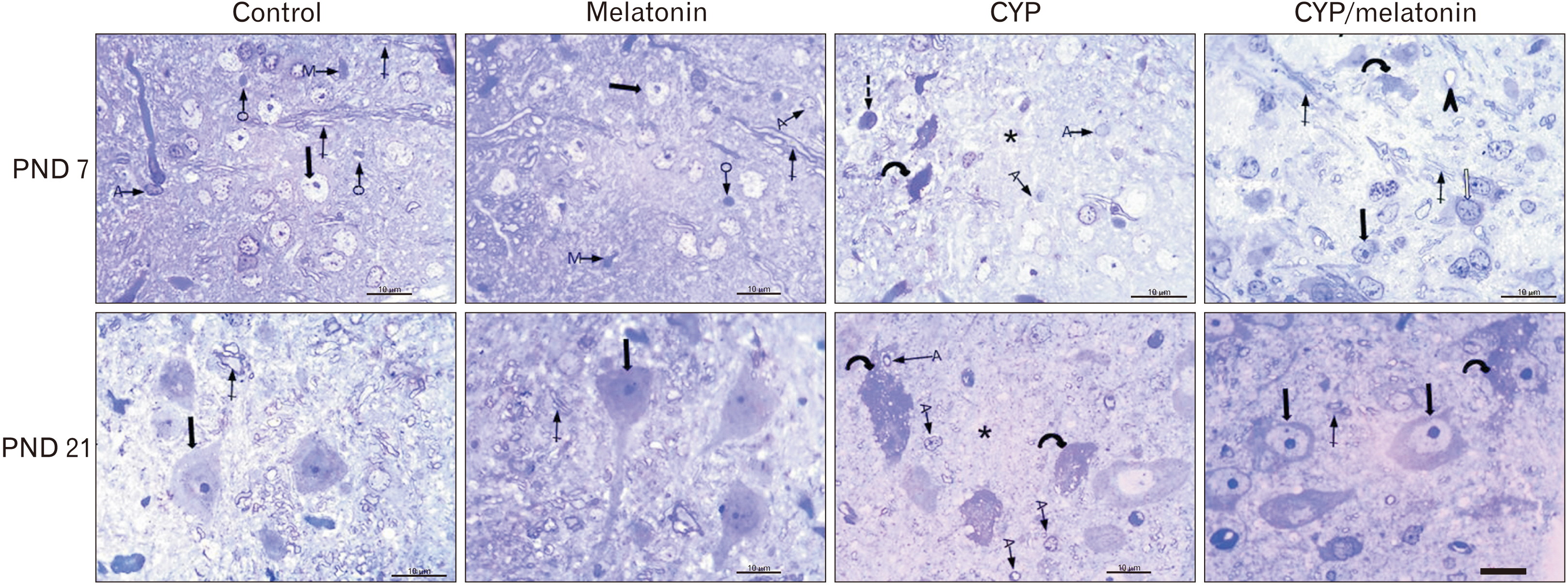
Fig. 2 Representative semi-thin blue-stained sections of the rat medulla oblongata of control and melatonin aged 7 days: showing normal neurons with vesicular nuclei and prominent nucleoli (black arrow), different neuroglia (A, O, and M) and myelinated axons (crossed arrows). CYP treated group of the same age showing some shrunken neuronal bodies with an irregular outline, hardly identified nuclei, and deeply stained vacuolated cytoplasm (dashed arrow). Some neurons appear to have deeply stained nuclei and cytoplasm (curved arrow)., Numerous fine vacuoles were detected in neurons, numerous A, and neuropil devoid of myelinated axons (star). CYP/melatonin treated group shows nearly normal neurons with prominent nucleoli (black arrow), neurons containing coarse clumps of heterochromatin (white arrow), myelinated axons (crossed arrow). Darkly stained neurons (curved arrow) and dilated blood vessels are still observed. Sections of the control and melatonin groups aged 21 days showing large multipolar neurons with large nucleus & prominent nucleolus (black arrow). The neuron is surrounded by dense neuropil containing more myelinated axons (crossed arrow). CYP treated group of the same age shows neurons with numerous fine vacuoles (curved arrows), a large number of A, and neuropil devoid of myelinated axons (star). CYP/melatonin treated group shows neurons with pale vesicular nuclei with prominent nucleoli (black arrows) and myelinated axons in the surrounding neuropil. However, few darkly stained vacuolated neurons are detected. A, astrocyte; CYP, cypermethrin; M, microglia; O, oligodendrocyte; PND, postnatal day. Scale bar=10 μm.

Fig. 3 An electron micrograph of a section in control and melatonin rat medulla oblongata aged 7 days shows motor neurons having rounded euchromatic N with regular nuclear membranes (arrow) and prominent Nu. The homogenous cytoplasm contains normal m and rough endoplasmic R. Myelinated f with preserved myelin sheath are also observed. CYP treated group of the same age displays an irregular N surrounded by an indented nuclear membrane (arrows). The cytoplasm contains dilated cisternae of rER (R) and dilated m with destructed cristae (m). Few myelinated f could be detected. CYP/melatonin treated group shows an apparently normal nerve cell with a regular N but the N still shows heterochromatin. The cytoplasm contains normal more or less normal RER (R) and m. However, a small area of V could be still detected in the cytoplasm. A section of the control and melatonin group aged 21 days shows a large neuron with a large N, the chromatin inside the N is euchromatin mainly and less amount of heterochromatin. The cytoplasm of the neuron contains large m, G, and multiple free ribosomes (arrow). The neuron is surrounded by many myelinated axons (f). CYP treated group of the same age displays markedly affected neurons. The N is shrunken, darkly electron-dense, and irregular in shape with indentation of its nuclear membrane (arrows). Many dispersed irregularly shaped chromatin aggregates are demonstrated throughout the N. The cell has an irregular outline with a shrinkage cytoplasm containing multiple ballooned m with disrupted cristae and dilated rER (R) and G. Medulla oblongata sections of CYP/melatonin treated group of the same age exhibit a euchromatic N, fine dispersed chromatin, and prominent n. More or less normal rER (R) and myelinated axons (f) could be detected. However, mild irregular N (arrow) and ballooned m are still detected (transmission electron microscope, ×17,500, Scale bar=1 μm). CYP, cypermethrin; f, fibers; G, Golgi apparatus; m, mitochondria; N, nucleus; n, nucleolus; Nu, nucleoli; PND, postnatal day; R, reticulum; V, vacuolation.

Fig. 4 Expression of MAP-2 in the medulla oblongata of the control, melatonin, CYP, CYP/melatonin groups at 7 and 21 PNDs. CYP administration dramatically decreased the expression of MAP-2 in rat medulla oblongata compared to the control group. Melatonin co-treatment significantly upregulated the expression of MAP-2. CYP, cypermethrin; MAP-2, microtubule-associated protein-2; PND, postnatal day. Scale bar=20 µm.
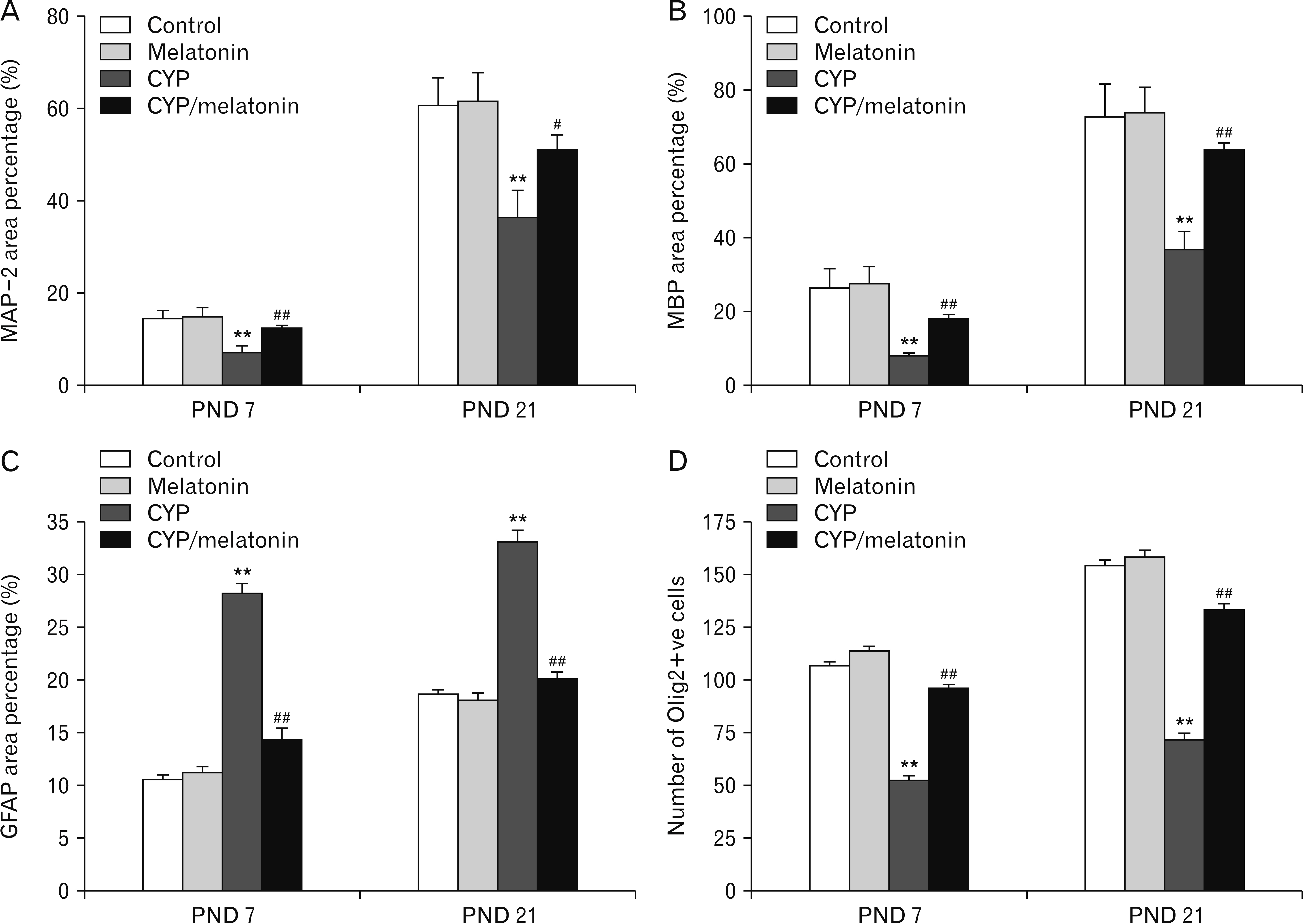
Fig. 5 Area percentage of (A) MAP-2, (B) MBP and (C) GFAP and (D) Olig2+ve cells in the medulla oblongata of the control, melatonin, CYP, CYP/melatonin groups at 7 and 21 PNDs. CYP, cypermethrin; GFAP, glial fibrillary acidic protein; MAP-2, microtubule-associated protein-2; MBP, myelin basic protein; Olig2, oligodendrocyte transcription factor; PND, postnatal day. **P<0.001, compared with the control group; and #P<0.05 and ##P<0.001, compared with the CYP group. data are expressed as means±standard deviation.
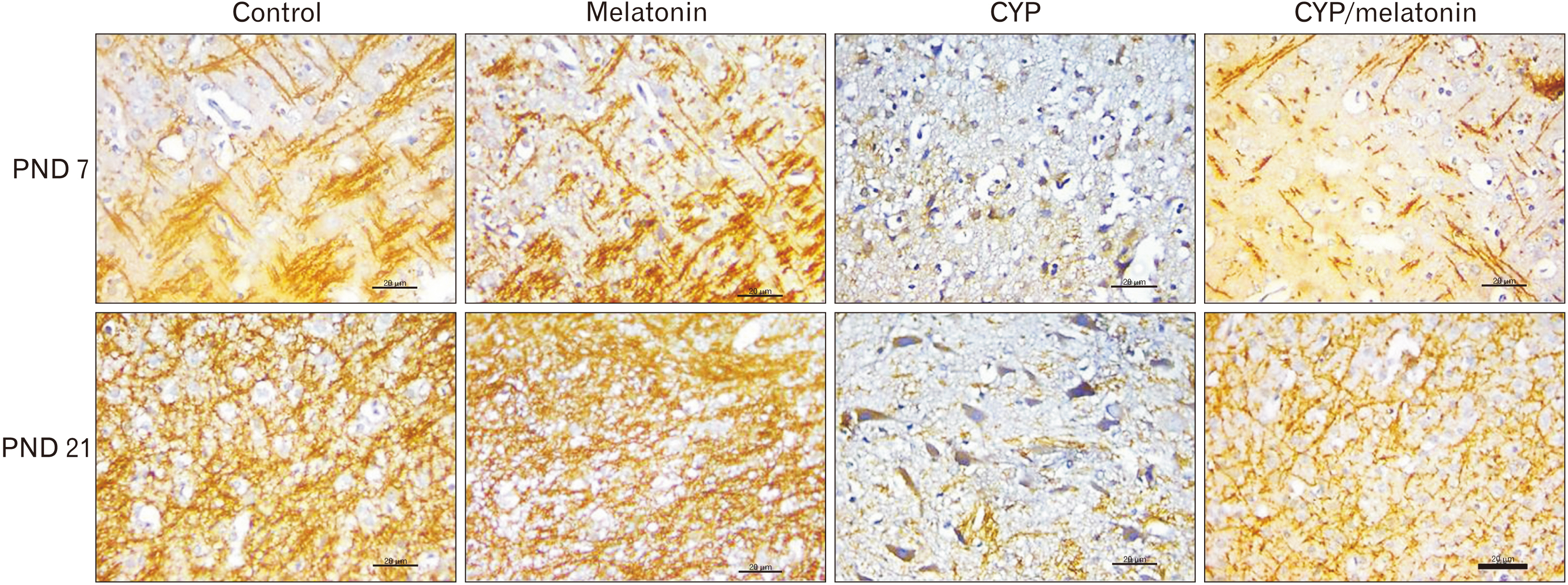
Fig. 6 Expression of MBP in the medulla oblongata of the control, melatonin, CYP, CYP/melatonin groups at 7 and 21 PNDs. CYP administration dramatically downregulated the expression of MBP in rat medulla oblongata compared to the control group. Melatonin co-treatment significantly increased the expression of MBP. CYP, cypermethrin; MBP, myelin basic protein; PND, postnatal day. Scale bar=20 µm.
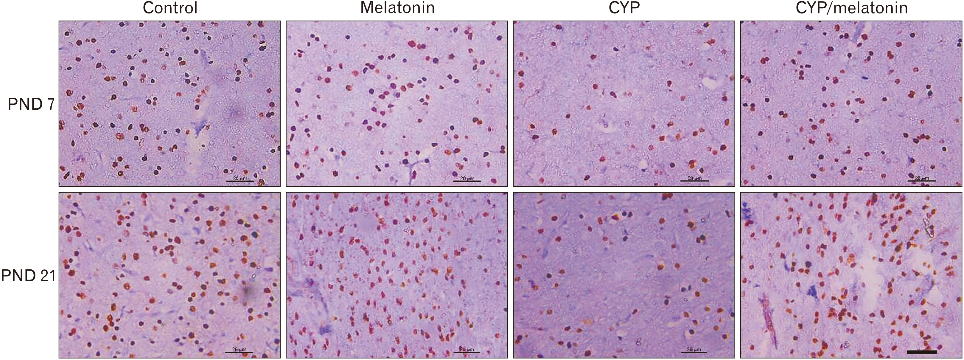
Fig. 7 Expression of Olig2 in the medulla oblongata of the control, melatonin, CYP, CYP/melatonin groups at 7 and 21 PNDs. CYP administration showed a reduction in the expression of Olig2+ve cells in rat medulla oblongata compared to the control group. Melatonin co-treatment increased the expression of Olig2+ve cells. CYP, cypermethrin; Olig2, oligodendrocyte transcription factor; PND, postnatal day. Scale bar=20 µm.
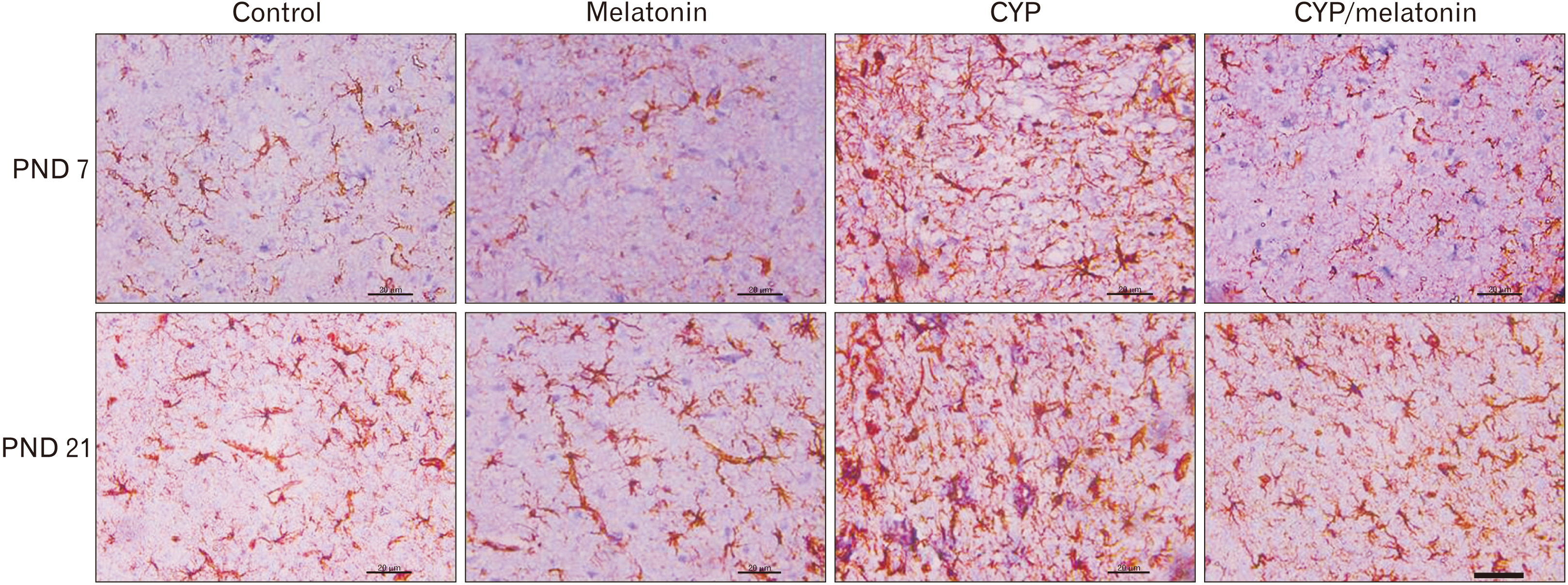
Fig. 8 Expression of GFAP in the medulla oblongata of the control, melatonin, CYP, CYP/melatonin groups at 7 and 21 PNDs. The administration of CYP showed an increase in the expression of GFAP in rat medulla oblongata compared to the control group. Melatonin treatment with CYP reduced the expression of GFAP. CYP, cypermethrin; GFAP, glial fibrillary acidic protein; PND, postnatal day. Scale bar=20 µm.
Reference
-
References
1. Sankar P, Telang AG, Manimaran A. 2011; Effect of piperine on cypermethrin-induced oxidative damage in rats. J Vet Sci Technol. 2:105. DOI: 10.4172/2157-7579.1000105.
Article2. Sankar P, Telang AG, Manimaran A. 2010; Curcumin protects against cypermethrin-induced genotoxicity in rats. Environ Toxicol Pharmacol. 30:289–91. DOI: 10.1016/j.etap.2010.07.005. PMID: 21787662.
Article3. Hussien HM, Abdou HM, Yousef MI. 2013; Cypermethrin induced damage in genomic DNA and histopathological changes in brain and haematotoxicity in rats: the protective effect of sesame oil. Brain Res Bull. 92:76–83. DOI: 10.1016/j.brainresbull.2011.10.020. PMID: 22085743.
Article4. Huang F, Liu Q, Xie S, Xu J, Huang B, Wu Y, Xia D. 2016; Cypermethrin induces macrophages death through cell cycle arrest and oxidative stress-mediated JNK/ERK signaling regulated apoptosis. Int J Mol Sci. 17:885. DOI: 10.3390/ijms17060885. PMID: 27322250. PMCID: PMC4926419.
Article5. Ferrari CKB. 2000; Free radicals, lipid peroxidation and antioxidants in apoptosis: implications in cancer, cardiovascular and neurological diseases. Biologia. 55:581–90.6. Sánchez A, Calpena AC, Clares B. 2015; Evaluating the oxidative stress in inflammation: role of melatonin. Int J Mol Sci. 16:16981–7004. DOI: 10.3390/ijms160816981. PMID: 26225957. PMCID: PMC4581180.
Article7. Wongprayoon P, Govitrapong P. 2015; Melatonin attenuates methamphetamine-induced neuroinflammation through the melatonin receptor in the SH-SY5Y cell line. Neurotoxicology. 50:122–30. DOI: 10.1016/j.neuro.2015.08.008. PMID: 26283214.
Article8. Kazemi M, Shokri S, Ganjkhani M, Ali R, Iraj JA. 2016; Modulation of axonal sprouting along rostro-caudal axis of dorsal hippocampus and no neuronal survival in parahippocampal cortices by long-term post-lesion melatonin administration in lithium-pilocarpine model of temporal lobe epilepsy. Anat Cell Biol. 49:21–33. DOI: 10.5115/acb.2016.49.1.21. PMID: 27051565. PMCID: PMC4819075.
Article9. Selevan SG, Kimmel CA, Mendola P. 2000; Identifying critical windows of exposure for children's health. Environ Health Perspect. 108 Suppl 3:451–5. DOI: 10.1289/ehp.00108s3451. PMID: 10852844. PMCID: PMC1637810.
Article10. Sayim F, Yavasoglu NÜK, Uyanikgil Y, Aktug H, Yavasoglu A, Turgut M. 2005; Neurotoxic effects of cypermethrin in wistar rats: a haematological, biochemical and histopathological study. J Health Sci. 51:300–307. DOI: 10.1248/jhs.51.300.
Article11. Issa NM, Al-Gholam MA. 2020; Apr. 17. The effect of N- acetylcysteine on the sensory retina of male albino rats exposed prenatally to cypermethrin. Folia Morphol (Warsz). [Epub]. https://doi.org/10.5603/FM.a2020.0043. DOI: 10.5603/FM.a2020.0043. PMID: 32301102.
Article12. Assayed ME, Khalaf AA, Salem HA. 2010; Protective effects of garlic extract and vitamin C against in vivo cypermethrin-induced cytogenetic damage in rat bone-marrow. Mutat Res. 702:1–7. DOI: 10.1016/j.mrgentox.2010.02.020. PMID: 20673810.13. Tain YL, Huang LT, Lin IC, Lau YT, Lin CY. 2010; Melatonin prevents hypertension and increased asymmetric dimethylarginine in young spontaneous hypertensive rats. J Pineal Res. 49:390–8. DOI: 10.1111/j.1600-079X.2010.00806.x. PMID: 20950359.
Article14. Huang C, Li X. 2014; Maternal cypermethrin exposure during the perinatal period impairs testicular development in C57BL male offspring. PLoS One. 9:e96781. DOI: 10.1371/journal.pone.0096781. PMID: 24810582. PMCID: PMC4014553.
Article15. Luo J, Chen G, Wei L, Qian H, Lai X, Wang D, Lv J, Yu X. 2014; Severe diffuse axon injury in chronic alcoholic rat medulla oblongata following a concussion blow. Alcohol Alcohol. 49:231–7. DOI: 10.1093/alcalc/agu009. PMID: 24595328.
Article16. Kim Suvarna, Layton CW, Bancroft JD. 2013. Bancroft's theory and practice of histological techniques. 7th ed. Churchill Livingstone;Oxford:17. Sharma P, Firdous S, Singh R. 2014; Neurotoxic effect of cypermethrin and protective role of resveratrol in Wistar rats. Int J Nutr Pharmacol Neurol Dis. 4:104–11. DOI: 10.4103/2231-0738.129598.
Article18. Elser BA, Kayali K, Dhakal R, O'Hare B, Wang K, Lehmler HJ, Stevens HE. 2020; Combined maternal exposure to cypermethrin and stress affect embryonic brain and placental outcomes in mice. Toxicol Sci. 175:182–96. DOI: 10.1093/toxsci/kfaa040. PMID: 32191333. PMCID: PMC7253216.
Article19. Dallegrave A, Pizzolato TM, Barreto F, Bica VC, Eljarrat E, Barceló D. 2018; Residue of insecticides in foodstuff and dietary exposure assessment of Brazilian citizens. Food Chem Toxicol. 115:329–35. DOI: 10.1016/j.fct.2018.03.028. PMID: 29574011.
Article20. Tarocco A, Caroccia N, Morciano G, Wieckowski MR, Ancora G, Garani G, Pinton P. 2019; Melatonin as a master regulator of cell death and inflammation: molecular mechanisms and clinical implications for newborn care. Cell Death Dis. 10:317. DOI: 10.1038/s41419-019-1556-7. PMID: 30962427. PMCID: PMC6453953.
Article21. Grewal KK, Sandhu GS, Kaur R, Brar RS, Sandhu HS. 2010; Toxic impacts of cypermethrin on behavior and histology of certain tissues of albino rats. Toxicol Int. 17:94–8. DOI: 10.4103/0971-6580.72679. PMID: 21170254. PMCID: PMC2997464.
Article22. Tiso M, Gangemi R, Bargellesi Severi A, Pizzolitto S, Fabbi M, Risso A. 1995; Spontaneous apoptosis in human thymocytes. Am J Pathol. 147:434–44. PMID: 7639336. PMCID: PMC1869823.23. Abdul-Hamid M, Moustafa N, Abd Alla Asran AEM, Mowafy L. 2017; Cypermethrin-induced histopathological, ultrastructural and biochemical changes in liver of albino rats: the protective role of propolis and curcumin. Beni-Suef Univ J Basic Appl Sci. 6:160–73. DOI: 10.1016/j.bjbas.2017.03.002.
Article24. Wakabayashi T. 2002; Megamitochondria formation - physiology and pathology. J Cell Mol Med. 6:497–538. DOI: 10.1111/j.1582-4934.2002.tb00452.x. PMID: 12611638. PMCID: PMC6741312.
Article25. Guo C, Sun L, Chen X, Zhang D. 2013; Oxidative stress, mitochondrial damage and neurodegenerative diseases. Neural Regen Res. 8:2003–14. DOI: 10.3969/j.issn.1673-5374.2013.21.009. PMID: 25206509. PMCID: PMC4145906.26. Alizadeh A, Dyck SM, Karimi-Abdolrezaee S. 2015; Myelin damage and repair in pathologic CNS: challenges and prospects. Front Mol Neurosci. 8:35. DOI: 10.3389/fnmol.2015.00035. PMID: 26283909. PMCID: PMC4515562.
Article27. Matesic DF, Lin RC. 1994; Microtubule-associated protein 2 as an early indicator of ischemia-induced neurodegeneration in the gerbil forebrain. J Neurochem. 63:1012–20. DOI: 10.1046/j.1471-4159.1994.63031012.x. PMID: 8051544.
Article28. Guo Y, Gong HS, Zhang J, Xie WL, Tian C, Chen C, Shi Q, Wang SB, Xu Y, Zhang BY, Dong XP. 2012; Remarkable reduction of MAP2 in the brains of scrapie-infected rodents and human prion disease possibly correlated with the increase of calpain. PLoS One. 7:e30163. DOI: 10.1371/journal.pone.0030163. PMID: 22272295. PMCID: PMC3260227.
Article29. Betancourt AM, Burgess SC, Carr RL. 2006; Effect of developmental exposure to chlorpyrifos on the expression of neurotrophin growth factors and cell-specific markers in neonatal rat brain. Toxicol Sci. 92:500–6. DOI: 10.1093/toxsci/kfl004. PMID: 16675515.
Article30. Chen X, Guo C, Kong J. 2012; Oxidative stress in neurodegenerative diseases. Neural Regen Res. 7:376–85. DOI: 10.3969/j.issn.1673-5374.2012.05.009. PMID: 25774178. PMCID: PMC4350122.31. Oksanen M, Lehtonen S, Jaronen M, Goldsteins G, Hämäläinen RH, Koistinaho J. 2019; Astrocyte alterations in neurodegenerative pathologies and their modeling in human induced pluripotent stem cell platforms. Cell Mol Life Sci. 76:2739–60. DOI: 10.1007/s00018-019-03111-7. PMID: 31016348. PMCID: PMC6588647.
Article32. Mohamed HK, Eltony SA. 2020; Effect of acute pentylenetetrazol injection induced epileptic seizures on rat dentate gyrus at different postnatal ages. Anat Cell Biol. 53:84–94. DOI: 10.5115/acb.19.083. PMID: 32274253. PMCID: PMC7118254.
Article33. Rao MV, Purohit AR. 2011; Neuroprotection by melatonin on mercury induced toxicity in the rat brain. Pharmacol Pharm. 2:375–85. DOI: 10.4236/pp.2011.24049.
Article34. Motallebzadeh E, Tameh AA, Zavareh SAT, Farhood B, Aliasgharzedeh A, Mohseni M. 2020; Apr. 23. Neuroprotective effect of melatonin on radiation-induced oxidative stress and apoptosis in the brainstem of rats. J Cell Physiol. [Epub]. https://doi.org/10.1002/jcp.29722. DOI: 10.1002/jcp.29722. PMID: 32324264.
Article35. Cheung RT. 2003; The utility of melatonin in reducing cerebral damage resulting from ischemia and reperfusion. J Pineal Res. 34:153–60. DOI: 10.1034/j.1600-079X.2003.00034.x. PMID: 12614473.
Article36. Cohen-Mansfield J, Garfinkel D, Lipson S. 2000; Melatonin for treatment of sundowning in elderly persons with dementia - a preliminary study. Arch Gerontol Geriatr. 31:65–76. DOI: 10.1016/S0167-4943(00)00068-6. PMID: 10989165.
Article37. Meléndez J, Maldonado V, Ortega A. 1996; Effect of melatonin on beta-tubulin and MAP2 expression in NIE-115 cells. Neurochem Res. 21:653–8. DOI: 10.1007/BF02527721. PMID: 8829136.38. Ghareghani M, Scavo L, Jand Y, Farhadi N, Sadeghi H, Ghanbari A, Mondello S, Arnoult D, Gharaghani S, Zibara K. 2019; Melatonin therapy modulates cerebral metabolism and enhances remyelination by increasing PDK4 in a mouse model of multiple sclerosis. Front Pharmacol. 10:147. DOI: 10.3389/fphar.2019.00147. PMID: 30873027. PMCID: PMC6403148.
Article39. Baydas G, Reiter RJ, Yasar A, Tuzcu M, Akdemir I, Nedzvetskii VS. 2003; Melatonin reduces glial reactivity in the hippocampus, cortex, and cerebellum of streptozotocin-induced diabetic rats. Free Radic Biol Med. 35:797–804. DOI: 10.1016/S0891-5849(03)00408-8. PMID: 14583344.
Article
- Full Text Links
-
- Actions
-
Cited
- CITED
-
- Close
- Share
-
- Similar articles
-
- The Effect of Melatonin Injection into Rat Brain Stem with Chronic Stress on Serotonergic Immunoreactivity
- Effect of melatonin on the onset of puberty in male juvenile rats
- The effect of treatment with tryptophan and/or reserpine on the serotonergic immunoreactivity in raphe nucleus of medulla oblongata and midbrain of the rats
- Immunocytochemical and ultrastructural study of localization of the putrescine in rat medulla oblongata
- Protective Effect of Melatonin on Neuropathy in Streptozotocin-Induced Diabetic Rats
