J Korean Med Sci.
2020 Dec;35(46):e413. 10.3346/jkms.2020.35.e413.
Establishment of a Nationwide Korean Imaging Cohort of Coronavirus Disease 2019
- Yoon SH
 1
1 - Ham SY2
- Nam BD3
- Chae KJ4
- Lee D5
- Yoo JY6
- Bak SH7
- Kim JY8
- Kim JH9
- Kim KB10
- Jung JI11
- Lim JK12
- Lee JE13
- Chung MJ14
- Lee YK15
- Kim YS16
- Jo JE17
- Lee SM18
- Kwon W19
- Park CM1
- Kim YH13
- Jeong YJ20
- Affiliations
-
- 1Department of Radiology, Seoul National University Hospital, Seoul National College of Medicine, Seoul, Korea
- 2Department of Radiology, Kangbuk Samsung Hospital, Seoul, Korea
- 3Department of Radiology, Soonchunhyang University Seoul Hospital, Soonchunhyang University College of Medicine, Seoul, Korea
- 4Department of Radiology, Research Institute of Clinical Medicine of Jeonbuk National UniversityBiomedical Research Institute of Jeonbuk National University Hospital, Jeonju, Korea
- 5Department of Radiology, Dankook University Hospital, Cheonan, Korea
- 6Department of Radiology, Chungbuk National University Hospital, Cheongju, Korea
- 7Department of Radiology, Kangwon National University Hospital, Kangwon National University School of Medicine, Chuncheon, Korea
- 8Department of Radiology, Keimyung University Dongsan Hospital, Keimyung University School of Medicine, Daegu, Korea
- 9Department of Radiology, Chungnam National University Hospital, College of Medicine, Daejeon, Korea
- 10Department of Radiology, Daegu Fatima Hospital, Daegu, Korea
- 11Department of Radiology, Seoul St. Mary's Hospital, College of Medicine, The Catholic University of Korea, Seoul, Korea
- 12Department of Radiology, Kyungpook National University Hospital, School of Medicine, Kyungpook National University, Daegu, Korea
- 13Department of Radiology, Chonnam National University Hospital, Gwangju, Korea
- 14Department of Radiology, Samsung Medical Center, Sungkyunkwan University School of Medicine, Seoul, Korea
- 15Department of Radiology, Seoul Medical Center, Seoul, Korea
- 16Department of Radiology, Yeungnam University Hospital, Yeungnam University College of Medicine, Daegu, Korea
- 17Department of Radiology, Busan Medical Center, Busan, Korea
- 18Department of Radiology and Research Institute of Radiology, Asan Medical Center, University of Ulsan College of Medicine, Seoul, Korea
- 19Department of Radiology, Ewha Womans University Seoul Hospital, Seoul, Korea
- 20Department of Radiology, Pusan National University Hospital, Pusan National University School of Medicine and Biomedical Research Institute, Busan, Korea
- KMID: 2509040
- DOI: http://doi.org/10.3346/jkms.2020.35.e413
Abstract
- Background
The Korean Society of Thoracic Radiology (KSTR) recently constructed a nation-wide coronavirus disease 2019 (COVID-19) database and imaging repository, referred to the Korean imaging cohort of COVID-19 (KICC-19) based on the collaborative efforts of its members. The purpose of this study was to provide a summary of the clinico-epidemiological data and imaging data of the KICC-19.
Methods
The KSTR members at 17 COVID-19 referral centers retrospectively collected imaging data and clinical information of consecutive patients with reverse transcription polymerase chain reaction-proven COVID-19 in respiratory specimens from February 2020 through May 2020 who underwent diagnostic chest computed tomography (CT) or radiograph in each participating hospital.
Results
The cohort consisted of 239 men and 283 women (mean age, 52.3 years; age range, 11–97 years). Of the 522 subjects, 201 (38.5%) had an underlying disease. The most common symptoms were fever (n = 292) and cough (n = 245). The 151 patients (28.9%) had lymphocytopenia, 86 had (16.5%) thrombocytopenia, and 227 patients (43.5%) had an elevated CRP at admission. The 121 (23.4%) needed nasal oxygen therapy or mechanical ventilation (n = 38; 7.3%), and 49 patients (9.4%) were admitted to an intensive care unit. Although most patients had cured, 21 patients (4.0%) died. The 465 (89.1%) subjects underwent a low to standard-dose chest CT scan at least once during hospitalization, resulting in a total of 658 CT scans. The 497 subjects (95.2%) underwent chest radiography at least once during hospitalization, which resulted in a total of 1,475 chest radiographs.
Conclusion
The KICC-19 was successfully established and comprised of 658 CT scans and 1,475 chest radiographs of 522 hospitalized Korean COVID-19 patients. The KICC-19 will provide a more comprehensive understanding of the clinical, epidemiological, and radiologic characteristics of patients with COVID-19.
Figure
-
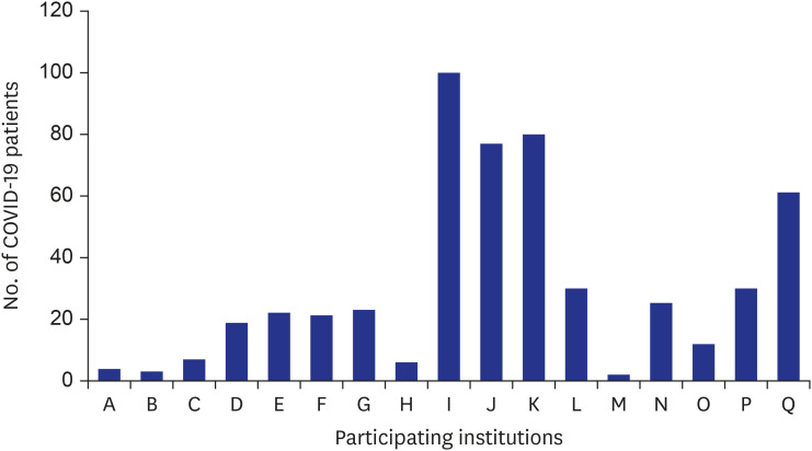
Fig. 1 The number of patients with COVID-19 gathered at each institution.COVID-19 = coronavirus disease 2019.
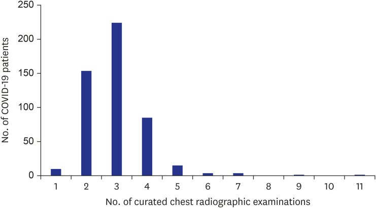
Fig. 2 Distribution of the number of curated chest radiographic examinations in the Korean imaging cohort of COVID-19.COVID-19 = coronavirus disease 2019.
Cited by 2 articles
-
Prognostic Implications of CT Feature Analysis in Patients with COVID-19: a Nationwide Cohort Study
Yeon Joo Jeong, Bo Da Nam, Jin Young Yoo, Kun-Il Kim, Hee Kang, Jung Hwa Hwang, Yun-Hyeon Kim, Kyung Soo Lee
J Korean Med Sci. 2021;36(8):e51. doi: 10.3346/jkms.2021.36.e51.Impact of COVID-19 Pandemic on Biomedical Publications and Their Citation Frequency
Sooyoung Park, Hyun Jeong Lim, Jaero Park, Yeon Hyeon Choe
J Korean Med Sci. 2022;37(40):e296. doi: 10.3346/jkms.2022.37.e296.
Reference
-
1. Kim H, Hong H, Yoon SH. Diagnostic performance of CT and reverse transcriptase polymerase chain reaction for coronavirus disease 2019: a meta-analysis. Radiology. 2020; 296(3):E145–E155. PMID: 32301646.2. Herpe G, Lederlin M, Naudin M, Ohana M, Chaumoitre K, Gregory J, et al. Efficacy of chest CT for COVID-19 pneumonia in France. Radiology. Forthcoming. 2020; DOI: 10.1148/radiol.2020202568.3. Zu ZY, Jiang MD, Xu PP, Chen W, Ni QQ, Lu GM, et al. Coronavirus disease 2019 (COVID-19): a perspective from china. Radiology. 2020; 296(2):E15–E25. PMID: 32083985.
Article4. Choi H, Qi X, Yoon SH, Park SJ, Lee KH, Kim JY, et al. Extension of coronavirus disease 2019 (COVID-19) on chest CT and implications for chest radiograph interpretation. Radiol Cardiothorac Imaging. 2020; 2:e200107.5. Wong HYF, Lam HYS, Fong AH, Leung ST, Chin TW, Lo CSY, et al. Frequency and distribution of chest radiographic findings in patients positive for COVID-19. Radiology. 2020; 296(2):E72–E78. PMID: 32216717.6. Toussie D, Voutsinas N, Finkelstein M, Cedillo MA, Manna S, Maron SZ, et al. Clinical and chest radiography features determine patient outcomes in young and middle-aged adults with COVID-19. Radiology. 2020; 297(1):E197–E206. PMID: 32407255.7. Schalekamp S, Huisman M, van Dijk RA, Boomsma MF, Freire Jorge PJ, de Boer WS, et al. Model-based prediction of critical illness in hospitalized patients with COVID-19. Radiology. Forthcoming. 2020; DOI: 10.1148/radiol.2020202723.8. Rubin GD, Ryerson CJ, Haramati LB, Sverzellati N, Kanne JP, Raoof S, et al. The role of chest imaging in patient management during the COVID-19 pandemic: a multinational consensus statement from the fleischner society. Radiology. 2020; 296(1):172–180. PMID: 32255413.
Article9. Yoon SH, Lee KH, Kim JY, Lee YK, Ko H, Kim KH, et al. Chest radiographic and CT findings of the 2019 novel coronavirus disease (COVID-19): analysis of nine patients treated in Korea. Korean J Radiol. 2020; 21(4):494–500. PMID: 32100485.
Article10. Jung HK, Kim JY, Lee MS, Lee JY, Park JS, Hyun M, et al. Characteristics of COVID-19 patients who progress to pneumonia on follow-up chest radiograph: 236 patients from a single isolated cohort in Daegu, South Korea. Korean J Radiol. 2020; 21(11):1265–1272. PMID: 32729278.
Article11. Park B, Park J, Lim JK, Shin KM, Lee J, Seo H, et al. Prognostic implication of volumetric quantitative ct analysis in patients with COVID-19: a multicenter study in Daegu, Korea. Korean J Radiol. 2020; 21(11):1256–1264. PMID: 32767868.
Article12. Hwang EJ, Kim H, Yoon SH, Goo JM, Park CM. Implementation of a deep learning-based computer-aided detection system for the interpretation of chest radiographs in patients suspected for COVID-19. Korean J Radiol. 2020; 21(10):1150–1160. PMID: 32729263.
Article13. Chon Y, Kim JY, Suh YJ, Lee JY, Park JS, Moon SM, et al. Adverse initial CT findings associated with poor prognosis of coronavirus disease. J Korean Med Sci. 2020; 35(34):e316. PMID: 32864912.
Article14. Lee SE, Kim YS. Clinical and radiological findings of coronavirus disease 2019 pneumonia: 51 adult patients from a single center in Daegu, South Korea. J Korean Soc Radiol. 2020; 81(3):591–603.
Article15. Rho JY, Yoon KH, Jeong S, Lee JH, Park C, Kim HW. Usefulness of mobile computed tomography in patients with coronavirus disease 2019 pneumonia: a case series. Korean J Radiol. 2020; 21(8):1018–1023. PMID: 32677386.
Article16. Yoon SH, Kim M. Anterior pulmonary ventilation abnormalities in COVID-19. Radiology. 2020; 297(2):E276–E277. PMID: 32787702.17. Shim SS. COVID-19 in Korea: what actions have been taken by radiologists to support the fight against the disease? J Korean Soc Radiol. 2020; 81(3):604–607.
Article18. Lee HJ, Moon JW, Woo JY, Kim YN. Clinical and radiologic findings of COVID-19 pneumonia: South Korean experience from three cases. J Korean Soc Radiol. 2020; 81(3):583–590.
Article19. Jin KN, Yoon SH, Park CH, Beck KS, Do KH, Yong HS. KSR/KSTR guidelines for the use of diagnostic imaging for COVID-19. J Korean Soc Radiol. 2020; 81(3):577–582.
Article20. Jeong YJ, Kim YH. Korean imaging cohort of COVID-19: potential role in education and research. J Korean Soc Radiol. 2020; 81(3):608–609.
Article21. Wang D, Hu B, Hu C, Zhu F, Liu X, Zhang J, et al. Clinical characteristics of 138 hospitalized patients with 2019 novel coronavirus-infected pneumonia in Wuhan, China. JAMA. 2020; 323(11):1061–1069. PMID: 32031570.
Article22. Wu C, Chen X, Cai Y, Xia J, Zhou X, Xu S, et al. Risk factors associated with acute respiratory distress syndrome and death in patients with coronavirus disease 2019 pneumonia in Wuhan, China. JAMA Intern Med. 2020; 180(7):934–943. PMID: 32167524.
Article23. Zhou F, Yu T, Du R, Fan G, Liu Y, Liu Z, et al. Clinical course and risk factors for mortality of adult inpatients with COVID-19 in Wuhan, China: a retrospective cohort study. Lancet. 2020; 395(10229):1054–1062. PMID: 32171076.
Article24. Yang X, Yu Y, Xu J, Shu H, Xia J, Liu H, et al. Clinical course and outcomes of critically ill patients with SARS-CoV-2 pneumonia in Wuhan, China: a single-centered, retrospective, observational study. Lancet Respir Med. 2020; 8(5):475–481. PMID: 32105632.
Article25. Hare SS, Rodrigues JC, Jacob J, Edey A, Devaraj A, Johnstone A, et al. A UK-wide British Society of Thoracic Imaging COVID-19 imaging repository and database: design, rationale and implications for education and research. Clin Radiol. 2020; 75(5):326–328. PMID: 32222251.
Article26. Kundu S, Elhalawani H, Gichoya JW, Kahn CE. How might AI and chest imaging help unravel COVID-19's mysteries? Radiol Artif Intell. 2020; Forthcoming. DOI: 10.1148/ryai.2020200053.27. Sung HK, Kim JY, Heo J, Seo H, Jang YS, Kim H, et al. Clinical course and outcomes of 3,060 patients with coronavirus disease 2019 in Korea, January-May 2020. J Korean Med Sci. 2020; 35(30):e280. PMID: 32743995.
Article
- Full Text Links
-
- Actions
-
Cited
- CITED
-
- Close
- Share
-
- Similar articles
-
- Clinical and Epidemiological Characteristics of Coronavirus Disease 2019 in the Early Stage of Outbreak
- COVID-19 infection and severe clinical outcomes in patients with kidney disease by vaccination status: a nationwide cohort study in Korea
- Independent Impact of Diabetes on the Severity of Coronavirus Disease 2019 in 5,307 Patients in South Korea: A Nationwide-Cohort Study (Diabetes Metab J 2020;44:737-46)
- Coronavirus Disease 2019-Liver InjuryLiterature Review and Guidelines Based on the Recommendations of Hepatological Societies
- Independent Impact of Diabetes on the Severity of Coronavirus Disease 2019 in 5,307 Patients in South Korea: A Nationwide Cohort Study (Diabetes Metab J 2020;44:737-46)
