KoreaMed, a service of the Korean Association of Medical Journal Editors (KAMJE), provides access to articles published in Korean medical, dental, nursing, nutrition and veterinary journals. KoreaMed records include links to full-text content in Synapse and publisher web sites.
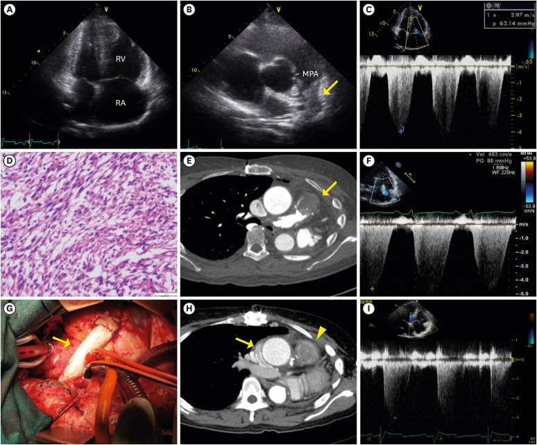
Figure 1 Various images obtained during long-term treatment. (A) Markedly enlarged right atrium and ventricle were observed on TTE. (B) A supra-valvular mass-like lesion was seen in MPA (yellow arrow). (C) Markedly elevated RVSP (94 mmHg) caused by the obstructive mass lesion. (D) Pathologic study showed grade 2 pulmonary artery leiomyosarcoma, characterized as a well-demarcated mass composed of fascicular growth and an 11/10 high power field mitotic count. (E) Chest CT scan reveals a 3 cm recurred leiomyosarcoma (yellow arrow) at the junction area between the RV and MPA. (F) Markedly elevated RVSP (100 mmHg) was observed. (G) A 16 mm Gore-tex conduit (yellow arrow) insertion between RV and right pulmonary artery was performed to relieve severe right ventricular pressure elevation. (H) Post-operative chest CT scan showed that right pulmonary artery was well enhanced by contrast through a patent Gore-tex conduit (yellow arrow). A remnant leiomyosarcoma at proximal MPA was seen (yellow arrowhead). (I) Decreased RVSP (36 mmHg) was observed on TTE after successful bypass surgery.CT = computed tomography; MPA = main pulmonary artery; RV = right ventricle; RVSP = right ventricular systolic pressure; TTE = transthoracic echocardiography.