A Case of Petrous Apex Meningocele: Unusual Presentation of Cerebrospinal Fluid Leak Following Adenoidectomy
- Affiliations
-
- 1Department of Otorhinolaryngology-Head and Neck Surgery, Samsung Medical Center, Sungkyunkwan University School of Medicine, Seoul, Korea
- 2Department of Neurosurgery, Samsung Medical Center, Sungkyunkwan University School of Medicine, Seoul, Korea
- KMID: 2508960
- DOI: http://doi.org/10.18787/jr.2020.00328
Abstract
- Cerebrospinal fluid (CSF) leak is possible and can be a cause of recurrent bacterial meningitis. Petrous apex meningocele (PAM) is mostly asymptomatic and is often found during incidental imaging tests. We experienced a case of CSF rhinorrhea with recurrent meningitis in bilateral PAM after adenoidectomy. This report highlights the diagnostic process of CSF leak, identification of leakage site, and surgical approach to petrous apex lesions.
Keyword
Figure
-
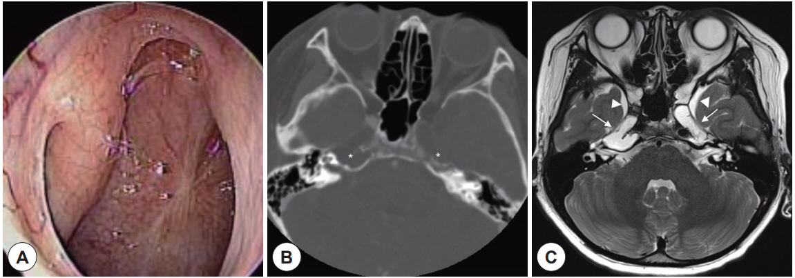
Fig. 1. Initial endoscopic and imaging findings. A: Scar is observed in nasopharynx, but there is no cerebrospinal fluid (CSF) leak. B: Osteolytic lesions (asterisks) in bilateral petrous apex are seen on axial view of CT. C: Axial T2-weighted MRI. Bilateral meningocele (arrow) of bright T2 high signal intensity like CSF in petrous apex extending to Meckel’s cave (arrowhead) and cavernous sinus.
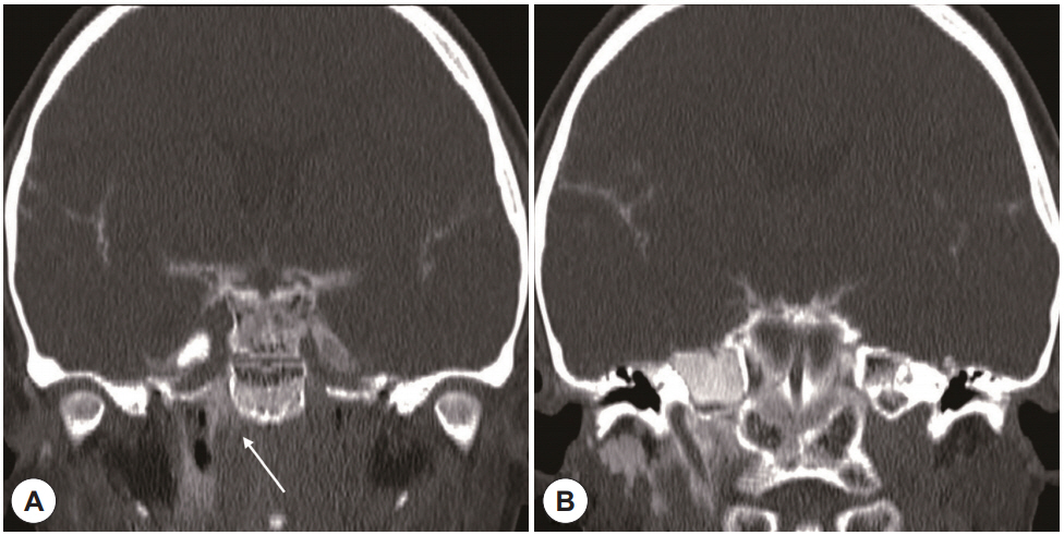
Fig. 2. CT cisternography after intrathecal contrast injection. A: Arrow shows leak of contrast through foramen lacerum. B: Diffuse contrast staining is shown around retropharyngeal and prevertebral space.
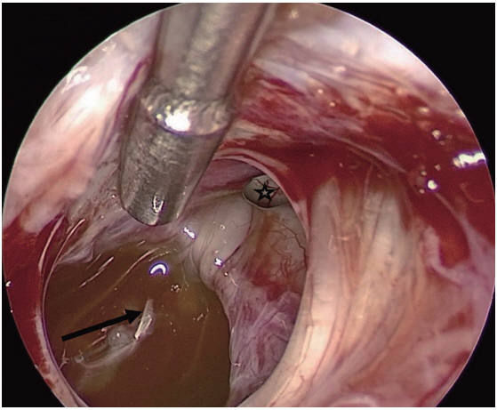
Fig. 3. Endoscopic view through transorbital approach to petrous apex. Arrow indicated petrous apex meningocele and asterisk indicated posterior fossa communicating with petrous apex meningocele. Cerebrospinal fluid (CSF) is stained with fluorescein dye.
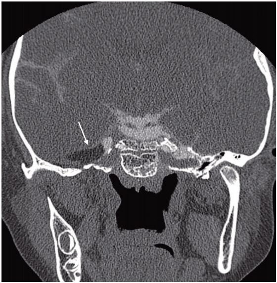
Fig. 4. CT cisternography after postoperative 1 week demonstrates Cerebrospinal fluid (CSF) leak which previously observed under right petrous apex is disappearing. The interposition of fat tissue (arrow) to petrous apex meningocele is seen.
Reference
-
References
1. Razek AA, Huang BY. Lesions of the petrous apex: classification and findings at CT and MR imaging. Radiographics. 2012; 32(1):151–73.2. Moore KR, Fischbein NJ, Harnsberger HR, Shelton C, Glastonbury CM, White DK, et al. Petrous apex cephaloceles. AJNR Am J Neuroradiol. 2001; 22(10):1867–71.3. Stark TA, McKinney AM, Palmer CS, Maisel RH, Truwit CL. Dilation of the subarachnoid spaces surrounding the cranial nerves with petrous apex cephaloceles in Usher syndrome. AJNR Am J Neuroradiol. 2009; 30(2):434–6.4. Alorainy IA. Petrous apex cephalocele and empty sella: is there any relation? Eur J Radiol. 2007; 62(3):378–84.5. Schuknecht B, Simmen D, Briner HR, Holzmann D. Nontraumatic skull base defects with spontaneous CSF rhinorrhea and arachnoid herniation: imaging findings and correlation with endoscopic sinus surgery in 27 patients. AJNR Am J Neuroradiol. 2008; 29(3):542–9.6. Raghavan U, Majumdar S, Jones NS. Spontaneous CSF rhinorrhoea from separate defects of the anterior and middle cranial fossa. J Laryngol Otol. 2002; 116(7):546–7.7. Stone JA, Castillo M, Neelon B, Mukherji SK. Evaluation of CSF leaks: high-resolution CT compared with contrast-enhanced CT and radionuclide cisternography. AJNR Am J Neuroradiol. 1999; 20(4):706–12.8. Chow JM, Goodman D, Mafee MF. Evaluation of CSF rhinorrhea by computerized tomography with metrizamide. Otolaryngol Head Neck Surg. 1989; 100(2):99–105.9. Shetty PG, Shroff MM, Sahani DV, Kirtane MV. Evaluation of highresolution CT and MR cisternography in the diagnosis of cerebrospinal fluid fistula. AJNR Am J Neuroradiol. 1998; 19(4):633–9.10. Johnson DB, Brennan P, Toland J, O’Dwyer AJ. Magnetic resonance imaging in the evaluation of cerebrospinal fluid fistulae. Clin Radiol. 1996; 51(12):837–41.11. Hervey-Jumper SL, Ghori AK, Quint DJ, Marentette LJ, Maher CO. Cerebrospinal fluid leak with recurrent meningitis following tonsillectomy. J Neurosurg Pediatr. 2010; 5(3):302–5.12. Di Somma A, Andaluz N, Cavallo LM, Topczewski TE, Frio F, Gerardi RM, et al. Endoscopic transorbital route to the petrous apex: a feasibility anatomic study. Acta Neurochir (Wien). 2018; 160(4):707–20.13. Topczewski TE, Di Somma A, Pineda J, Ferres A, Torales J, Reyes L, et al. Endoscopic transorbital route to the petrous apex: a feasibility anatomic study. Acta Neurochir (Wien). 2018. p.1-13.
- Full Text Links
-
- Actions
-
Cited
- CITED
-
- Close
- Share
-
- Similar articles
-
- Petrous Apex Cephalocele: Report of Two Cases and Review of the Literature
- Intracranial Hypotension Associated with Meningocele
- A Case of Frontoethmoidal Mucopyocele Combined with Cerebrospinal Fluid Leak and Complicated Tension Pneumocephalus after Marsupialization
- Surgical Anatomy for the Infracochlear Approach to the Petrous Apex
- Traumatic Cerebrospinal Fluid Leak: Diagnosis and Management
