A Case of Frontoethmoidal Mucopyocele Combined with Cerebrospinal Fluid Leak and Complicated Tension Pneumocephalus after Marsupialization
- Affiliations
-
- 1Department of Otorhinolaryngology-Head & Neck Surgery, Pusan National University Yangsan Hospital, Yangsan, Korea. rohhj@pusan.ac.kr
- KMID: 2412919
- DOI: http://doi.org/10.18787/jr.2018.25.1.38
Abstract
- After the trauma of frontoethmoidal sinus, post-traumatic mucocele may occur. Surgical removal of the lesions rarely produces cerebrospinal fluid (CSF) leakage and even delayed tension pneumocephalus. We experienced a case of fronto-ethmoid mucocele complicated with peri-operative CSF leakage and post-operative tension pneumocephalus which was improved by conservative treatment. It is imperative to take into account the potential for tension pneumocephalus when a patient suffers from severe headache after sinus surgery.
MeSH Terms
Figure
-
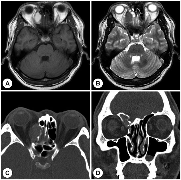
Fig. 1 Preoperative images. Brain MRI axial images show right frontoethmoidal homogenous lesion with high signal intensity (SI) on T1 weighted image (WI) (A) and intermediate SI on T2 WI (B). Non-enhanced paranasal sinus (PNS) CT shows of right frontoethmoidal sinus expansile lesion with erosion of lamina papyracea which is mildly compressing the superior oblique muscle on axial image (C) and broken lateral lamella (arrowhead) on coronal image (D).
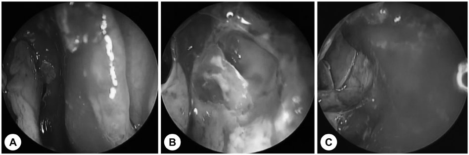
Fig. 2 Intraoperative endoscopic findings. The pus inside the mucocele is removed (A). Cerebrospinal fluid (CSF) leakage is observed at lateral lamella (B). CSF leakage site is repaired with perpendicular plate of ethmoid bone (C).
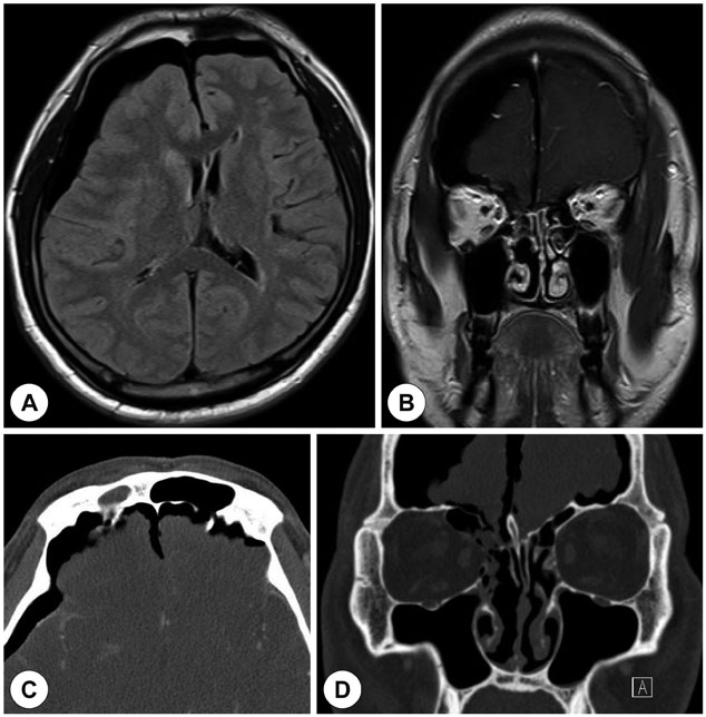
Fig. 3 The 10th postoperative images. Brain MRI T1 WI show pneumocephalus at frontal convexity compressing right frontal lobe, a left midline shift of brain and mild meningeal enhancement on axial (A) and coronal (B) image. Post-contrast PNS CT shows air collection at right frontotemporal region and a small bony defect at lateral lamella on axial (C) and coronal (D) image.
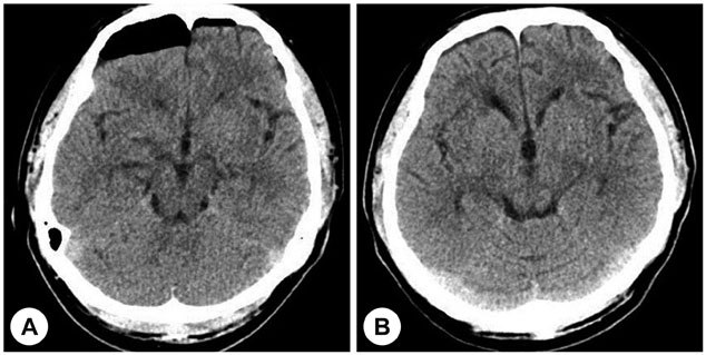
Fig. 4 The 15th and 23rd postoperative images. The 15th axial brain CT shows decreased air collection at frontal region and corrected midline shift (A). The 23rd axial brain CT shows no air collection at frontal region (B).
Cited by 1 articles
-
A Case of Brain Herniation and Cerebrospinal Fluid Leakage during Endoscopic Marsupialization of Ethmoid Sinus Mucocele
Song Jae Lee, Sang Gyu Park, Hae Won Choi, Kyung Rae Kim
Korean J Otorhinolaryngol-Head Neck Surg. 2020;63(9):427-431. doi: 10.3342/kjorl-hns.2019.00542.
Reference
-
1. Pero CD, Nuss DW. Transnasal endoscopic removal of orbital, ethmoid sinus, and anterior skull base foreign body with mucocele formation. Skull Base. 2008; 18:417–422.
Article2. Guy WM, Brissett AE. Contemporary management of traumatic fractures of the frontal sinus. Otolaryngol Clin North Am. 2013; 46(5):733–748.
Article3. Pizzo LJ, Mishler KE. Frontoethmoid mucocele with CSF leak. Ear Nose Throat J. 1984; 63(11):571–573.4. Chou S, Ning M, Buonanno F. Focal intraparenchymal tension pneumocephalus. Neurology. 2006; 67(8):1485.
Article5. Rao V, Fredriksli O, Gulati S. Post-traumatic epidural tension pneumocephalus: a case report. J Med Case Rep. 2015; 9:151.
Article6. Lee HC, Park IH, Choi CJ, Lee HM. A case of endonasal, endoscopic repair of posttraumatic delayed pneumocephalus. J Rhinol. 2010; 17(1):41–44.7. Chi BJ, Bang SH, Chang WP. A case of traumatic subarachnoid pneumocephalus: as a complication of intranasal ethmoidectomy. Korean J Otolaryngol-Head Neck Surg. 1991; 34(2):359–365.8. Markham JW. The clinical features of pneumocephalus based upon a survey of 284 cases with report of 11 additional cases. Acta Neurochir (Wien). 1967; 16(1):1–78.
Article9. Naseem M, Hood J, Devasthali R. Traumatic pneumocephalus caused by a stab wound to the neck. AJNR Am J Neuroradiol. 1986; 7(1):174–175.10. Singh R, Hazarika P, Nayak DR, Balakrishnan R, Hazarika M, Singh A. Endoscopic repair of cerebrospinal fluid rhinorrhea-Manipal experience. Indian J Otolaryngol Head Neck Surg. 2009; 61(1):14–18.
Article11. Babl FE, Arnett AM, Barnett E, Brancato JC, Kharasch SJ, Janecka IP. Atraumatic pneumocephalus: A case report and review of the literature. Pediatr Emerg Care. 1999; 15(2):106–109.12. Walker FO, Vern BA. The mechanism of pneumocephalus formation in patients with CSF fistulas. J Neurol Neurosurg Psychiatry. 1986; 49:203–205.
Article13. Goldman LW. Principles of CT and CT technology. J Nucl Med Technol. 2007; 35(3):115–128.
Article14. Aksoy F, Dogan R, Ozturan O, Tugrul S, Yildirim YS. Tension pneumocephlus: an extremely small defect leading to an exteremely serious problem. Am J Otolaryngol. 2013; 34(6):749–752.
Article15. DelGaudio JM, Ingley AP. Treatment of pneumocephalus after endoscopic sinus and microscopic skull base surgery. Am J Otolaryngol. 2010; 31(4):226–230.
Article
- Full Text Links
-
- Actions
-
Cited
- CITED
-
- Close
- Share
-
- Similar articles
-
- Delayed Tension Pneumocephalus Following Intraoperative Cerebrospinal Fluid Leakage Repair
- Proper Management of Posttraumatic Tension Pneumocephalus
- Two Cases of Delayed Tension Pneumocephalus
- Tension Pneumocephalus after Transsphenoidal Surgery for a Giant Pituitary Tumor: Case Report
- Tension Pneumocephalus after Transsphenoidal Surgery: Report of Two Cases
