Delayed Arteriovenous Fistula after Reverse Flow Sural Island Flap in Lower Leg: A Case Report
- Affiliations
-
- 1Department of Transplantation and Vascular Surgery, Wonkwang University Hospital, Iksan, Korea
- 2Department of Orthopedic Surgery, Wonkwang University Hospital, Iksan, Korea
- KMID: 2508946
- DOI: http://doi.org/10.12790/ahm.20.0049
Abstract
- Delayed arteriovenous (AV) fistula after soft tissue reconstruction with flap surgery is a rare complication. Here, we describe a case of delayed AV fistula formation after 4 years of reverse flow sural island flap surgery in the lower leg. The patient had swelling, tenderness, and color change to dark purple on previous flap area and foot, similar to cellulitis. Thrill and murmur were observed. AV fistula was formed around the previous vascular pedicle area, as revealed in angiography. We excised the right posterior tibial artery-saphenous vein fistula. The patient is having satisfactory progress since the surgery. We suggest that AV fistula was caused by enhanced angiogenesis and vascular damage.
Keyword
Figure
-
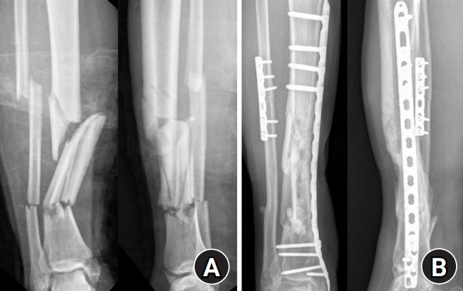
Fig. 1. (A) Initially open fractures of the tibia and fibula were observed. (B) The fractures showed complete union in the 2 years after the surgery.

Fig. 2. After 4 years, the patient visited with swelling, color change, and an ulcer on foot.
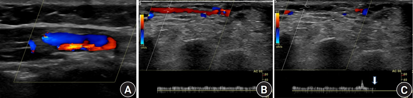
Fig. 3. Ultrasound examination. (A) Arteriovenous fistula was detected around the previous neurovascular pedicle area. (B) A bruit was detected with the classical machinery-like murmur. (C) It was disappeared when we compressed the proximal lesion (arrow).
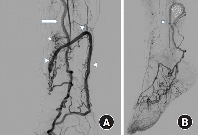
Fig. 4. Selective angiography. (A) In selective angiography, the posterior tibial artery (arrow) was connected to the small saphenous vein (arrowheads) and formation arteriovenous fistula (asterisk). (B) The small saphenous vein was dilated and its drainage was refluxed into (curved arrow) the veins of the foot with multiple communicating vessels (arrowhead).
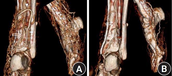
Fig. 5. In preoperative computed tomography (CT) angiography and immediate postoperative CT angiography, arteriovenous fistula and the small saphenous vein were disappeared and venous dilatation was decreased (A), compared preoperative CT angiography (B). The posterior tibial artery (arrows), the small saphenous vein (arrowhead).
Reference
-
1. Ninković M, Sućur D, Starović B, Marković S. Arteriovenous fistulae after free flap surgery in a replanted hand. J Hand Surg Br. 1992; 17:657–9.2. Maher SM, Rabee HM, Takrouri MS, Al-Salman MM. Traumatic arteriovenous fistula. Ann Thorac Surg. 1997; 63:1792–4.3. Collin TW, Morris P, Sassoon E, O’Neill T. Arteriovenous fistula in a free pectoralis minor flap. Eur J Plast Surg. 2003; 26:367–9.
Article4. Nikfarjam J, Taub PJ, Patel A, Rose E. Arteriovenous fistula following radial forearm free flap. J Reconstr Microsurg. 2011; 27:295–8.
Article5. Mylankal KJ, Johnson B, Ettles DF. Iatrogenic arteriovenous fistula as a cause for leg ulcers: a case report. Ann Vasc Dis. 2011; 4:139–42.
Article6. Sukop A, Kufa R, Dušková M. Late arteriovenous fistula after finger replantation. Eur J Plast Surg. 2006; 28:421–5.
Article7. Komai Y, Nakano A, Niimi H. Capillary angiogenesis and remodeling induced in rat limb by arteriovenous shunting. Clin Hemorheol Microcirc. 2005; 32:199–208.8. Mork C, Asker CL, Salerud EG, Kvernebo K. Microvascular arteriovenous shunting is a probable pathogenetic mechanism in erythromelalgia. J Invest Dermatol. 2000; 114:643–6.9. Touho H, Furuoka N, Ohnishi H, Komatsu T, Karasawa J. Traumatic arteriovenous fistula treated by superselective embolisation with microcoils: case report. Neuroradiology. 1995; 37:65–7.
Article
- Full Text Links
-
- Actions
-
Cited
- CITED
-
- Close
- Share
-
- Similar articles
-
- Versatility of Delayed Reverse Sural Flap for Reconstruction of the Distal Lower Extremity in High-Risk Patients
- Reconstruction of Regions Below the Knee Using Island Flaps
- The Reverse Sural Artery Flap for Soft Tissue Defect of Foot and Ankle in Diabetic Patient
- Sural Artery Flap for the Treatment of Osteomyelitis of the Lower Leg
- Reconstruction of the Soft Tissue Defect of the Lower Leg by Distally Based Superficial Sural Artery Fasciocutaneous Island Flap Using Supercharged Vein
