Healthc Inform Res.
2020 Oct;26(4):321-327. 10.4258/hir.2020.26.4.321.
Distribution and Characteristics of Pancreatic Volume Using Computed Tomography Volumetry
- Affiliations
-
- 1Department of Family Medicine, Chungbuk National University Hospital, Cheongju, Korea
- 2Department of Biomedical Engineering, Medical Devices R&D Center, Gachon University Gil Medical Center, Incheon, Korea
- 3Department of Surgery, Gachon University Gil Medical Center, Incheon, Korea
- KMID: 2508624
- DOI: http://doi.org/10.4258/hir.2020.26.4.321
Abstract
Objectives
Changes in the pancreatic volume (PV) are useful as potential clinical markers for some pancreatic-related diseases. The objective of this study was to measure the volume of the pancreas using computed tomography (CT) volumetry and to evaluate the relationships between sex, age, body mass index (BMI), and sarcopenia.
Methods
We retrospectively analyzed the abdominal CT scans of 1,003 subjects whose ages ranged between 10 and 90 years. The pancreas was segmented manually to define the region of interest (ROI) based on CT images, and then the PVs were measured by counting the voxels in all ROIs within the pancreas boundary. Sarcopenia was identified by examination of CT images that determined the crosssectional area of the skeletal muscle around the third lumbar vertebra.
Results
The mean volume of the pancreas was 62.648 ± 19.094 cm3. The results indicated a negative correlation between the PV and age. There was a positive correlation between the PV and BMI for both sexes, females, and males (r = 0.343, p < 0.001; r = 0.461, p < 0.001; and r = 0.244, p < 0.001, respectively). Additionally, there was a positive correlation between the PV and sarcopenia for females (r = 0.253, p < 0.001) and males (r = 0.200, p < 0.001).
Conclusions
CT pancreas volumetry results may help physicians follow up or predict conditions of the pancreas after interventions for pancreatic-related disease in the future.
Figure
-
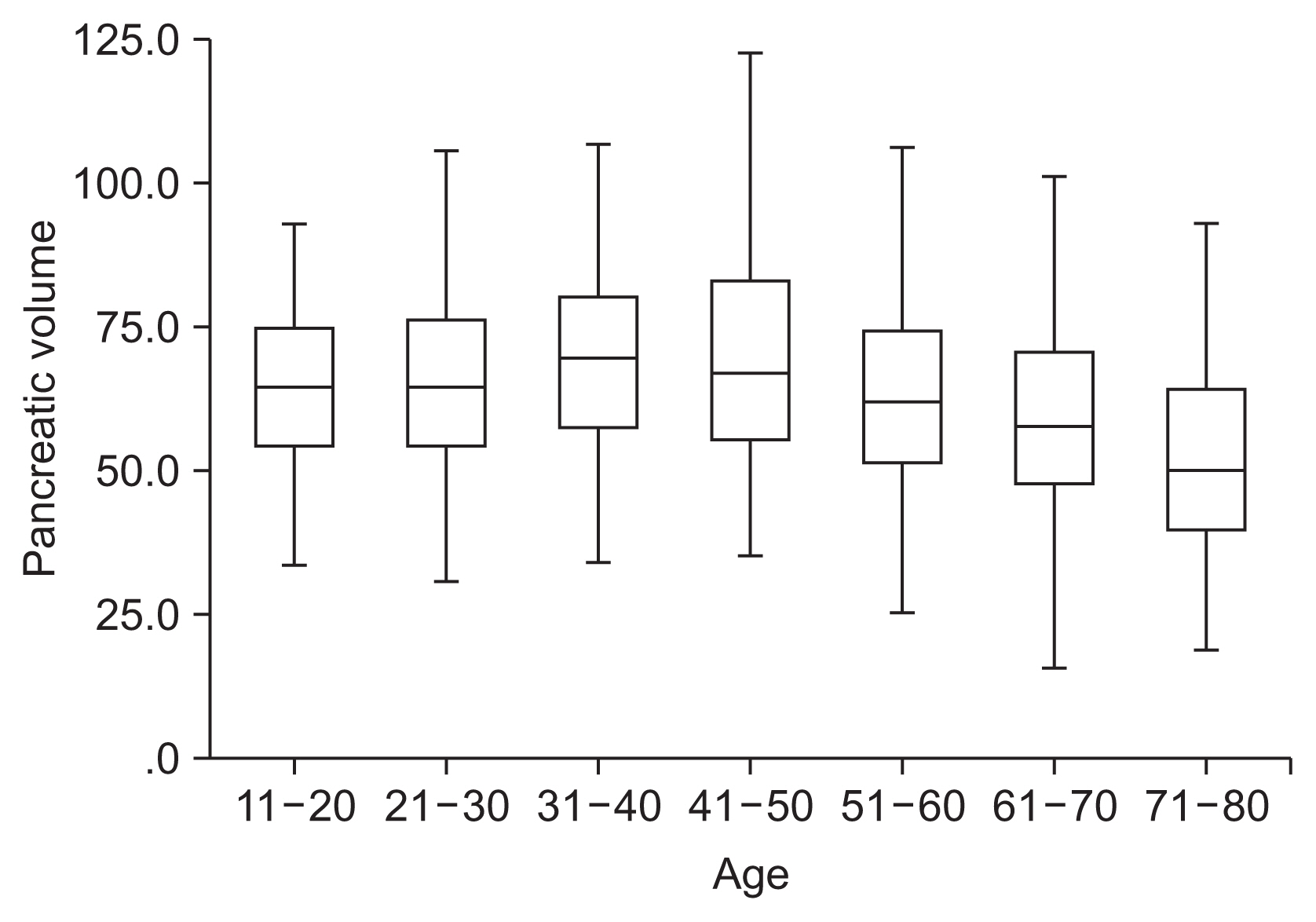
Figure 1 Box-and-whisker plots of pancreatic volume with respect to age. Boxes indicate median and 25th–75th percentile ranges.
Reference
-
References
1. Djuric-Stefanovic A, Masulovic D, Kostic J, Randjic K, Saranovic D. CT volumetry of normal pancreas: correlation with the pancreatic diameters measurable by the cross-sectional imaging, and relationship with the gender, age, and body constitution. Surg Radiol Anat. 2012; 34(9):811–7.
Article2. Saisho Y. Pancreas volume and fat deposition in diabetes and normal physiology: consideration of the interplay between endocrine and exocrine pancreas. Rev Diabet Stud. 2016; 13(2–3):132–47.
Article3. Williams AJ, Thrower SL, Sequeiros IM, Ward A, Bickerton AS, Triay JM, et al. Pancreatic volume is reduced in adult patients with recently diagnosed type 1 diabetes. J Clin Endocrinol Metab. 2012; 97(11):E2109–13.
Article4. Goda K, Sasaki E, Nagata K, Fukai M, Ohsawa N, Hahafusa T. Pancreatic volume in type 1 und type 2 diabetes mellitus. Acta diabetologica. 2001; 38:145–9.
Article5. Miyamoto R, Oshiro Y, Sano N, Inagawa S, Ohkohchi N. Remnant pancreatic volume as an indicator of poor prognosis in pancreatic cancer patients after resection. Pancreatology. 2019; 19(5):716–21.
Article6. Hirata K, Nakata B, Amano R, Yamazoe S, Kimura K, Hirakawa K. Predictive factors for change of diabetes mellitus status after pancreatectomy in preoperative diabetic and nondiabetic patients. J Gastrointest Surg. 2014; 18(9):1597–603.
Article7. Caglar V, Kumral B, Uygur R, Alkoc OA, Ozen OA, Demirel H. Study of volume, weight and size of normal pancreas, spleen and kidney in adults autopsies. Forensic Med Anat Res. 2014; 2:63–9.8. Geraghty EM, Boone JM, McGahan JP, Jain K. Normal organ volume assessment from abdominal CT. Abdom Imaging. 2004; 29(4):482–90.
Article9. Saisho Y, Butler AE, Meier JJ, Monchamp T, Allen-Auerbach M, Rizza RA, et al. Pancreas volumes in humans from birth to age one hundred taking into account sex, obesity, and presence of type-2 diabetes. Clin Anat. 2007; 20(8):933–42.
Article10. Lee JS, Kim YS, Kim EY, Jin W. Prognostic significance of CT-determined sarcopenia in patients with advanced gastric cancer. PLoS One. 2018; 13(8):e0202700.
Article11. Matsuda Y. Age-related pathological changes in the pancreas. Front Biosci (Elite Ed). 2018; 10:137–42.
Article12. Chan MY, Chok KS. Sarcopenia in pancreatic cancer: effects on surgical outcomes and chemotherapy. World J Gastrointest Oncol. 2019; 11(7):527–37.13. Lim S, Bae JH, Chun EJ, Kim H, Kim SY, Kim KM, et al. Differences in pancreatic volume, fat content, and fat density measured by multidetector-row computed tomography according to the duration of diabetes. Acta Diabetol. 2014; 51(5):739–48.
Article14. Ronneberger O, Fischer P, Brox T. U-Net: convolutional networks for biomedical image segmentation. Navab N, Hornegger J, Wells W, Frangi A, editors. Medical image computing and computer-assisted intervention. Cham, Switzerland: Springer;2015. p. 234–41.
Article15. WHO Expert Consultation. Appropriate body-mass index for Asian populations and its implications for policy and intervention strategies. Lancet. 2004; 363(9403):157–63.16. Kou K, Saisho Y, Jinzaki M, Itoh H. Relationship between body mass index and pancreas volume in Japanese people. JOP. 2014; 15(6):626–7.17. Caglar V, Songur A, Yagmurca M, Acar M, Toktas M, Gonul Y. Age-related volumetric changes in pancreas: a stereological study on computed tomography. Surg Radiol Anat. 2012; 34(10):935–41.
Article18. Mu’ti A, Paramita S. Relationship of pancreatic volumes using CT scan in indonesian adults with age, sex, and body mass index. Folia Medica Indonesiana. 2020; 56(1):31–5.
Article19. de la Grandmaison GL, Clairand I, Durigon M. Organ weight in 684 adult autopsies: new tables for a Caucasoid population. Forensic Sci Int. 2001; 119(2):149–54.
Article20. Prado CM, Heymsfield SB. Lean tissue imaging: a new era for nutritional assessment and intervention. JPEN J Parenter Enteral Nutr. 2014; 38(8):940–53.21. Cruz-Jentoft AJ, Bahat G, Bauer J, Boirie Y, Bruyere O, Cederholm T, et al. Sarcopenia: revised European consensus on definition and diagnosis. Age Ageing. 2019; 48(1):16–31.
Article22. Zamboni M, Gattazzo S, Rossi AP. Myosteatosis: a relevant, yet poorly explored element of sarcopenia. Eur Geriatr Med. 2019; 10(1):5–6.
Article23. Baumgartner RN. Body composition in healthy aging. Ann N Y Acad Sci. 2000; 904:437–48.
Article24. Butler AE, Janson J, Bonner-Weir S, Ritzel R, Rizza RA, Butler PC. Beta-cell deficit and increased beta-cell apoptosis in humans with type 2 diabetes. Diabetes. 2003; 52(1):102–10.25. Oya J, Nakagami T, Yamamoto Y, Fukushima S, Takeda M, Endo Y, et al. Effects of age on insulin resistance and secretion in subjects without diabetes. Intern Med. 2014; 53(9):941–7.
Article26. Sakata N, Egawa S, Rikiyama T, Yoshimatsu G, Masuda K, Ohtsuka H, et al. Computed tomography reflected endocrine function of the pancreas. J Gastrointest Surg. 2011; 15(3):525–32.
Article27. Sakai S, Tanimoto K, Imbe A, Inaba Y, Shishikura K, Tanimoto Y, et al. Decreased β-cell function is associated with reduced skeletal muscle mass in Japanese subjects without diabetes. PloS One. 2016; 11(9):e0162603.
Article28. Balzano G, Dugnani E, Gandolfi A, Scavini M, Pasquale V, Aleotti F, et al. Effect of diabetes on survival after resection of pancreatic adenocarcinoma: a prospective, observational study. PLoS One. 2016; 11(11):e0166008.
Article
- Full Text Links
-
- Actions
-
Cited
- CITED
-
- Close
- Share
-
- Similar articles
-
- Comparison of Predicted Postoperative Lung Function in Pneumonectomy Using Computed Tomography and Lung Perfusion Scans
- Evaluation of Computer Aided Volumetry for Simulated Small Pulmonary Nodules on Computed Tomography
- A study of Parameters in Spiral CT Volumetry Using Balloon Phantoms
- Effect of the High-Pitch Mode in Dual-Source Computed Tomography on the Accuracy of Three-Dimensional Volumetry of Solid Pulmonary Nodules: A Phantom Study
- Calculation of Standard Liver Volume of Korean Adults
