Investig Magn Reson Imaging.
2020 Mar;24(1):61-65. 10.13104/imri.2020.24.1.61.
Langerhans Cell Histiocytosis of the Rib of an Adult Female Patient: a Case Report
- Affiliations
-
- 1Department of Radiology, Chungbuk National University Hospital, Cheongju, Korea
- 2Department of Pathology, College of Medicine, Chungbuk National University, Cheongju, Korea
- KMID: 2508220
- DOI: http://doi.org/10.13104/imri.2020.24.1.61
Abstract
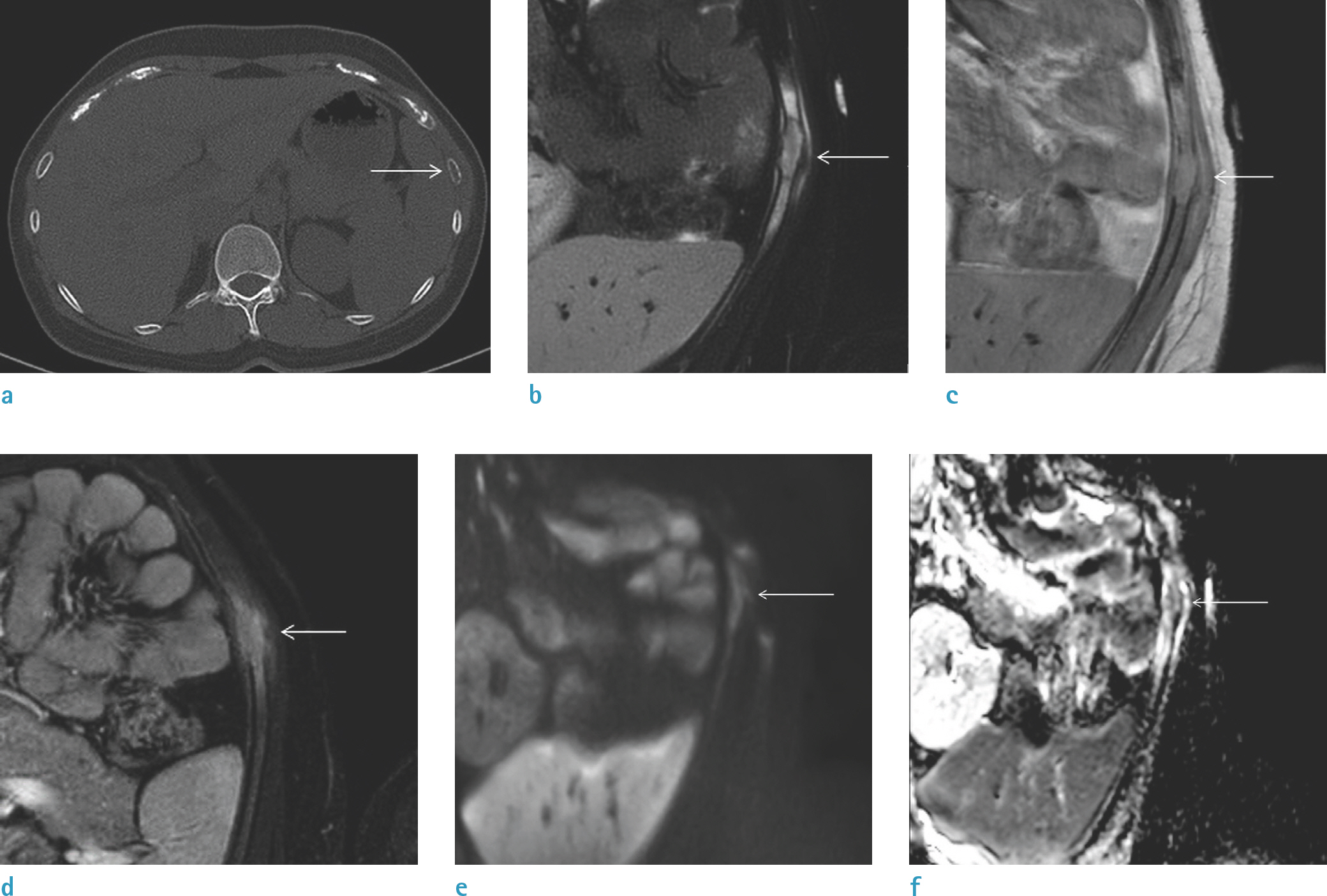
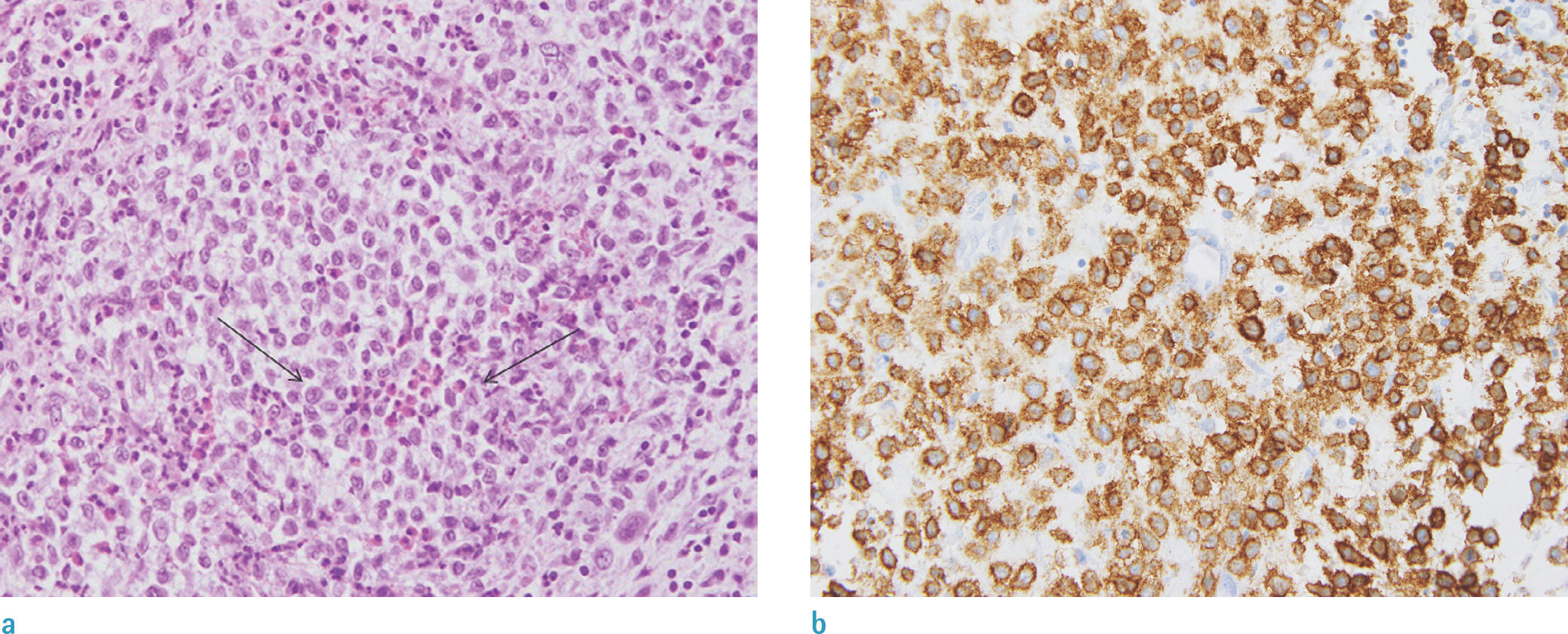
- Langerhans cell histiocytosis (LCH) is generally considered a childhood disease that exhibits various nonspecific clinical and radiological manifestations that mimic infection or malignancy. Here, we present a case of LCH involving the rib in an adult patient. CT and MRI revealed an expansile lytic lesion with periosteal reaction on the left 8th rib, suggesting a malignant bone tumor. Surgical resection was performed and histopathological examination was consistent with LCH. Owing to its rare occurrence in adults and nonspecific aggressive features, LCH should be included in the differential diagnosis of an aggressive-appearing rib lesion in both adults and children.
Keyword
Figure
Reference
-
1.Kim SH., Choi MY. Langerhans cell histiocytosis of the rib in an adult: a case report. Case Rep Oncol. 2016. 9:83–88.
Article2.Favara BE., Feller AC., Pauli M, et al. Contemporary classification of histiocytic disorders. The WHO Committee on Histiocytic/Reticulum Cell Proliferations. Reclassification Working Group of the Histiocyte Society. Med Pediatr Oncol. 1997. 29:157–166.3.Zaveri J., La Q., Yarmish G., Neuman J. More than just Langerhans cell histiocytosis: a radiologic review of histiocytic disorders. Radiographics. 2014. 34:2008–2024.
Article4.Baumgartner I., von Hochstetter A., Baumert B., Luetolf U., Follath F. Langerhans‘-cell histiocytosis in adults. Med Pediatr Oncol. 1997. 28:9–14.
Article5.Stull MA., Kransdorf MJ., Devaney KO. Langerhans cell histiocytosis of bone. Radiographics. 1992. 12:801–823.
Article6.Wester SM., Beabout JW., Unni KK., Dahlin DC. Langerhans’ cell granulomatosis (histiocytosis X) of bone in adults. Am J Surg Pathol. 1982. 6:413–426.7.Stockschlaeder M., Sucker C. Adult Langerhans cell histiocytosis. Eur J Haematol. 2006. 76:363–368.
Article8.Islinger RB., Kuklo TR., Owens BD, et al. Langerhans’ cell histiocytosis in patients older than 21 years. Clin Orthop Relat Res. 2000. 231–235.
Article9.Samet J., Weinstein J., Fayad LM. MRI and clinical features of Langerhans cell histiocytosis (LCH) in the pelvis and extremities: can LCH really look like anything? Skeletal Radiol. 2016. 45:607–613.
Article10.Girschikofsky M., Arico M., Castillo D, et al. Management of adult patients with Langerhans cell histiocytosis: recommendations from an expert panel on behalf of Euro-Histio-Net. Orphanet J Rare Dis. 2013. 8:72.
Article
- Full Text Links
- Actions
-
Cited
- CITED
-
- Close
- Share
- Similar articles
-
- Spontaneous Pneumothorax due to Pulmonary Invasion in Multisystemic Langerhans Cell Histiocytosis: A case report
- Adult Onset of Langerhans Cell Histiocytosis in the Rib: Report of 2 cases
- A Case of Pulmonary Langerhans Cell Histiocytosis with Pneumothorax
- Adult Langerhans Cell Histiocytosis of the Spine Presenting with Neurological Deficits: A Case Report
- Two Cases of Langerhans' Cell Histiocytosis: Report of Two Cases


