Recent Radiation Reduction Strategies for Neurointerventionists
- Affiliations
-
- 1Department of Radiology, St. Vincent’s Hospital, The Catholic University of Korea, Suwon, Korea
- KMID: 2508071
- DOI: http://doi.org/10.5469/neuroint.2020.00346
Figure
-
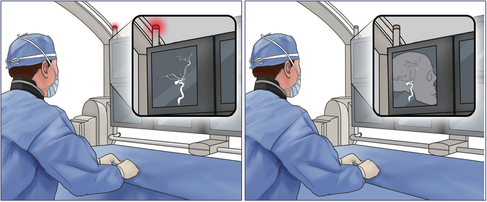
Fig. 1. Optimize the fluoroscopy settings. The optimization of fluoroscopy settings such as frame rate, filter options, automatic exposure control, focal spot, current, and peak potential according to each specific procedure is critical. Not every vascular analysis warrants 2D or 3D digital subtraction angiography. Likewise, not every fluoroscopy projection warrants the same pulse rate, focal spot, and frame rates.
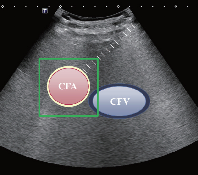
Fig. 2. Use ultrasonography for localization. Ultrasonography is considered an adjunct modality for arterial or venous access to avoid unnecessary fluoroscopy projection and wire navigation. Whether it is used to perform a direct puncture into a venous malformation or to gain femoral artery or venous access, ultrasonography can be an invaluable tool to achieve an accurate puncture and localization without radiation. CFA, common femoral artery; CFV, common femoral vein.

Fig. 3. Reduce the craniocaudal angle. Smaller craniocaudal angulation or true posteroanterior projection may result in less radiation exposure than the conventional projection.
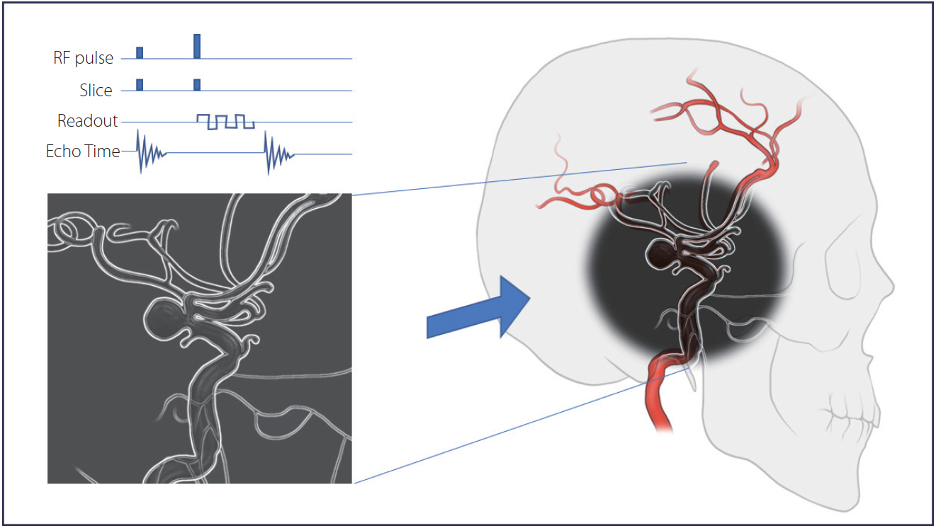
Fig. 4. Take advantage of fusion technology. Fusion of 3D magnetic resonance angiography (MRA) with 2D digital subtraction angiography may also reduce the radiation dose. Modern fluoroscopy can import and overlay images from high-resolution MRA and use them as a real-time “virtual” roadmap. RF, radiofrequency.
Reference
-
1. Riabroi K, Khanungwanitkul K, Wattanapongpitak P, Krisanachinda A, Hongsakul K. Patient radiation dose in neurointerventional radiologic procedure: a tertiary care experience. Neurointervention. 2018; 13:110–116.
Article2. Visweswaran S, Joseph S, Hegde V, Annalakshmi O, Jose MT, Perumal V. DNA damage and gene expression changes in patients exposed to low-dose X-radiation during neuro-interventional radiology procedures. Mutat Res. 2019; 844:54–61.
Article3. Vano E, Fernandez JM, Sanchez RM, Martinez D, Ibor LL, Gil A, et al. Patient radiation dose management in the follow-up of potential skin injuries in neuroradiology. AJNR Am J Neuroradiol. 2013; 34:277–282.
Article4. Song Y, Han S, Kim BJ, Oh SH, Kim JS, Kim TI, et al. Low-dose fluoroscopy protocol for diagnostic cerebral angiography. Neurointervention. 2020; 15:67–73.
Article5. van der Marel K, Vedantham S, van der Bom IM, Howk M, Narain T, Ty K, et al. Reduced patient radiation exposure during neurodiagnostic and interventional X-ray angiography with a new imaging platform. AJNR Am J Neuroradiol. 2017; 38:442–449.
Article6. Kim DJ, Park MK, Jung DE, Kang JH, Kim BM. Radiation dose reduction without compromise to image quality by alterations of filtration and focal spot size in cerebral angiography. Korean J Radiol. 2017; 18:722–728.
Article7. Slattery MM, Goh GS, Power S, Given MF, McGrath FP, Lee MJ. Comparison of ultrasound-guided and fluoroscopy-assisted antegrade common femoral artery puncture techniques. Cardiovasc Intervent Radiol. 2015; 38:579–582.
Article8. Stone P, Campbell J, Thompson S, Walker J. A prospective, randomized study comparing ultrasound versus fluoroscopic guided femoral arterial access in noncardiac vascular patients. J Vasc Surg. 2020; 72:259–267.
Article9. Song Y, Kim Y, Han S, Kim TI, Choi JH, Maeng JY, et al. Estimated radiation dose according to the craniocaudal angle in cerebral digital subtraction angiography: patient and phantom study. J Neuroradiol. 2019; 46:345–350.
Article10. Jang DK, Stidd DA, Schafer S, Chen M, Moftakhar R, Lopes DK. Monoplane 3D overlay roadmap versus conventional biplane 2D roadmap technique for neurointervenional procedures. Neurointervention. 2016; 11:105–113.
Article
- Full Text Links
-
- Actions
-
Cited
- CITED
-
- Close
- Share
-
- Similar articles
-
- CT radiation dose and radiation reduction strategies
- Strategies of computed tomography radiation dose reduction: justification and optimization
- Molecular markers associated with radiation resistance and new molecular agents
- Application of radiation technology in vaccines development
- Radiation exposure from Chest CT: Issues and Strategies
