Incidence of hypoplastic posterior communicating artery and fetal posterior cerebral artery in Andhra population of India: a retrospective 3-Tesla magnetic resonance angiographic study
- Affiliations
-
- 1Department of of Anatomy, Narayana Medical College, Nellore, Andhra Pradesh, India
- 2Department of of Radiology, Narayana Medical College & General Hospital, Nellore, Andhra Pradesh, India
- KMID: 2507639
- DOI: http://doi.org/10.5115/acb.20.066
Abstract
- The posterior communicating arteries (PCoA) are important component of collateral circulation between the anterior and posterior part of circle of Willis (CW). The hypoplasia or aplasia of PCoA will reflect on prognosis of the neurological diseases. Precise studies of the incidence of hypoplastic PCoA in Andhra Pradesh population of India are hitherto unreported, since the present study was undertaken. Two hundred and thirty one magnetic resonance angiography (MRA) images were analyzed to identify the hypoplasia of PCoA and presence of fetal type of posterior cerebral artery (f-PCA) in patients with different neurological symptoms. All the patients underwent 3.0T MRI exposure. The results were statistically analysed. A total of 63 (27.3%) PCoA hypoplasia and 13 cases with f-PCA (5.6%) cases were identified. The hypoplastic PCoA was noted more in males than females (P<0.05) and right side hypoplasia was common than the left (P<0.04); bilateral hypoplasia of PCoA was seen in 37 cases out of 63 and is significant. The hypoplastic cases of the present study also were associated with variations of anterior cerebral arteries and one case was having vertebral artery hypoplasia. Incidence of PCoA as unilateral or bilateral with other associated anomalies of CW is more prone to develop stroke, migraine and cognitive dysfunction. Knowledge of these variations in the PCoA plays a pivotal role in diagnoses of neurological disorders and in neurovascular surgeries and angiographic point of view.
Keyword
Figure
-

Fig. 1 MRA images showing (A) hypoplasia of right side PCoA (arrowhead) with left fetal type of posterior cerebral artery (arrow); (B) left side PCoA hypoplasia (arrowhead) with hypoplastic left A1 segment of anterior cerebral artery (asterisk). MRA, magnetic resonance angiography; PCoA, posterior communicating artery.
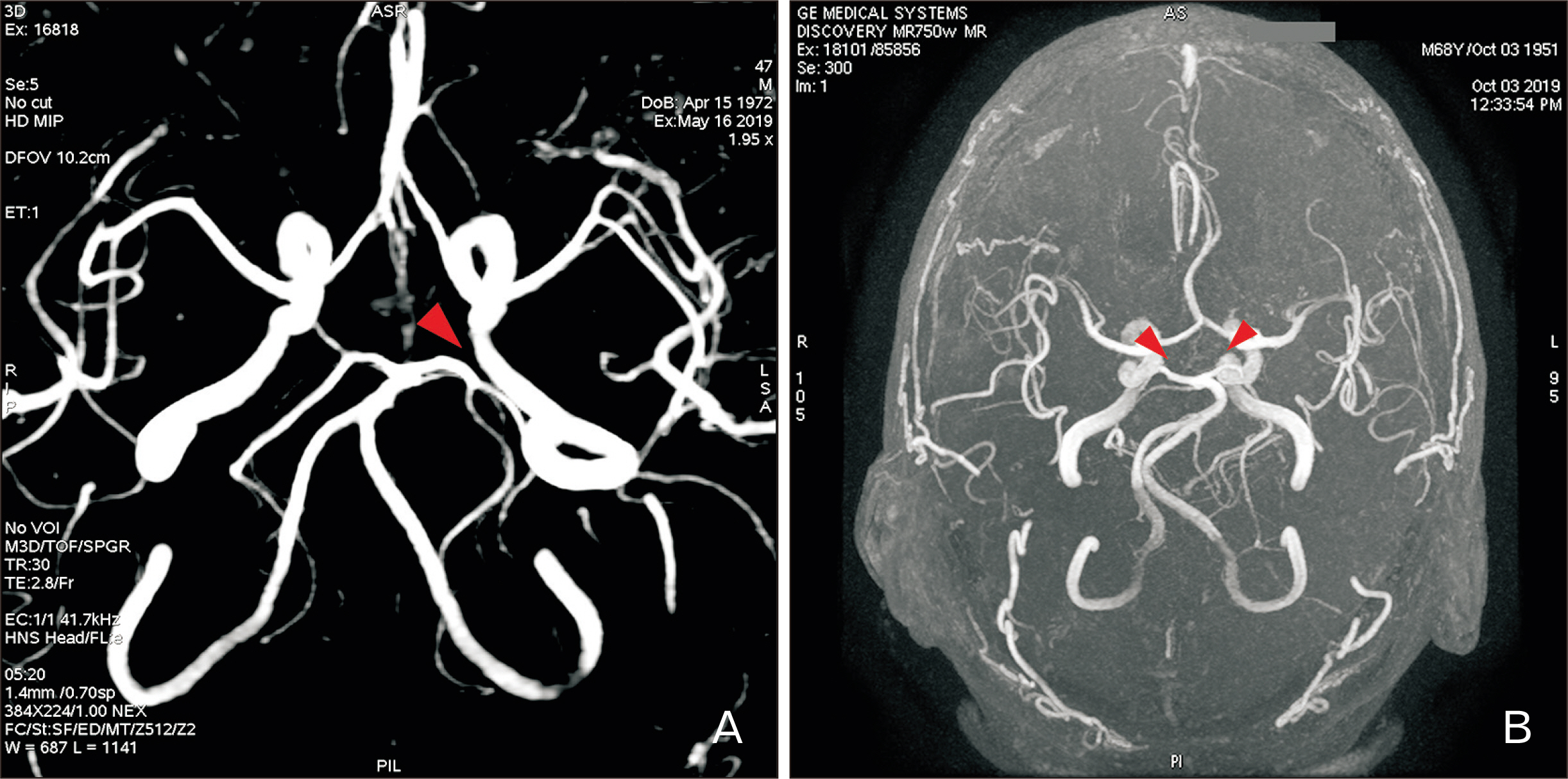
Fig. 2 MRA images showing (A) left side PCoA hypoplasia (arrowhead); (B) bilateral PCoA hypoplasia (arrowheads). MRA, magnetic resonance angiography; PCoA, posterior communicating artery.
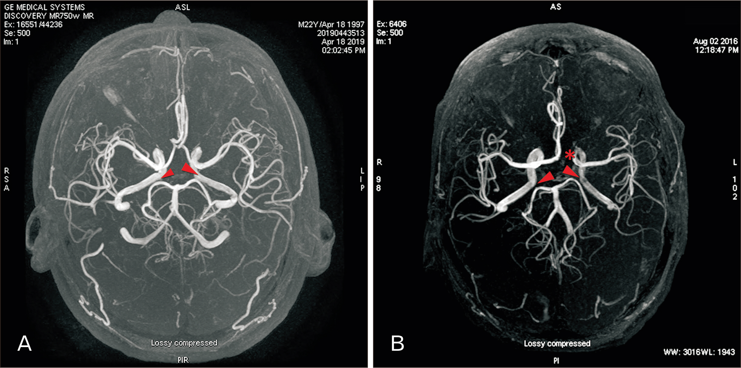
Fig. 3 MRA images showing (A) bilateral PCoA hypoplasia (arrowheads); (B) bilateral PCoA hypoplasia (arrowheads) with hypoplastic A1 segment of anterior cerebral artery (asterisk). MRA, magnetic resonance angiography; PCoA, posterior communicating artery.
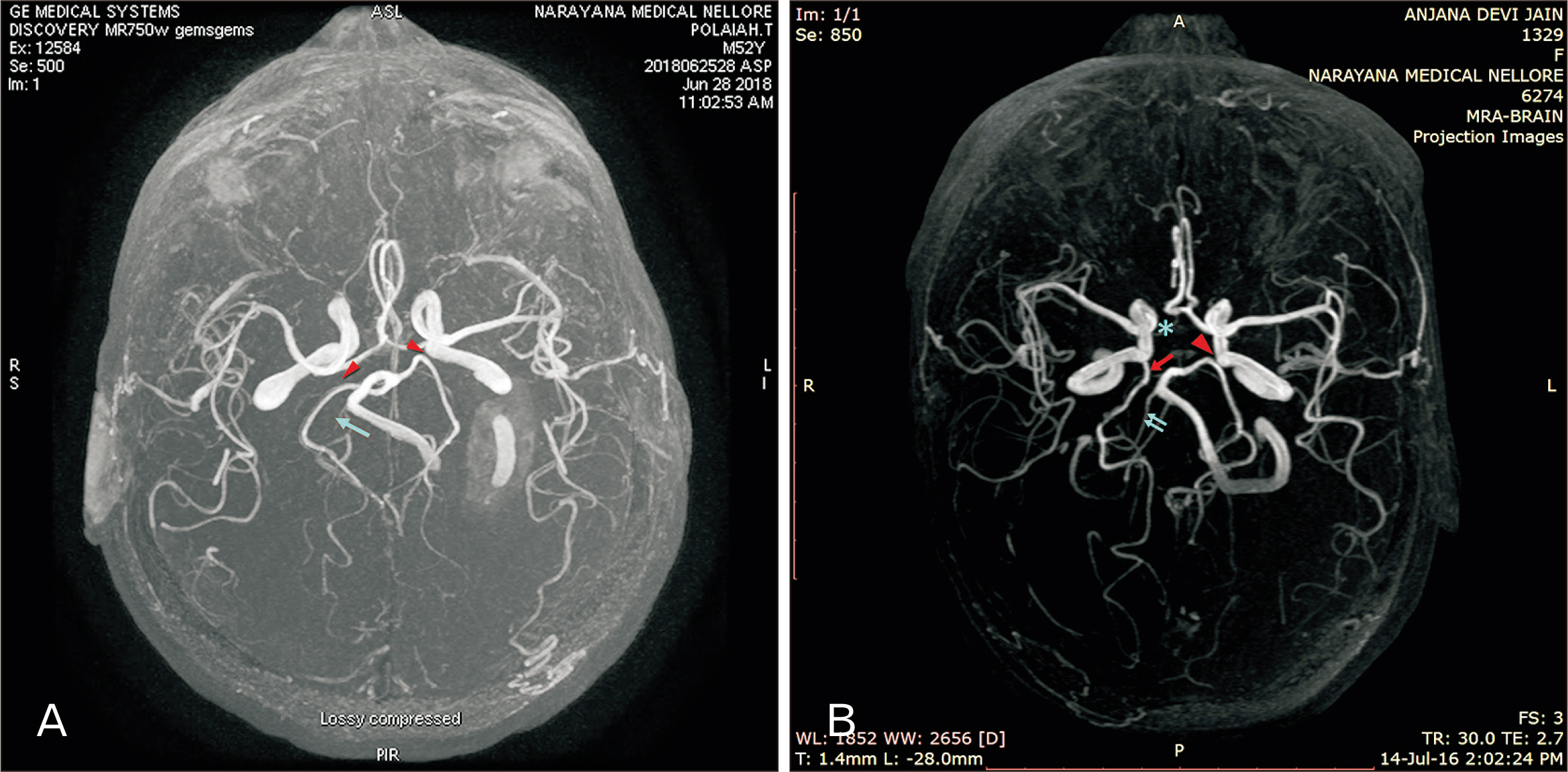
Fig. 4 MRA images showing (A) bilateral PCoA hypoplasia (arrowheads) with hypoplastic right vertebral artery (arrow); (B) left side PCoA hypoplasia (arrowhead) with right fetal type of posterior cerebral artery (arrow); this case also presented hypoplastic right A1 segment of anterior cerebral artery (asterisk) and hypoplastic right vertebral artery (double arrows). MRA, magnetic resonance angiography; PCoA, posterior communicating artery.
Reference
-
References
1. Eftekhar B, Dadmehr M, Ansari S, Ghodsi M, Nazparvar B, Ketabchi E. 2006; Are the distributions of variations of circle of Willis different in different populations? - Results of an anatomical study and review of literature. BMC Neurol. 6:22. DOI: 10.1186/1471-2377-6-22. PMID: 16796761. PMCID: PMC3680999.
Article2. Bouthillier A, van Loveren HR, Keller JT. 1996; Segments of the internal carotid artery: a new classification. Neurosurgery. 38:425–32. discussion 432–3. DOI: 10.1227/00006123-199603000-00001. PMID: 8837792. PMCID: PMC7411175.
Article3. Roopashree R. 2013; A anatomical study on relationship between posterior cerebral artery and posterior communicating artery. Int J Anat Radiol Surg. 2:9–12.4. Krabbe-Hartkamp MJ, van der Grond J, de Leeuw FE, de Groot JC, Algra A, Hillen B, Breteler MM, Mali WP. 1998; Circle of Willis: morphologic variation on three-dimensional time-of-flight MR angiograms. Radiology. 207:103–11. DOI: 10.1148/radiology.207.1.9530305. PMID: 9530305. PMCID: PMC4683875.
Article5. Feldman BA. 2013; Embryology and variations of cerebral arteries- a pictorial review. European Soc Radiol. 1–33. doi: 10.1594/ecr2013/C-2520.6. Chuang YM, Liu CY, Pan PJ, Lin CP. 2008; Posterior communicating artery hypoplasia as a risk factor for acute ischemic stroke in the absence of carotid artery occlusion. J Clin Neurosci. 15:1376. DOI: 10.1016/j.jocn.2008.02.002. PMID: 18945618.
Article7. Pentyala S, Sankar KD, Bhanu PS, Kumar NSS. 2019; Magnetic resonance angiography of hypoplastic A1 segment of anterior cerebral artery at 3.0-Tesla in Andhra Pradesh population of India. Anat Cell Biol. 52:43. DOI: 10.5115/acb.2019.52.1.43. PMID: 30984451.
Article8. Kapoor K, Singh B, Dewan LI. 2008; Variations in the configuration of the circle of Willis. Anat Sci Int. 83:96. DOI: 10.1111/j.1447-073X.2007.00216.x. PMID: 18507619.
Article9. Conijn MM, Hendrikse J, Zwanenburg JJ, Takahara T, Geerlings MI, Mali WP, Luijten PR. 2009; Perforating arteries originating from the posterior communicating artery: a 7.0-Tesla MRI study. Eur Radiol. 19:2986–92. DOI: 10.1007/s00330-009-1485-4. PMID: 19533146. PMCID: PMC2778782.
Article10. Zwanenburg JJ, Hendrikse J, Takahara T, Visser F, Luijten PR. 2008; MR angiography of the cerebral perforating arteries with magnetization prepared anatomical reference at 7 T: comparison with time-of-flight. J Magn Reson Imaging. 28:1519. DOI: 10.1002/jmri.21591. PMID: 19025959.11. Ardakani SK, Dadmehr M, Nejat F, Ansari S, Eftekhar B, Tajik P, El Khashab M, Yazdani S, Ghodsi M, Mahjoub F, Monajemzadeh M, Nazparvar B, Abdi-Rad A. 2008; The cerebral arterial circle (circulus arteriosus cerebri): an anatomical study in fetus and infant samples. Pediatr Neurosurg. 44:388–92. DOI: 10.1159/000149906. PMID: 18703885.12. Neau JP, Bogousslavsky J. 1996; The syndrome of posterior choroidal artery territory infarction. Ann Neurol. 39:779. DOI: 10.1002/ana.410390614. PMID: 8651650.13. Wardlaw JM, Dennis MS, Warlow CP, Sandercock PA. 2001; Imaging appearance of the symptomatic perforating artery in patients with lacunar infarction: occlusion or other vascular pathology? Ann Neurol. 50:208. DOI: 10.1002/ana.1082. PMID: 11506404.
Article14. Chuang YM, Huang YC, Hu HH, Yang CY. 2006; Toward a further elucidation: role of vertebral artery hypoplasia in acute ischemic stroke. Eur Neurol. 55:193. DOI: 10.1159/000093868. PMID: 16772715.
Article15. Fischer U, Arnold M, Nedeltchev K, Brekenfeld C, Ballinari P, Remonda L, Schroth G, Mattle HP. 2005; NIHSS score and arteriographic findings in acute ischemic stroke. Stroke. 36:2121–5. DOI: 10.1161/01.STR.0000182099.04994.fc. PMID: 16151026.
Article16. Vilimas A, Barkauskas E, Vilionskis A, Rudzinskaite J, Morkunaite R. 2003; Vertebral artery hypoplasia: importance for stroke development, the role of posterior communicating artery, possibility for surgical and conservative treatment. Acta Med Litu. 10:110–4.17. Park JH, Kim JM, Roh JK. 2007; Hypoplastic vertebral artery: frequency and associations with ischaemic stroke territory. J Neurol Neurosurg Psychiatry. 78:954. DOI: 10.1136/jnnp.2006.105767. PMID: 17098838. PMCID: PMC2117863.
Article18. De Silva KR, Silva R, De Silva C, Gunasekera WS, Dias P, Jayesekera RW. 2010; Comparison of the configuration of the posterior bifurcation of the posterior communicating artery between fetal and adult brains: a study of a Sri Lankan population. Ann Indian Acad Neurol. 13:198. DOI: 10.4103/0972-2327.70886. PMID: 21085531. PMCID: PMC2981758.
Article19. Sahni D, Jit I, Lal V. 2007; Variations and anomalies of the posterior communicating artery in Northwest Indian brains. Surg Neurol. 68:449. DOI: 10.1016/j.surneu.2006.11.047. PMID: 17905073.
Article20. Krishnamurthy A, Nayak SR, Ganesh Kumar C, Jetti R, Prabhu LV, Ranade AV, Rai R. 2008; Morphometry of posterior cerebral artery: embryological and clinical significance. Rom J Morphol Embryol. 49:43–5. PMID: 18273501.21. Menshawi K, Mohr JP, Gutierrez J. 2015; A functional perspective on the embryology and anatomy of the cerebral blood supply. J Stroke. 17:144. DOI: 10.5853/jos.2015.17.2.144. PMID: 26060802. PMCID: PMC4460334.
Article22. Caldemeyer KS, Carrico JB, Mathews VP. 1998; The radiology and embryology of anomalous arteries of the head and neck. AJR Am J Roentgenol. 170:197. DOI: 10.2214/ajr.170.1.9423632. PMID: 9423632.
Article23. Abrahams JM, Hurst RW, Bagley LJ, Zager EL. 1999; Anterior choroidal artery supply to the posterior cerebral artery distribution: embryological basis and clinical implications. Neurosurgery. 44:1308. DOI: 10.1227/00006123-199906000-00083. PMID: 10371631.
Article24. Lasjaunias P, Berenstein A, ter Brugge KG. Lasjaunias P, Berenstein A, ter Brugge KG, editors. 2001. Intradural Arteries. Clinical vascular anatomy and variations. 2nd ed. Springer;Berlin: p. 479–629. DOI: 10.1007/978-3-662-10172-8_6.
Article25. Jayaraman MV, Mayo-Smith WW. 2004; Multi-detector CT angiography of the intra-cranial circulation: normal anatomy and pathology with angiographic correlation. Clin Radiol. 59:690. DOI: 10.1016/j.crad.2003.12.004. PMID: 15262542.
Article26. Padge DH. 1948; The development of the cranial arteries in the human embryo. Contrib Embryol. 32:205–61.27. Parmar H, Sitoh YY, Hui F. 2005; Normal variants of the intracranial circulation demonstrated by MR angiography at 3T. Eur J Radiol. 56:220. DOI: 10.1016/j.ejrad.2005.05.005. PMID: 15950421.
Article28. Hoksbergen AW, Fülesdi B, Legemate DA, Csiba L. 2000; Collateral configuration of the circle of Willis: transcranial color-coded duplex ultrasonography and comparison with postmortem anatomy. Stroke. 31:1346. DOI: 10.1161/01.STR.31.6.1346. PMID: 10835455.29. Al-Hussain SM, Shoter AM, Bataina ZM. 2001; Circle of Willis in adults. Saudi Med J. 22:895.30. Zhou W, Lu M, Li J, Chen F, Hu Q, Yang S. 2019; Functional posterior communicating artery of patients with posterior circulation ischemia using phase contrast magnetic resonance angiography. Exp Ther Med. 17:337. DOI: 10.3892/etm.2018.6897. PMID: 30651800. PMCID: PMC6307428.
Article31. Gunnal SA, Farooqui MS, Wabale RN. 2015; Study of posterior cerebral artery in human cadaveric brain. Anat Res Int. 2015:681903. DOI: 10.1155/2015/681903. PMID: 26413321. PMCID: PMC4561305.
Article32. Tuncer MC, Akgül YH, Karabulut Ö. 2010; MR angiography imaging of absence vertebral artery causing of pulsatile tinnitus: a case report. Int J Morphol. 28:357–63. DOI: 10.4067/S0717-95022010000200003.
Article33. Vasuki AKM, Devi MN, Jamuna M. 2015; Absent intracranial part of right vertebral artery-a case report. Innov J Med Health Sci. 5:6–8.34. Kang DW, Chu K, Ko SB, Kwon SJ, Yoon BW, Roh JK. 2002; Lesion patterns and mechanism of ischemia in internal carotid artery disease: a diffusion-weighted imaging study. Arch Neurol. 59:1577. DOI: 10.1001/archneur.59.10.1577. PMID: 12374495.
- Full Text Links
-
- Actions
-
Cited
- CITED
-
- Close
- Share
-
- Similar articles
-
- Magnetic resonance angiography of hypoplastic A1 segment of anterior cerebral artery at 3.0-Tesla in Andhra Pradesh population of India
- Clinical Analysis of the Pattern of Anterior-Posterior Circulation in Patients with Posterior Communicating Artery Aneurysm
- Variations and anomalies of the circle of Willis in Korean: Cerebral digital subtraction angiogram studies in 200 cases
- Transposition of Anterior Choroidal Artery and Posterior Communicating Artery Origin
- Fusiform “True” Posterior Communicating Artery Aneurysm with Basilar Artery Occlusion: A Case Report
