Korean J Ophthalmol.
2020 Apr;34(2):113-120. 10.3341/kjo.2019.0105.
Repeatability of Manual Measurement of Foveal Avascular Zone Area in Optical Coherence Tomography Angiography Images in High Myopia
- Affiliations
-
- 1Department of Ophthalmology, Konyang University College of Medicine, Daejeon, Korea.
- KMID: 2507400
- DOI: http://doi.org/10.3341/kjo.2019.0105
Abstract
- Purpose
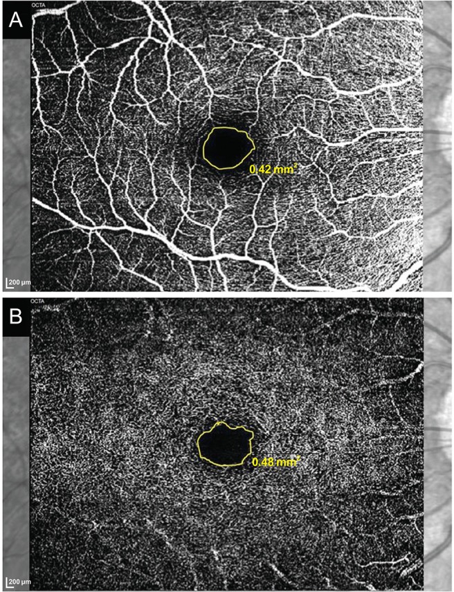
To analyze the repeatability of manual measurement of foveal avascular zone (FAZ) area in an optical coherence tomography angiography (OCTA) image in high myopia.
Methods
This study comprised patients with high myopia and controls. Two consecutive FAZ areas of the superficial and deep capillary plexus were obtained using OCTA. The intraclass correlation coefficient (ICC) and coefficient of variation (CV) were assessed, and univariate and multivariate generalized linear mixed models were conducted to identify factors related to repeatability.
Results
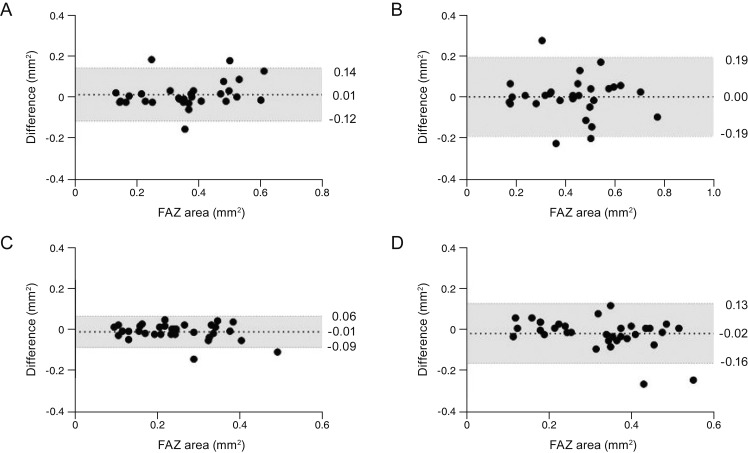
Thirty eyes with high myopia and 34 eyes of healthy subjects were included in the study. The mean age in high myopia and control subjects was 55.5 and 60.8 years, respectively, the mean spherical equivalent was −9.98 and −0.55 diopters, and the mean axial length was 28.0 and 23.9 mm. The ICCs of FAZ area of the superficial capillary plexus (SCP) were 0.891 and 0.919, while the CVs were 8.8% and 8.5%. In measurement of the deep capillary plexus, the ICCs were 0.788 and 0.907, while the CVs were 11.2% and 11.0%, which were acceptable but exhibited lower repeatability than those of SCP. Multivariate analyses showed that older age (p = 0.030) and greater axial length (p = 0.005) were significantly associated with lower repeatability of SCP FAZ area measurements. In addition, greater axial length (p = 0.044) was a significant factor for lower repeatability of deep capillary plexus FAZ area measurements.
Conclusions
Manual measurement of FAZ area using OCTA exhibited relatively good repeatability for high myopia. Age and axial length affected repeatability and should be considered when analyzing FAZ areas in high myopia patients.
Figure
Reference
-
1. Katz J, Tielsch JM, Sommer A. Prevalence and risk factors for refractive errors in an adult inner city population. Invest Ophthalmol Vis Sci. 1997; 38:334–340. PMID: 9040465.2. Wang Q, Klein BE, Klein R, Moss SE. Refractive status in the Beaver Dam Eye Study. Invest Ophthalmol Vis Sci. 1994; 35:4344–4347. PMID: 8002254.3. Wong TY, Foster PJ, Hee J, et al. Prevalence and risk factors for refractive errors in adult Chinese in Singapore. Invest Ophthalmol Vis Sci. 2000; 41:2486–2494. PMID: 10937558.4. Saw SM, Gazzard G, Shih-Yen EC, Chua WH. Myopia and associated pathological complications. Ophthalmic Physiol Opt. 2005; 25:381–391. PMID: 16101943.
Article5. Hayashi K, Ohno-Matsui K, Shimada N, et al. Long-term pattern of progression of myopic maculopathy: a natural history study. Ophthalmology. 2010; 117:1595–1611. PMID: 20207005.6. Curtin BJ. The posterior staphyloma of pathologic myopia. Trans Am Ophthalmol Soc. 1977; 75:67–86. PMID: 613534.7. Ohno-Matsui K, Yoshida T, Futagami S, et al. Patchy atrophy and lacquer cracks predispose to the development of choroidal neovascularisation in pathological myopia. Br J Ophthalmol. 2003; 87:570–573. PMID: 12714395.
Article8. Spaide RF, Klancnik JM Jr, Cooney MJ. Retinal vascular layers imaged by fluorescein angiography and optical coherence tomography angiography. JAMA Ophthalmol. 2015; 133:45–50. PMID: 25317632.
Article9. Pilotto E, Frizziero L, Crepaldi A, et al. Repeatability and reproducibility of foveal avascular zone area measurement on normal eyes by different optical coherence tomography angiography instruments. Ophthalmic Res. 2018; 59:206–211. PMID: 29421813.
Article10. Sung MS, Lee TH, Heo H, Park SW. Association between optic nerve head deformation and retinal microvasculature in high myopia. Am J Ophthalmol. 2018; 188:81–90. PMID: 29421295.
Article11. Balaratnasingam C, Inoue M, Ahn S, et al. Visual acuity is correlated with the area of the foveal avascular zone in diabetic retinopathy and retinal vein occlusion. Ophthalmology. 2016; 123:2352–2367. PMID: 27523615.
Article12. Kwon J, Choi J, Shin JW, et al. Alterations of the foveal avascular zone measured by optical coherence tomography angiography in glaucoma patients with central visual field defects. Invest Ophthalmol Vis Sci. 2017; 58:1637–1645. PMID: 28297029.
Article13. Guo J, She X, Liu X, Sun X. Repeatability and reproducibility of foveal avascular zone area measurements using AngioPlex spectral domain optical coherence tomography angiography in healthy subjects. Ophthalmologica. 2017; 237:21–28. PMID: 28088800.
Article14. Al-Sheikh M, Tepelus TC, Nazikyan T, Sadda SR. Repeatability of automated vessel density measurements using optical coherence tomography angiography. Br J Ophthalmol. 2017; 101:449–452. PMID: 27450146.
Article15. Carpineto P, Mastropasqua R, Marchini G, et al. Reproducibility and repeatability of foveal avascular zone measurements in healthy subjects by optical coherence tomography angiography. Br J Ophthalmol. 2016; 100:671–676. PMID: 26377414.
Article16. Corvi F, Pellegrini M, Erba S, et al. Reproducibility of vessel density, fractal dimension, and foveal avascular zone using 7 different optical coherence tomography angiography devices. Am J Ophthalmol. 2018; 186:25–31. PMID: 29169882.
Article17. Lei J, Durbin MK, Shi Y, et al. Repeatability and reproducibility of superficial macular retinal vessel density measurements using optical coherence tomography angiography en face images. JAMA Ophthalmol. 2017; 135:1092–1098. PMID: 28910435.
Article18. Manalastas PIC, Zangwill LM, Saunders LJ, et al. Reproducibility of optical coherence tomography angiography macular and optic nerve head vascular density in glaucoma and healthy eyes. J Glaucoma. 2017; 26:851–859. PMID: 28858159.
Article19. Lee MW, Kim KM, Lim HB, et al. Repeatability of vessel density measurements using optical coherence tomography angiography in retinal diseases. Br J Ophthalmol. 2018; 7. 04. [Epub]. DOI: 10.1136/bjophthalmol-2018-312516.
Article20. Amoroso F, Miere A, Semoun O, et al. Optical coherence tomography angiography reproducibility of lesion size measurements in neovascular age-related macular degeneration (AMD). Br J Ophthalmol. 2018; 102:821–826. PMID: 28855197.
Article21. He J, Chen Q, Yin Y, et al. Association between retinal microvasculature and optic disc alterations in high myopia. Eye (Lond). 2019; 33:1494–1503. PMID: 31019262.
Article22. Vurgese S, Panda-Jonas S, Jonas JB. Scleral thickness in human eyes. PLoS One. 2012; 7:e29692. PMID: 22238635.
Article23. Spaide RF, Fujimoto JG, Waheed NK. Image artifacts in optical coherence angiography. Retina. 2015; 35:2163–2180. PMID: 26428607.24. Durbin MK, An L, Shemonski ND, et al. Quantification of retinal microvascular density in optical coherence tomographic angiography images in diabetic retinopathy. JAMA Ophthalmol. 2017; 135:370–376. PMID: 28301651.
Article
- Full Text Links
- Actions
-
Cited
- CITED
-
- Close
- Share
- Similar articles
-
- Optical Coherence Tomography-angiography: Comparison of the Foveal Avascular Zone between Diabetic Retinopathy and Normal Subjects
- Macular Microvasculature Features of the Eyes of Healthy Korean Children Using Optical Coherence Tomography Angiography
- Efficacy of Optical Coherence Tomography Angiography in Measuring the Foveal Avascular Zone Area in Patients with Branch Retinal Vein Occlusion
- Evaluation of Foveal Avascular Zone and Capillary Plexuses in Diabetic Patients by Optical Coherence Tomography Angiography
- Foveal Microvascular Changes Based on Optical Coherence Tomography Angiography in Mild Nonproliferative Diabetic Retinopathy


