J Korean Med Sci.
2020 Sep;35(37):e332. 10.3346/jkms.2020.35.e332.
Air and Environmental Contamination Caused by COVID-19 Patients: a MultiCenter Study
- Affiliations
-
- 1Department of Infectious Diseases, Chonnam National University Medical School, Gwangju, Korea
- 2Department of Laboratory Medicine, Chonnam National University Medical School, Gwangju, Korea
- 3Department of Infectious Disease, Keimyung University Dongsan Hospital, Daegu, Korea
- 4Office for Infection Control, Chonnam National University Hwasun Hospital, Hwasun, Korea
- KMID: 2506565
- DOI: http://doi.org/10.3346/jkms.2020.35.e332
Abstract
- Background
The purpose of this study was to determine the extent of air and surface contamination of severe acute respiratory syndrome coronavirus-2 (SARS-CoV-2) in four health care facilities with hospitalized coronavirus disease 2019 (COVID-19) patients.
Methods
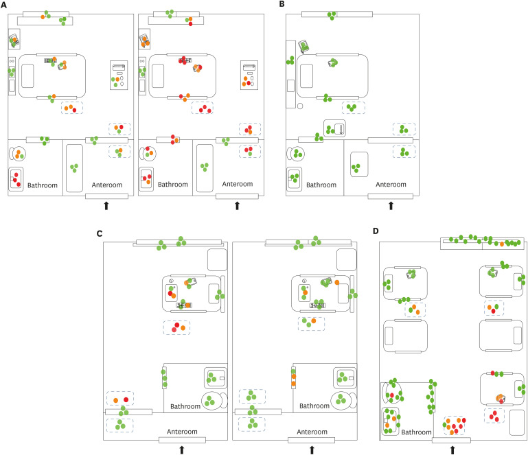
We investigated air and environmental contamination in the rooms of eight COVID-19 patients in four hospitals. Some patients were in negative-pressure rooms, and others were not. None had undergone aerosol-generating procedures. On days 0, 3, 5, and 7 of hospitalization, the surfaces in the rooms and anterooms were swabbed, and air samples were collected 2 m from the patient and from the anterooms.
Results
All 52 air samples were negative for SARS-CoV-2 RNA. Widespread surface contamination of SARS-CoV-2 RNA was observed. In total, 89 of 320 (27%) environmental surface samples were positive for SARS-CoV-2 RNA. Surface contamination of SARSCoV-2 RNA was common in rooms without surface disinfection and in rooms sprayed with disinfectant twice a day. However, SARS-CoV-2 RNA was not detected in a room cleaned with disinfectant wipes on a regular basis.
Conclusion
Our data suggest that remote (> 2 m) airborne transmission of SARS-CoV-2 from hospitalized COVID-19 patients is uncommon when aerosol-generating procedures have not been performed. Surface contamination was widespread, except in a room routinely cleaned with disinfectant wipes.
Keyword
Figure
Reference
-
1. World Health Organization. Situation report. Updated 2020. Accessed June 1, 2020. https://www.who.int/docs/default-source/coronaviruse/situation-reports/20200601-covid-19-sitrep-133.pdf?sfvrsn=9a56f2ac_4.2. Liu Y, Gayle AA, Wilder-Smith A, Rocklöv J. The reproductive number of COVID-19 is higher compared to SARS coronavirus. J Travel Med. 2020; 27(2):taaa021. PMID: 32052846.
Article3. Centers for Disease Control and Prevention. Interim infection prevention and control recommendations for patients with suspected or confirmed coronavirus disease 2019 (COVID-19) in healthcare settings. Updated 2020. Accessed June 1, 2020. https://www.cdc.gov/coronavirus/2019-ncov/hcp/infection-control-recommendations.html.4. Chia PY, Coleman KK, Tan YK, Ong SWX, Gum M, Lau SK, et al. Detection of air and surface contamination by SARS-CoV-2 in hospital rooms of infected patients. Nat Commun. 2020; 11(1):2800. PMID: 32472043.
Article5. Guo ZD, Wang ZY, Zhang SF, Li X, Li L, Li C, et al. Aerosol and surface distribution of severe acute respiratory syndrome coronavirus 2 in hospital wards, Wuhan, China, 2020. Emerg Infect Dis. 2020; 26(7):1583–1591. PMID: 32275497.
Article6. van Doremalen N, Bushmaker T, Morris DH, Holbrook MG, Gamble A, Williamson BN, et al. Aerosol and surface stability of SARS-CoV-2 as compared with SARS-CoV-1. N Engl J Med. 2020; 382(16):1564–1567. PMID: 32182409.
Article7. Liu Y, Ning Z, Chen Y, Guo M, Liu Y, Gali NK, et al. Aerodynamic analysis of SARS-CoV-2 in two Wuhan hospitals. Nature. 2020; 582(7813):557–560. PMID: 32340022.
Article8. Bahl P, Doolan C, de Silva C, Chughtai AA, Bourouiba L, MacIntyre CR. Airborne or droplet precautions for health workers treating COVID-19? J Infect Dis. 2020.9. Setti L, Passarini F, De Gennaro G, Barbieri P, Perrone MG, Borelli M, et al. Airborne transmission route of COVID-19: why 2 meters/6 feet of inter-personal distance could not be enough. Int J Environ Res Public Health. 2020; 17(8):2932.
Article10. Ong SWX, Tan YK, Chia PY, Lee TH, Ng OT, Wong MSY, et al. Air, surface environmental, and personal protective equipment contamination by severe acute respiratory syndrome coronavirus 2 (SARS-CoV-2) from a symptomatic patient. JAMA. 2020; 323(16):1610–1612. PMID: 32129805.
Article11. Yung CF, Kam KQ, Wong MSY, Maiwald M, Tan YK, Tan BH, et al. Environment and personal protective equipment tests for SARS-CoV-2 in the isolation room of an infant with infection. Ann Intern Med. 2020; 173(3):240–242. PMID: 32236490.
Article12. Cheng VC, Wong SC, Chan VW, So SY, Chen JH, Yip CC, et al. Air and environmental sampling for SARS-CoV-2 around hospitalized patients with coronavirus disease 2019 (COVID-19). Infect Control Hosp Epidemiol. 2020.
Article
- Full Text Links
- Actions
-
Cited
- CITED
-
- Close
- Share
- Similar articles
-
- COVID-19 Vaccination for Pilots and Air Traffic Controllers
- Psychological Predictors of Anxious Responses to the COVID-19 Pandemic: Evidence from Pakistan
- COVID-19 and Cancer: Questions to Be Answered
- COVID-19 and Aviation Medical Examination
- Aviation Operators’ Response Plan to Overcome the Crisis in the Aviation Industry Caused by COVID-19


