J Korean Foot Ankle Soc.
2020 Sep;24(3):120-123. 10.14193/jkfas.2020.24.3.120.
Plantar Keratosis Induced by Heterotopic Ossification under the Medial Sesamoid Bone: A Case Report
- Affiliations
-
- 1Departments of Orthopaedic Surgery, Ilsan Paik Hospital, Inje University College of Medicine, Goyang, Korea
- 2Departments of Orthopaedic Pathology, Ilsan Paik Hospital, Inje University College of Medicine, Goyang, Korea
- KMID: 2506529
- DOI: http://doi.org/10.14193/jkfas.2020.24.3.120
Abstract
- Heterotopic ossification is the formation of extra-skeletal bone in the muscle and soft tissues, and an osteoma is a benign bone-forming tumor composed of compact or mature trabecular bone limited almost exclusively to the craniofacial bones. This paper reports an extremely rare case of heterotopic ossification mimicking an osteoma that occurred independently at the plantar side of the medial sesamoid bone. The patient was a 46-year-old male with a three-month history of pain and a hard mass on the plantar aspect of the left forefoot sole. After excising the lesion, the patient’s symptoms were relieved, and no pain or complications occurred. This paper discusses this exceedingly rare case of heterotopic ossification around the medial sesamoid bone with a review of the relevant literature.
Figure
-
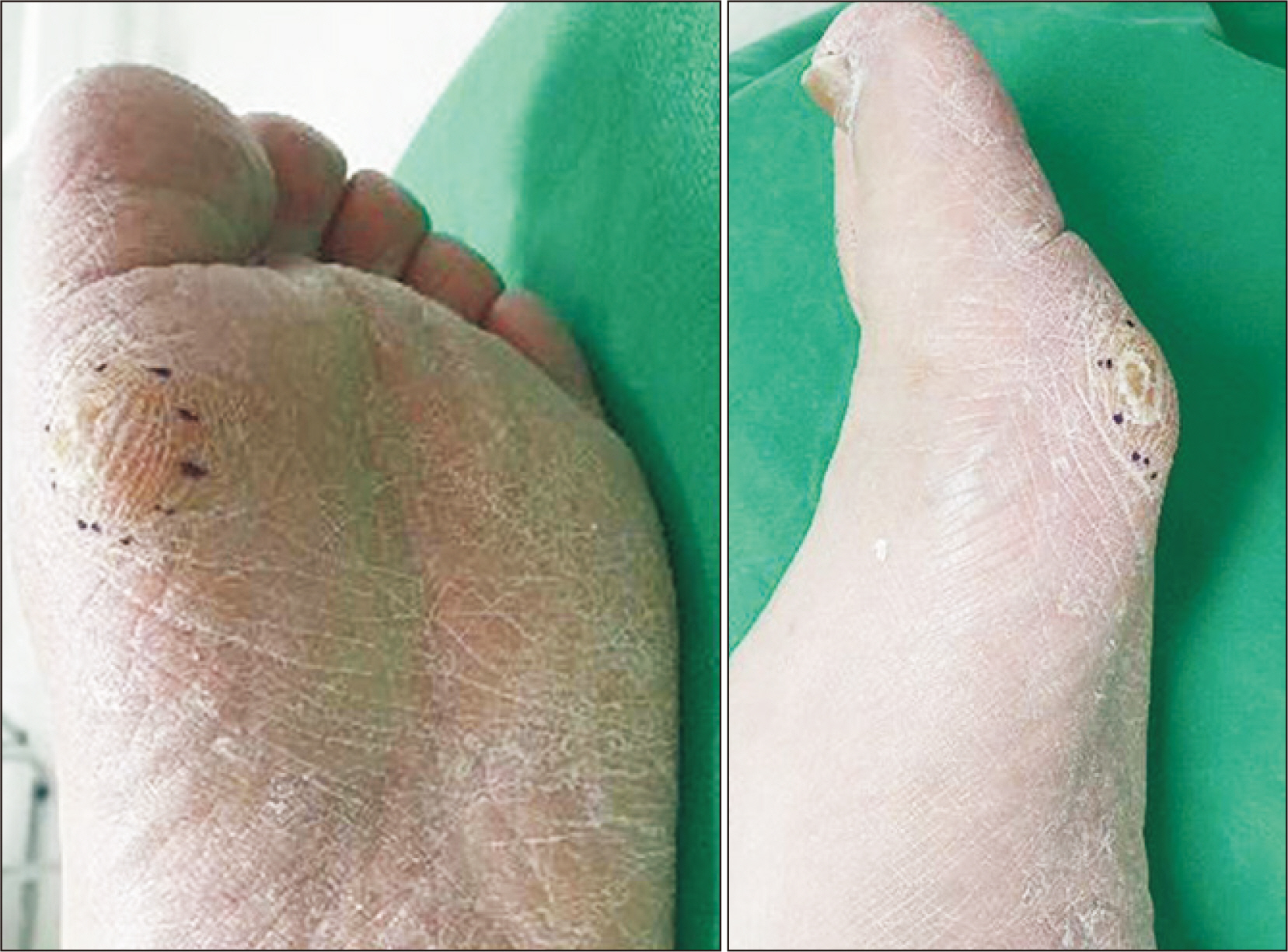
Figure. 1 Clinical photographs show plantar keratosis at the plantar side of the medial sesamoid bone.

Figure. 2 Preoperative weight-bearing anteroposterior (A) and lateral (B) radiographs present the bony mass lesion under medial sesamoid. (C) On sesamoid axial image, bony mass lesion is more markedly observed.

Figure. 3 Sagittal (A) and axial (B) computed tomographic images show the bony mass lesion separated from the medial sesamoid bone.

Figure. 4 A 10×9×6 mm3 sized oval bony mass was excised.

Figure. 5 (A) A round exostotic tumorous lesion was removed (H&E stain scan view). (B) The mass was composed of compact lamellar bone with intraosseous Haversian systems (H&E stain, 10x).
Reference
-
1. Meyers C, Lisiecki J, Miller S, Levin A, Fayad L, Ding C, et al. 2019; Heterotopic ossification: a comprehensive review. JBMR Plus. 3:e10172. doi: 10.1002/jbm4.10172. DOI: 10.1002/jbm4.10172. PMID: 31044187. PMCID: PMC6478587.
Article2. Motamedi K, Seeger LL. 2011; Benign bone tumors. Radiol Clin North Am. 49:1115–34. v. doi: 10.1016/j.rcl.2011.07.002. DOI: 10.1016/j.rcl.2011.07.002. PMID: 22024291.
Article3. Nakashima Y, Haga N, Kitoh H, Kamizono J, Tozawa K, Katagiri T, et al. 2010; Deformity of the great toe in fibrodysplasia ossificans progressiva. J Orthop Sci. 15:804–9. doi: 10.1007/s00776-010-1542-5. DOI: 10.1007/s00776-010-1542-5. PMID: 21116899.
Article4. Ouchi K, Hakozaki M, Kikuchi SI, Yabuki S, Konno SI. 2017; Osteochondroma of the tibial sesamoid: a case report and review of the literature. J Foot Ankle Surg. 56:628–31. doi: 10.1053/j.jfas.2016.10.007. DOI: 10.1053/j.jfas.2016.10.007. PMID: 28215361.5. Mowad SC Sr, Zichichi S, Mullin R. 1995; Osteochondroma of the tibial sesamoid. J Am Podiatr Med Assoc. 85:765–6. doi: 10.7547/87507315-85-12-765. DOI: 10.7547/87507315-85-12-765. PMID: 8592289.
Article6. Okada A, Hatori M, Hashimoto Y, Lee E. 2012; Painful extraskeletal osteochondroma under the tarsal sesamoid: a case report and review of literature. Eur J Orthop Surg Traumatol. 22(Suppl 1):215–20. doi: 10.1007/s00590-011-0857-z. DOI: 10.1007/s00590-011-0857-z. PMID: 26662780.
Article7. Nora FE, Dahlin DC, Beabout JW. 1983; Bizarre parosteal osteochondromatous proliferations of the hands and feet. Am J Surg Pathol. 7:245–50. doi: 10.1097/00000478-198304000-00003. DOI: 10.1097/00000478-198304000-00003. PMID: 6837834.
Article8. Torreggiani WC, Munk PL, Al-Ismail K, O’Connell JX, Nicolaou S, Lee MJ, et al. 2001; MR imaging features of bizarre parosteal osteochondromatous proliferation of bone (Nora’s lesion). Eur J Radiol. 40:224–31. doi: 10.1016/s0720-048x(01)00362-x. DOI: 10.1016/S0720-048X(01)00362-X.
Article9. Harty JA, Kelly P, Niall D, O’Keane JC, Stephens MM. 2000; Bizarre parosteal osteochondromatous proliferation (Nora’s lesion) of the sesamoid: a case report. Foot Ankle Int. 21:408–12. doi: 10.1177/107110070002100509. DOI: 10.1177/107110070002100509. PMID: 10830660.
Article10. Noguchi M, Ikoma K, Matsumoto N, Nagasawa K. 2004; Bizarre parosteal osteochondromatous proliferation of the sesamoid: an unusual hallux valgus deformity. Foot Ankle Int. 25:503–6. doi: 10.1177/107110070402500710. DOI: 10.1177/107110070402500710. PMID: 15319109.
Article11. Dobas DC, Silvers MD. 1977; The frequency of partite sesamoids of the first metatarsophalangeal joint. J Am Podiatry Assoc. 67:880–2. doi: 10.7547/87507315-67-12-880. DOI: 10.7547/87507315-67-12-880. PMID: 925322.
Article
- Full Text Links
-
- Actions
-
Cited
- CITED
-
- Close
- Share
-
- Similar articles
-
- Intractable Plantar Keratoses due to Interphalangeal Sesamoid Bone of the Hallux (A Case Report)
- Heterotopic Ossification of the Elbow after Medial Epicondylectomy
- Osteomyelitis in Heterotopic Ossification after Trochanteric Pressure Sore Reconstruction: A Case Report
- Heterotopic Ossification Mimics Neurogenic Tumor: A Case Report
- Heterotopic Ossification Combined with Infection in the Hand: A Case Report
