Leiomyosarcoma of the jaw: case series
- Affiliations
-
- 1Department of Oral and Maxillofacial Surgery, Seoul National University Dental Hospital, Seoul, Korea
- 2Clinical Translational Research Center for Dental Science, Seoul National University Dental Hospital, Seoul, Korea
- 3Oral Cancer Center, Seoul National University Dental Hospital, Seoul, Korea
- 4Department of Oral and Maxillofacial Surgery, Inje University Busan Paik Hospital, Busan, Korea
- 5Department of Oral Pathology, Seoul National University Dental Hospital, Seoul, Korea
- 6Dental Research Institute, Seoul National University, Seoul, Korea
- KMID: 2505449
- DOI: http://doi.org/10.5125/jkaoms.2020.46.4.275
Abstract
Objectives
Leiomyosarcoma is a malignant neoplasm that affects smooth muscle tissue and it is very rare in the field of oral and maxillofcial surgery. The purpose of this study was to obtain information on diagnosis of and treatment methods for leiomyosarcoma by retrospectively reviewing of the cases.
Patients and Methods
The study included nine patients who were diagnosed with leiomyosarcoma in the Department of Oral and Maxillofacial Surgery at Seoul National University Dental Hospital. The subjects were analyzed with respect to sex, age, clinical features, primary site of disease, treatment method, recurrence, and metastasis.
Results
Particular clinical features included pain, edema, mouth-opening limitations, dysesthesia, and enlarged lymph nodes. All cases except one were surgically treated, and recurrence was found in two cases. Four of nine patients were followed up without recurrence and one patient underwent additional surgery due to recurrence.
Conclusion
In our case series, notable symptoms included pain, edema, mouth-opening limitations, and dysesthesia; however, it was difficult to label these as specific symptoms of leiomyosarcoma. Considering the aggressive characteristics of the disease and poor prognosis, surgical treatment is necessary with careful consideration of postoperative radiotherapy and chemotherapy.
Keyword
Figure
-
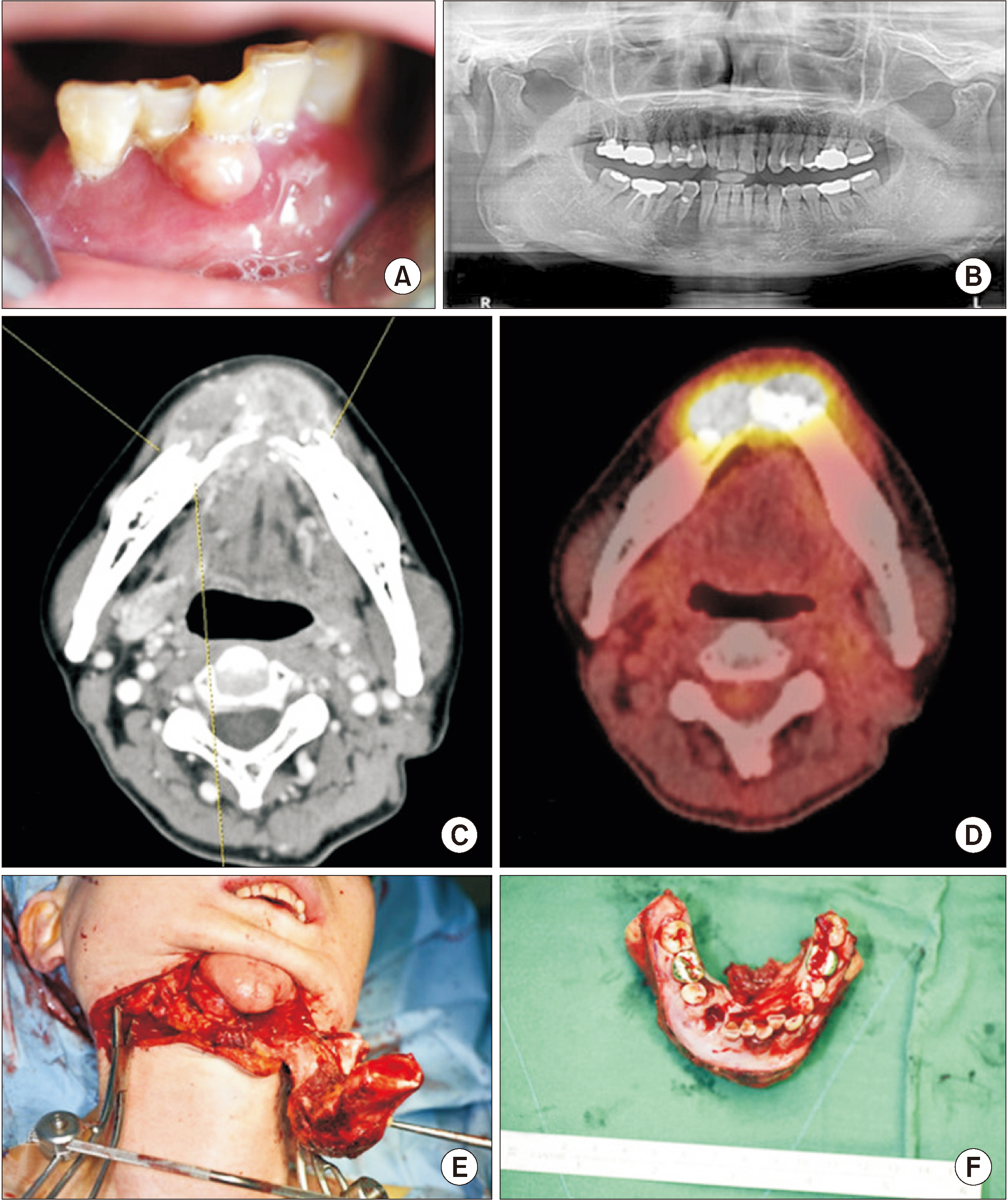
Fig. 1 A 55-year-old female patient. A. Pericoronal gingival swelling, and tooth mobility was shown. B. Panorama showing radiopaque lesion on the #43 to #45 area. C. Enhanced computed tomographic image of #33 to #46; destruction of the alveolar bone was observed, and the continuity of some lingual cortical bone disappeared. Soft tissue bulging, which was thought to be granulation tissue, was observed as a continuity loss site. Permeable osteolysis at the border of the lesion. D. F-18 flurorodeoxyglucose positron emission tomography scan showing a hypermetabolic lesion in the lower-anterior gingival area. E, F. Partial mandibulectomy was performed.

Fig. 2 A 23-year-old female patient. A. Microscopic image shows fascicles of spindle-shaped cells with blunt-ended cigar-shaped nuclei. B. Smooth muscle actin(+) positive cytoplasm in spindle-shaped tumor cells. C. Microscopic image of recurrent tumor shows similar-spindle shaped cells similar to panel A of Fig. 2 with increased cellularity and mitosis; some areas of tumor show rounded cells with eosinophilic cytoplasm. H&E and immunohistochemical staining, A: ×100; B and C: ×200.
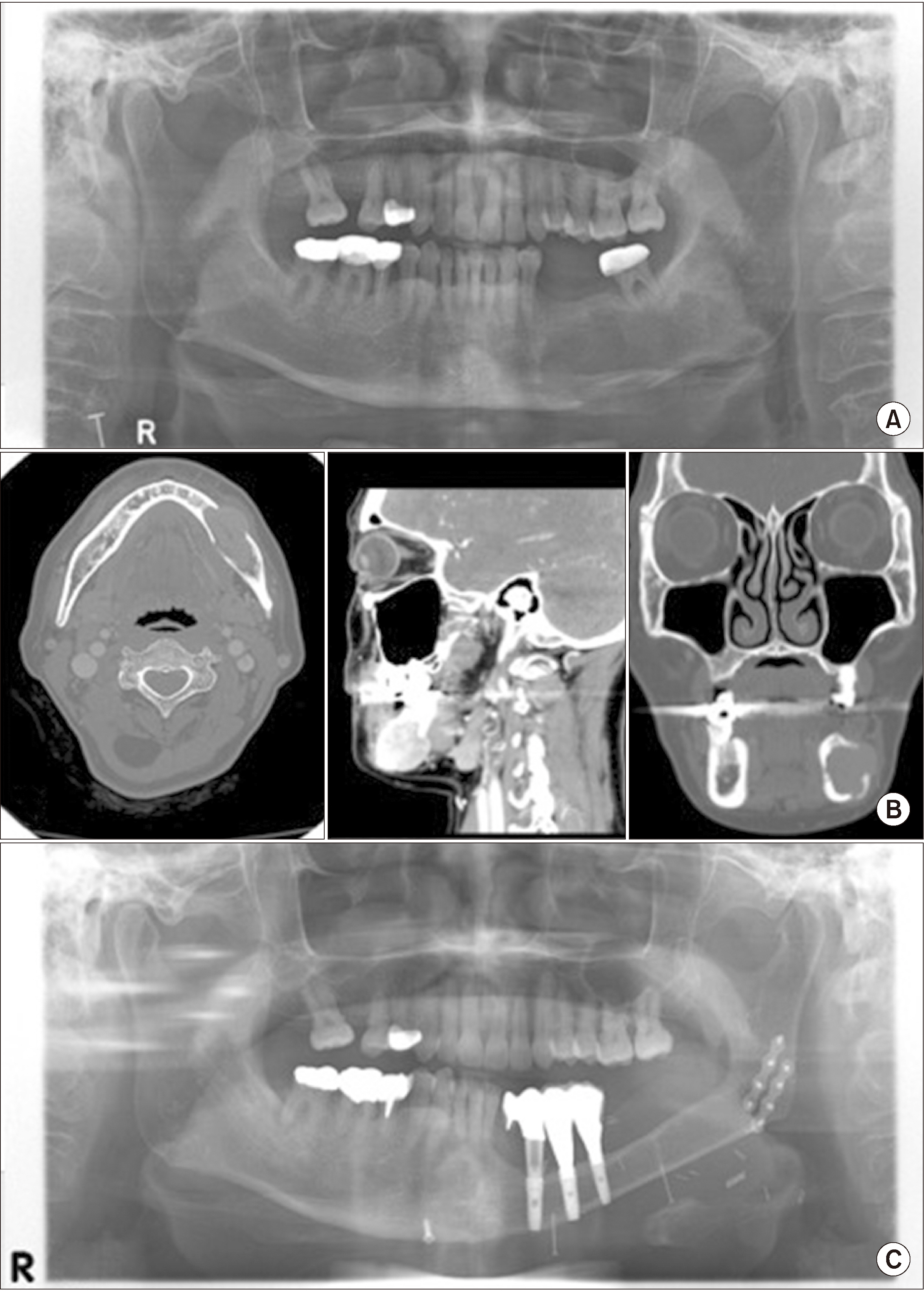
Fig. 3 A 48-year-old female patient. A. Preoperative panoramic image. Unclear radiography was observed at the left mandibular site; the border of the lesion was from #34 posterior to the left mandibular site. The alveolar crest was intact but cortical bone loss was observed at the lower mandible. B. Enhanced computerized tomographic image; a large, enhancing lesion was observed at #34 to #36, filling the marrow of the left mandible, partially perforating the cortical bone and swelling outwards, making buccal contact at the lower buccinator muscle and pushing the platysma muscle outwards. There is also lateral contact at the mylohyoid muscle and the anterior belly of the digastric muscle as well as anterior contact at the edge of sublingual gland. The mandibular canal was within the lesion, surrounded by the enhancing lesion. C. Postoperative panoramic image. Two years postoperatively, dental implants and prostheses are attached at the fibular region and there is no evidence of recurrence.
Reference
-
References
1. Fernandez Sanroman J, Alonso del Hoyo JR, Diaz FJ, Gil-Diez JL, Monje F, Naval L, et al. 1992; Sarcomas of the head and neck. Br J Oral Maxillofac Surg. 30:115–8. https://doi.org/10.1016/0266-4356(92)90081-s . DOI: 10.1016/0266-4356(92)90081-S.
Article2. Farman AG. 1975; Benign smooth muscle tumours. S Afr Med J. 49:1333–40. DOI: 10.1136/thx.19.2.185. PMID: 1154203.3. Wertheimer-Hatch L, Hatch GF 3rd, Hatch BS KF, Davis GB, Blanchard DK, Foster RS Jr Jr, et al. 2000; Tumors of the oral cavity and pharynx. World J Surg. 24:395–400. https://doi.org/10.1007/s002689910064 . DOI: 10.1007/s002689910064. PMID: 10706911.
Article4. Carter LC, Aguirre A, Boyd B, DeLacure MD. 1999; Primary leiomyosarcoma of the mandible in a 7-year-old girl: report of a case and review of the literature. Oral Surg Oral Med Oral Pathol Oral Radiol Endod. 87:477–84. https://doi.org/10.1016/s1079-2104(99)70248-9 . DOI: 10.1016/S1079-2104(99)70248-9.
Article5. Lo Muzio L, Favia G, Mignogna MD, Piattelli A, Maiorano E. 2000; Primary intraoral leiomyosarcoma of the tongue: an immunohistochemical study and review of the literature. Oral Oncol. 36:519–24. https://doi.org/10.1016/s1368-8375(00)00044-0 . DOI: 10.1016/S1368-8375(00)00044-0.
Article6. Schenberg ME, Slootweg PJ, Koole R. 1993; Leiomyosarcomas of the oral cavity. Report of four cases and review of the literature. J Craniomaxillofac Surg. 21:342–7. https://doi.org/10.1016/s1010-5182(05)80495-0 . DOI: 10.1016/S1010-5182(05)80495-0.
Article7. Yan B, Li Y, Pan J, Xia H, Li LJ. 2010; Primary oral leiomyosarcoma: a retrospective clinical analysis of 20 cases. Oral Dis. 16:198–203. https://doi.org/10.1111/j.1601-0825.2009.01635.x . DOI: 10.1111/j.1601-0825.2009.01635.x. PMID: 20374505.
Article8. Vilos GA, Rapidis AD, Lagogiannis GD, Apostolidis C. 2005; Leiomyosarcomas of the oral tissues: clinicopathologic analysis of 50 cases. J Oral Maxillofac Surg. 63:1461–77. https://doi.org/10.1016/j.joms.2005.06.018 . DOI: 10.1016/j.joms.2005.06.018. PMID: 16182914.
Article9. Izumi K, Maeda T, Cheng J, Saku T. 1995; Primary leiomyosarcoma of the maxilla with regional lymph node metastasis. Report of a case and review of the literature. Oral Surg Oral Med Oral Pathol Oral Radiol Endod. 80:310–9. https://doi.org/10.1016/s1079-2104(05)80389-0 . DOI: 10.1016/S1079-2104(05)80389-0.
Article10. Nikitakis NG, Lopes MA, Bailey JS, Blanchaert RH Jr Jr, Ord RA, Sauk JJ. 2002; Oral leiomyosarcoma: review of the literature and report of two cases with assessment of the prognostic and diagnostic significance of immunohistochemical and molecular markers. Oral Oncol. 38:201–8. https://doi.org/10.1016/s1368-8375(01)00047-1 . DOI: 10.1016/S1368-8375(01)00047-1.
Article11. Sumida T, Hamakawa H, Otsuka K, Tanioka H. 2001; Leiomyosarcoma of the maxillary sinus with cervical lymph node metastasis. J Oral Maxillofac Surg. 59:568–71. https://doi.org/10.1053/joms.2001.22691 . DOI: 10.1053/joms.2001.22691. PMID: 11326387.
Article12. Allen CM, Neville B, Damm DD, Marsh W. 1993; Leiomyosarcoma metastatic to the oral region. Report of three cases. Oral Surg Oral Med Oral Pathol. 76:752–6. https://doi.org/10.1016/0030-4220(93)90047-8 . DOI: 10.1016/0030-4220(93)90047-8.
Article13. Dry SM, Jorgensen JL, Fletcher CD. 2000; Leiomyosarcomas of the oral cavity: an unusual topographic subset easily mistaken for nonmesenchymal tumours. Histopathology. 36:210–20. https://doi.org/10.1046/j.1365-2559.2000.00814.x . DOI: 10.1046/j.1365-2559.2000.00814.x. PMID: 10692022.
Article14. Kim SM, Myoung H, Choung PH, Kim MJ, Lee SK, Lee JH. 2009; Metastatic leiomyosarcoma in the oral cavity: case report with protein expression profiles. J Craniomaxillofac Surg. 37:454–60. https://doi.org/10.1016/j.jcms.2009.06.010 . DOI: 10.1016/j.jcms.2009.06.010. PMID: 19664933.
Article15. Arlen M, Higinbotham NL, Huvos AG, Marcove RC, Miller T, Shah IC. 1971; Radiation-induced sarcoma of bone. Cancer. 28:1087–99. https://doi.org/10.1002/1097-0142(1971)28:5<1087::aid-cncr2820280502>3.0.co;2-f . DOI: 10.1002/1097-0142(1971)28:5<1087::AID-CNCR2820280502>3.0.CO;2-F. PMID: 5288429.
Article16. Cahan WG, Woodard HQ, Higinbotham NL, Stewart FW, Coley BL. 1998; Sarcoma arising in irradiated bone: report of eleven cases. Cancer. 82:8–34. https://doi.org/10.1002/(sici)1097-0142(19980101)82:1<8::aid-cncr3>3.0.co;2-w . DOI: 10.1002/(SICI)1097-0142(19980101)82:1<8::AID-CNCR3>3.0.CO;2-W. PMID: 9428476.
Article17. Ko E. 2019; Primary oral leiomyosarcoma: a systematic review and update. J Oral Pathol Med. 48:780–7. https://doi.org/10.1111/jop.12858 . DOI: 10.1111/jop.12858. PMID: 30958581.
Article18. Amarapala H, Tilakaratne WM. 2006; Leiomyosarcoma of the oral cavity: report of seven cases and review of literature. Oral Oncol Extra. 42:14–7. https://doi.org/10.1016/j.ooe.2005.08.001 . DOI: 10.1016/j.ooe.2005.08.001.
Article19. Ethunandan M, Stokes C, Higgins B, Spedding A, Way C, Brennan P. 2007; Primary oral leiomyosarcoma: a clinico-pathologic study and analysis of prognostic factors. Int J Oral Maxillofac Surg. 36:409–16. https://doi.org/10.1016/j.ijom.2006.12.015 . DOI: 10.1016/j.ijom.2006.12.015. PMID: 17395428.
Article20. Montgomery E, Goldblum JR, Fisher C. 2002; Leiomyosarcoma of the head and neck: a clinicopathological study. Histopathology. 40:518–25. https://doi.org/10.1046/j.1365-2559.2002.01412.x . DOI: 10.1046/j.1365-2559.2002.01412.x. PMID: 12047762.
Article21. Akcam T, Oysul K, Birkent H, Gerek M, Yetiser S. 2005; Leiomyosarcoma of the head and neck: report of two cases and review of the literature. Auris Nasus Larynx. 32:209–12. https://doi.org/10.1016/j.anl.2005.01.012 . DOI: 10.1016/j.anl.2005.01.012. PMID: 15917182.
Article22. Crossman T, Ward P, Herold J. 2008; Leiomyosarcoma of the tongue: a case report. Br J Oral Maxillofac Surg. 46:e69–70. https://doi.org/10.1016/j.bjoms.2008.03.008 . DOI: 10.1016/j.bjoms.2008.03.008. PMID: 18448216.
Article23. Misra A, Mistry N, Grimer R, Peart F. 2009; The management of soft tissue sarcoma. J Plast Reconstr Aesthet Surg. 62:161–74. https://doi.org/10.1016/j.bjps.2008.08.018 . DOI: 10.1016/j.bjps.2008.08.018. PMID: 19036655.
Article24. Ries Centeno C, Nadini F, Adam R, Godoy H, Reichart PA. 2006; Primary leiomyosarcoma of the mandible. Oral Oncol Extra. 42:40–5. https://doi.org/10.1016/j.ooe.2005.08.008 . DOI: 10.1016/j.ooe.2005.08.008.
Article25. Rodini CO, Pontes FS, Pontes HA, Santos PS, Magalhães MG, Pinto DS Jr Jr. 2007; Oral leiomyosarcomas: report of two cases with immunohistochemical profile. Oral Surg Oral Med Oral Pathol Oral Radiol Endod. 104:e50–5. https://doi.org/10.1016/j.tripleo.2007.05.005 . DOI: 10.1016/j.tripleo.2007.05.005. PMID: 17706443.
Article26. Rapidis AD, Gakiopoulou H, Stavrianos SD, Vilos GA, Faratzis G, Douzinas EE, et al. 2005; Sarcomas of the head and neck. Results from the treatment of 25 patients. Eur J Surg Oncol. 31:177–82. https://doi.org/10.1016/j.ejso.2004.09.020 . DOI: 10.1016/j.ejso.2004.09.020. PMID: 15698735.
Article27. Yadav J, Bakshi J, Chouhan M, Modi R. 2013; Head and neck leiomyosarcoma. Indian J Otolaryngol Head Neck Surg. 65(Suppl 1):1–5. https://doi.org/10.1007/s12070-011-0305-8 . DOI: 10.1007/s12070-011-0305-8. PMID: 24427607. PMCID: PMC3718933.
Article28. Suárez-Alén F, Otero-Rey E, Peñamaría-Mallón M, García-García A, Blanco-Carrión A. 2015; Oral leiomyosarcoma: the importance of early diagnosis. Gerodontology. 32:314–7. https://doi.org/10.1111/ger.12126 . DOI: 10.1111/ger.12126. PMID: 24698388.
Article29. Schütz A, Smeets R, Driemel O, Hakim SG, Kosmehl H, Hanken H, et al. 2013; Primary and secondary leiomyosarcoma of the oral and perioral region--clinicopathological and immunohistochemical analysis of a rare entity with a review of the literature. J Oral Maxillofac Surg. 71:1132–42. https://doi.org/10.1016/j.joms.2012.12.011 . DOI: 10.1016/j.joms.2012.12.011. PMID: 23434173.
Article30. Mitsudo K, Tohnai I, Fujimoto Y, Sawaki Y, Sugimura T, Nishiguchi H, et al. 2006; Leiomyosarcoma of the maxilla: effective chemotherapy with docetaxel (DOC) and cisplatin (CDDP) using superselective intra-arterial infusion via superficial temporal artery. Oral Oncol Extra. 42:258–62. https://doi.org/10.1016/j.ooe.2006.05.001 . DOI: 10.1016/j.ooe.2006.05.001.
Article
