The Distinct Role of Tcfs and Lef1 in the Self-Renewal or Differentiation of Mouse Embryonic Stem Cells
- Affiliations
-
- 1Department of Life Science, University of Seoul, Seoul, Korea
- 2Asan Institute for Life Sciences, Seoul, Korea
- 3Eli and Edythe Broad Center for Regenerative Medicine and Stem Cell Research at USC, Keck School of Medicine, University of Southern California, Los Angeles, CA, USA
- KMID: 2504332
- DOI: http://doi.org/10.15283/ijsc20044
Abstract
- Background and Objectives
Tcfs and Lef1 are DNA-binding transcriptional factors in the canonical Wnt signaling pathway. In the absence of β-catenin, Tcfs and Lef1 generally act as transcriptional repressors with co-repressor proteins such as Groucho, CtBP, and HIC-5. However, Tcfs and Lef1 turn into transcriptional activators during the interaction with β-catenin. Therefore, the activity of Tcfs and Lef1 is regulated by β-catenin. However, the intrinsic role of Tcfs and Lef1 has yet to be examined. The purpose of this study was to determine whether Tcfs and Lef1 play differential roles in the regulation of self-renewal and differentiation of mouse ES cells.
Methods and Results
Interestingly, the expression of Tcfs and Lef1 was dynamically altered under various differentiation conditions, such as removal of LIF, EB formation and neuronal differentiation in N2B27 media, suggesting that the function of each Tcf and Lef1 may vary in ES cells. Ectopic expression of Tcf1 or the dominant negative form of Lef1 (Lef1-DN) contributes to ES cells to self-renew in the absence of leukemia inhibitory factor (LIF), whereas ectopic expression of Tcf3, Lef1 or Tcf1-DN did not support ES cells to self-renew. Ectopic expression of either Lef1 or Lef1-DN blocked neuronal differentiation, suggesting that the transient induction of Lef1 was necessary for the initiation and progress of differentiation. ChIP analysis shows that Tcf1 bound to Nanog promoter and ectopic expression of Tcf1 enhanced the transcription of Nanog.
Conclusions
The overall data suggest that Tcf1 plays a critical role in the maintenance of stemness whereas Lef1 is involved in the initiation of differentiation.
Keyword
Figure
-

Fig. 1 The pattern of Tcfs and Lef1 expression in ES cells. (A) A6P10 ES cells were differentiated in the absence of LIF (left), in N2B27 medium without LIF (middle), or via EB formation without LIF (right). The differentiated ES cells were harvested on the days indicated. The amount of Tcfs and Lef1 transcripts was measured by RT-PCR analysis. The bands for Tcfs and Lef1 represent wild-type transcripts including β-catenin-binding domain. Self-renewal marker (Nanog), neural precursor markers (Sox1 and Fgf5) for N2B27 differentiation and mesodermal markers (Brachyury, Nkx2.5, αMHC) for EB differentiation were used to confirm proper differentiation; 18s rRNA was used as a loading control. (B) The endogenous protein levels of Tcfs/Lef1 were detected by western blotting. β-actin was used as a loading control.
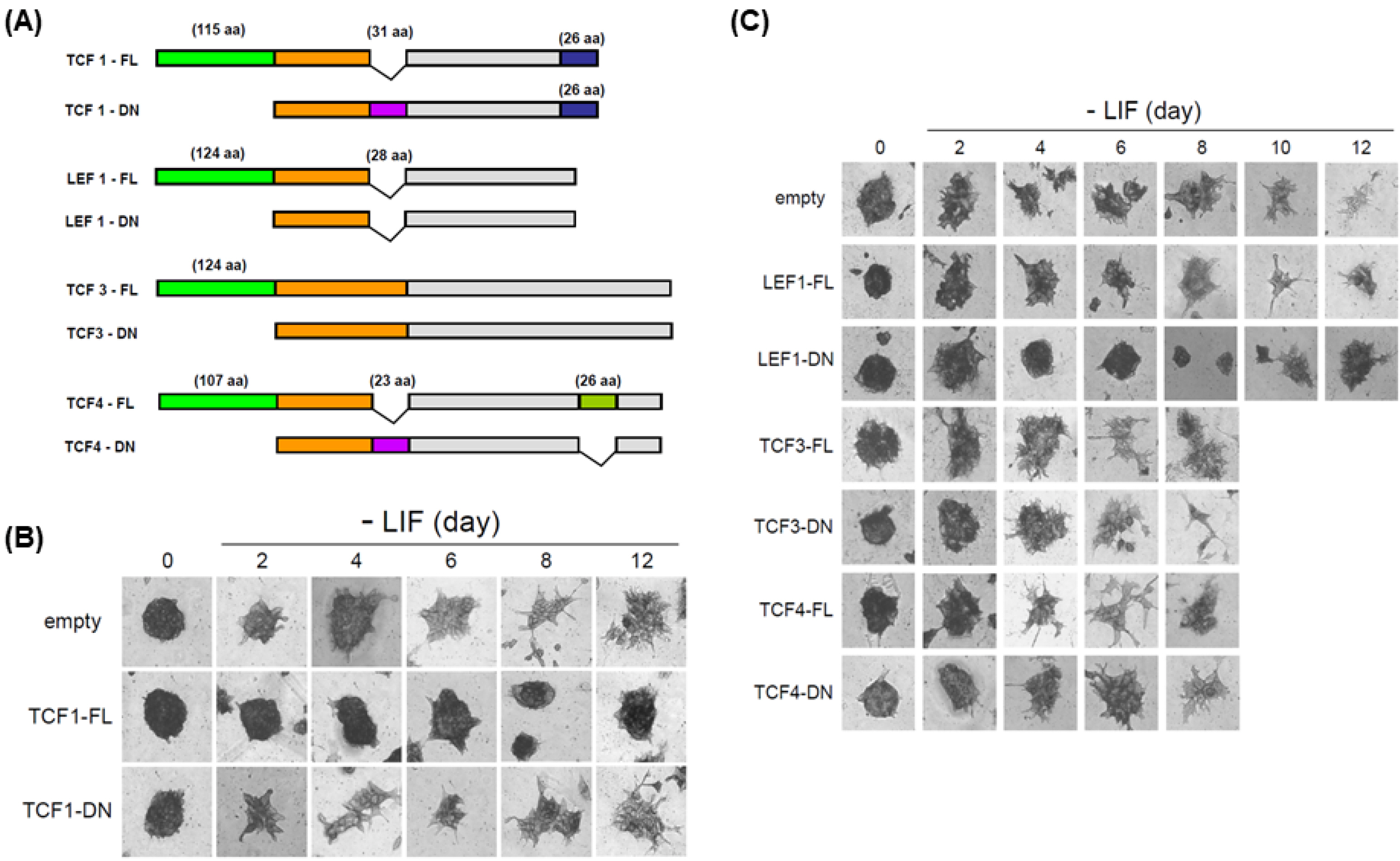
Fig. 2 The effect of ectopic expression of Tcfs and Lef1 after LIF depletion. (A) A schematic diagram of Tcfs and Lef1 constructs (FL, full length; and DN, dominant negative). (B, C) A6P10 ES cells were transfected with control vector, Tcf1 (FL or DN), Lef1 (FL or DN), Tcf3 (FL or DN), or Tcf4 (FL or DN) and then selected using Zeocin. Selected ES cells were stained with alkaline phosphatase (AP), a marker of self-renewal, on the days indicated after LIF removal.

Fig. 3 Ectopic expression of Tcf1 maintains self-renewal and delays differentiation of ES cells. (A) mRNA expressions for Oct4, Sox2 and Nanog at ES cells stage and Day 4 of differentiation upon TCF1 overexpression were determined by using qPCR. RNA was isolated from cells expressing empty vector (EV) or Tcf1-FL (TCF1) on specified stage. GAPDH was used for normalization. (B) The amount of Nanog transcript was measured by RT-PCR analysis. RNA was isolated from ES cells expressing control vector or Tcf1-FL on the days specified. 18s rRNA was used as a loading control. (C) ChIP assay was performed to examine the binding of Tcf1, Lef1, and Tcf3 on Nanog promoter. Samples for ChIP were isolated from ES cells grown with LIF and from ES cells expressing control vector or Tcf1-FL cultured for 2 days without LIF. For immunoprecipitiation, endogenous antibody against Tcf1, Lef1, or Tcf3 was used. (D) To differentiate into neural precursors, 46C ES cells expressing control vector, Tcf1-FL, or Tcf1-DN were plated for 6 days in N2B27 medium. GFP fluorescence showed that 46C ES cells expressing control vector or Tcf1-DN, but not Tcf1-FL effectively differentiated into neural precursor (upper panel). The amount of Nanog or Sox1 transcript on the days indicated was measured via RT-PCR analysis (lower panel). β-actin was used as a loading control. (E) RNA samples were isolated from A6P10 ES cells expressing control vector or Tcf1-FL on the days indicated after EB formation. Self-renewal (Nanog), mesoderm (Brachyury), and cardiomyocyte (Islet1, αMHC) markers were used for RT-PCR analysis.
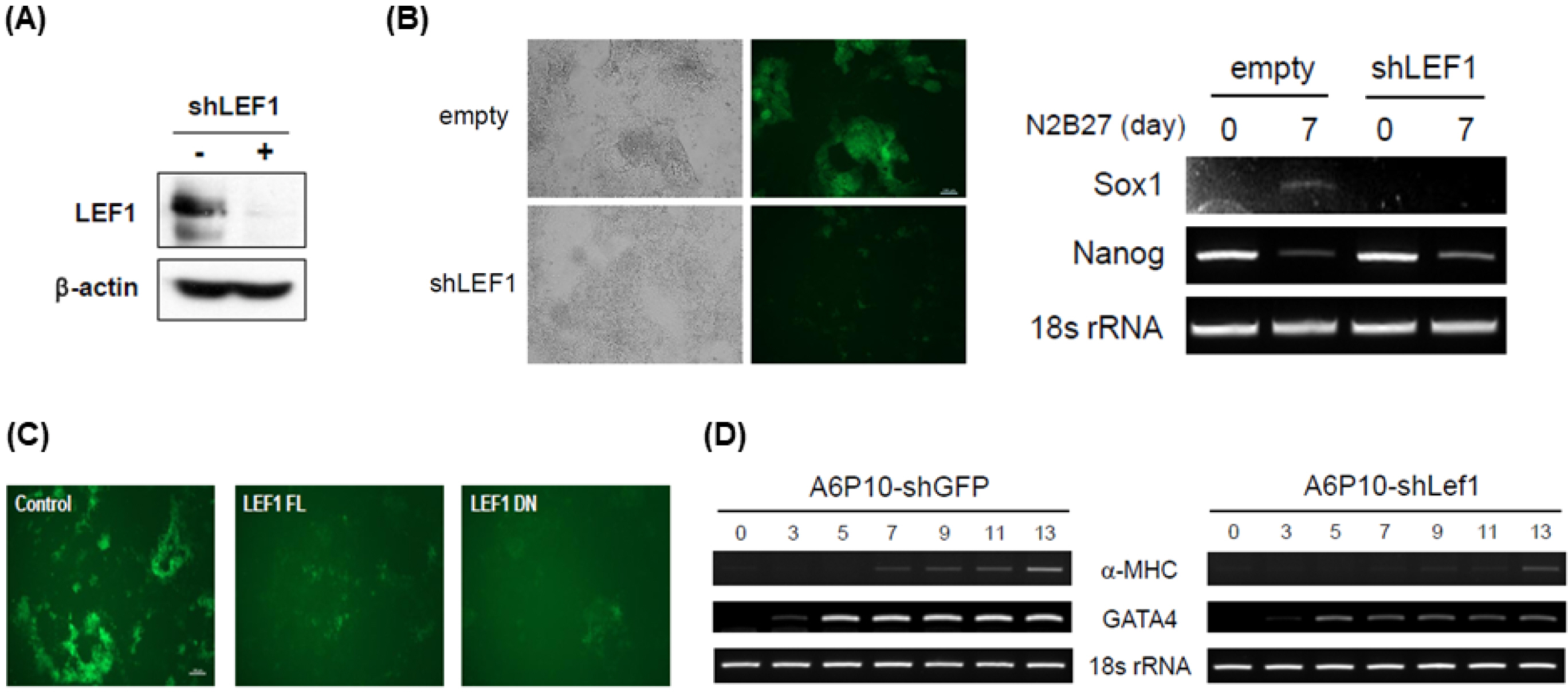
Fig. 4 Transient expression of Lef1 is required for proper differentiation. (A) A6P10 ES cells were infected with retroviruses expressing control or Lef1 shRNA, followed by selection using puromycin. After 2 days without LIF, the selected ES cells were harvested and analyzed by western blotting with anti-Lef1 antibody. β-actin was used as a loading control. (B) 46C ES cells expressing control shRNA or Lef1 shRNA were plated for 7 days in N2B27 medium. GFP fluorescence indicating a neural precursor was not induced in shLef1-46C cells (left panel). The amount of Nanog or Sox1 transcript was measured by RT-PCR analysis (right panel). (C) 46C ES cells were transfected with control vector, Lef1-FL, or Lef1-DN and then selected using Zeocin. 46C ES cells expressing each vector were plated for 7 days in N2B27 medium and GFP fluorescence was captured. (D) RNA samples were isolated from A6P10 ES cells expressing shGFP or shLef1 on the days indicated post-EB formation. Cardiomyocyte (α-MHC) and endoderm (GATA4) markers and loading control (18s rRNA) were used for RT-PCR analysis.

Fig. 5 A model describing the role of Tcfs and Lef1s in ES cells.
Reference
-
References
1. Reubinoff BE, Pera MF, Fong CY, Trounson A, Bongso A. 2000; Embryonic stem cell lines from human blastocysts: somatic differentiation in vitro. Nat Biotechnol. 18:399–404. DOI: 10.1038/74447. PMID: 10748519.
Article2. Thomson JA, Itskovitz-Eldor J, Shapiro SS, Waknitz MA, Swiergiel JJ, Marshall VS, Jones JM. 1998; Embryonic stem cell lines derived from human blastocysts. Science. 282:1145–1147. DOI: 10.1126/science.282.5391.1145. PMID: 9804556.
Article3. Smith AG. 2001; Embryo-derived stem cells: of mice and men. Annu Rev Cell Dev Biol. 17:435–462. DOI: 10.1146/annurev.cellbio.17.1.435. PMID: 11687496.
Article4. Dahéron L, Opitz SL, Zaehres H, Lensch MW, Andrews PW, Itskovitz-Eldor J, Daley GQ. 2004; LIF/STAT3 signaling fails to maintain self-renewal of human embryonic stem cells. Stem Cells. 22:770–778. DOI: 10.1634/stemcells.22-5-770. PMID: 15342941.
Article5. Nichols J, Zevnik B, Anastassiadis K, Niwa H, Klewe-Nebenius D, Chambers I, Schöler H, Smith A. 1998; Formation of pluripotent stem cells in the mammalian embryo depends on the POU transcription factor Oct4. Cell. 95:379–391. DOI: 10.1016/S0092-8674(00)81769-9.
Article6. Avilion AA, Nicolis SK, Pevny LH, Perez L, Vivian N, Lovell-Badge R. 2003; Multipotent cell lineages in early mouse development depend on SOX2 function. Genes Dev. 17:126–140. DOI: 10.1101/gad.224503. PMID: 12514105. PMCID: PMC195970.
Article7. Chambers I, Colby D, Robertson M, Nichols J, Lee S, Tweedie S, Smith A. 2003; Functional expression cloning of Nanog, a pluripotency sustaining factor in embryonic stem cells. Cell. 113:643–655. DOI: 10.1016/S0092-8674(03)00392-1. PMID: 12787505.
Article8. Mitsui K, Tokuzawa Y, Itoh H, Segawa K, Murakami M, Takahashi K, Maruyama M, Maeda M, Yamanaka S. 2003; The homeoprotein Nanog is required for maintenance of pluripotency in mouse epiblast and ES cells. Cell. 113:631–642. DOI: 10.1016/S0092-8674(03)00393-3. PMID: 12787504.
Article9. Niwa H, Miyazaki J, Smith AG. 2000; Quantitative expression of Oct-3/4 defines differentiation, dedifferentiation or self-renewal of ES cells. Nat Genet. 24:372–376. DOI: 10.1038/74199. PMID: 10742100.
Article10. Reya T, Clevers H. 2005; Wnt signalling in stem cells and cancer. Nature. 434:843–850. DOI: 10.1038/nature03319. PMID: 15829953.
Article11. Merrill BJ. 2012; Wnt pathway regulation of embryonic stem cell self-renewal. Cold Spring Harb Perspect Biol. 4:a007971. DOI: 10.1101/cshperspect.a007971. PMID: 22952393. PMCID: PMC3428775.
Article12. Aubert J, Dunstan H, Chambers I, Smith A. 2002; Functional gene screening in embryonic stem cells implicates Wnt antagonism in neural differentiation. Nat Biotechnol. 20:1240–1245. DOI: 10.1038/nbt763. PMID: 12447396.
Article13. Haegele L, Ingold B, Naumann H, Tabatabai G, Ledermann B, Brandner S. 2003; Wnt signalling inhibits neural differentiation of embryonic stem cells by controlling bone morphogenetic protein expression. Mol Cell Neurosci. 24:696–708. DOI: 10.1016/S1044-7431(03)00232-X. PMID: 14664819.
Article14. Kielman MF, Rindapää M, Gaspar C, van Poppel N, Breukel C, van Leeuwen S, Taketo MM, Roberts S, Smits R, Fodde R. 2002; Apc modulates embryonic stem-cell differentiation by controlling the dosage of beta-catenin signaling. Nat Genet. 32:594–605. DOI: 10.1038/ng1045. PMID: 12426568.
Article15. Sato N, Meijer L, Skaltsounis L, Greengard P, Brivanlou AH. 2004; Maintenance of pluripotency in human and mouse embryonic stem cells through activation of Wnt signaling by a pharmacological GSK-3-specific inhibitor. Nat Med. 10:55–63. DOI: 10.1038/nm979. PMID: 14702635.
Article16. Kirby LA, Schott JT, Noble BL, Mendez DC, Caseley PS, Peterson SC, Routledge TJ, Patel NV. 2012; Glycogen synthase kinase 3 (GSK3) inhibitor, SB-216763, promotes pluripotency in mouse embryonic stem cells. PLoS One. 7:e39329. DOI: 10.1371/journal.pone.0039329. PMID: 22745733. PMCID: PMC3383737.
Article17. Dravid G, Ye Z, Hammond H, Chen G, Pyle A, Donovan P, Yu X, Cheng L. 2005; Defining the role of Wnt/beta-catenin signaling in the survival, proliferation, and self-renewal of human embryonic stem cells. Stem Cells. 23:1489–1501. DOI: 10.1634/stemcells.2005-0034. PMID: 16002782.
Article18. Kwon C, Arnold J, Hsiao EC, Taketo MM, Conklin BR, Srivastava D. 2007; Canonical Wnt signaling is a positive regulator of mammalian cardiac progenitors. Proc Natl Acad Sci USA. 104:10894–10899. DOI: 10.1073/pnas.0704044104. PMID: 17576928. PMCID: PMC1904134.
Article19. Lindsley RC, Gill JG, Kyba M, Murphy TL, Murphy KM. 2006; Canonical Wnt signaling is required for development of embryonic stem cell-derived mesoderm. Development. 133:3787–3796. DOI: 10.1242/dev.02551. PMID: 16943279.
Article20. Naito AT, Shiojima I, Akazawa H, Hidaka K, Morisaki T, Kikuchi A, Komuro I. 2006; Developmental stage-specific biphasic roles of Wnt/beta-catenin signaling in cardiomyogenesis and hematopoiesis. Proc Natl Acad Sci USA. 103:19812–19817. DOI: 10.1073/pnas.0605768103. PMID: 17170140. PMCID: PMC1750922.
Article21. Otero JJ, Fu W, Kan L, Cuadra AE, Kessler JA. 2004; Beta-catenin signaling is required for neural differentiation of embryonic stem cells. Development. 131:3545–3557. DOI: 10.1242/dev.01218. PMID: 15262888.
Article22. Kim H, Kim S, Song Y, Kim W, Ying QL, Jho EH. 2015; Dual function of Wnt signaling during neuronal differentiation of mouse embryonic stem cells. Stem Cells Int. 2015:459301. DOI: 10.1155/2015/459301. PMID: 25945099. PMCID: PMC4402205.
Article23. Cadigan KM, Waterman ML. 2012; TCF/LEFs and Wnt signaling in the nucleus. Cold Spring Harb Perspect Biol. 4:a007906. DOI: 10.1101/cshperspect.a007906. PMID: 23024173. PMCID: PMC3536346.
Article24. Arce L, Yokoyama NN, Waterman ML. 2006; Diversity of LEF/TCF action in development and disease. Oncogene. 25:7492–7504. DOI: 10.1038/sj.onc.1210056. PMID: 17143293.
Article25. Hoppler S, Kavanagh CL. 2007; Wnt signalling: variety at the core. J Cell Sci. 120(Pt 3):385–393. DOI: 10.1242/jcs.03363. PMID: 17251379.
Article26. Hurlstone A, Clevers H. 2002; T-cell factors: turn-ons and turn-offs. EMBO J. 21:2303–2311. DOI: 10.1093/emboj/21.10.2303. PMID: 12006483. PMCID: PMC126013.27. Cole MF, Johnstone SE, Newman JJ, Kagey MH, Young RA. 2008; Tcf3 is an integral component of the core regulatory circuitry of embryonic stem cells. Genes Dev. 22:746–755. DOI: 10.1101/gad.1642408. PMID: 18347094. PMCID: PMC2275428.
Article28. Pereira L, Yi F, Merrill BJ. 2006; Repression of Nanog gene transcription by Tcf3 limits embryonic stem cell self-renewal. Mol Cell Biol. 26:7479–7491. DOI: 10.1128/MCB.00368-06. PMID: 16894029. PMCID: PMC1636872.
Article29. Tam WL, Lim CY, Han J, Zhang J, Ang YS, Ng HH, Yang H, Lim B. 2008; T-cell factor 3 regulates embryonic stem cell pluripotency and self-renewal by the transcriptional control of multiple lineage pathways. Stem Cells. 26:2019–2031. DOI: 10.1634/stemcells.2007-1115. PMID: 18467660. PMCID: PMC2692055.
Article30. Yi F, Pereira L, Merrill BJ. 2008; Tcf3 functions as a steady-state limiter of transcriptional programs of mouse embryonic stem cell self-renewal. Stem Cells. 26:1951–1960. DOI: 10.1634/stemcells.2008-0229. PMID: 18483421. PMCID: PMC2743928.
Article31. Zhang Y, Zhu Z, Ding H, Wan S, Zhang X, Li Y, Ji J, Wang X, Zhang M, Ye SD. 2020; β-catenin stimulates Tcf7l1 degradation through recruitment of casein kinase 2 in mouse embryonic stem cells. Biochem Biophys Res Commun. 524:280–287. DOI: 10.1016/j.bbrc.2020.01.074. PMID: 31987502.
Article32. Ye S, Zhang T, Tong C, Zhou X, He K, Ban Q, Liu D, Ying QL. 2017; Depletion of Tcf3 and Lef1 maintains mouse embryonic stem cell self-renewal. Biol Open. 6:511–517. DOI: 10.1242/bio.022426. PMID: 28288968. PMCID: PMC5399551.
Article33. Ying QL, Stavridis M, Griffiths D, Li M, Smith A. 2003; Convertsion of embryonic stem cells into neuroectodermal precursors in adherent monoculture. Nat Biotechnol. 21:183–186. DOI: 10.1038/nbt780. PMID: 12524553.
Article
- Full Text Links
-
- Actions
-
Cited
- CITED
-
- Close
- Share
-
- Similar articles
-
- Pja2 Inhibits Wnt/β-catenin Signaling by Reducing the Level of TCF/LEF1
- HSP60 is required for stemness and proper differentiation of mouse embryonic stem cells
- Current Concepts of Stem Cell Therapy
- Cell Biological Characteristics of Adult Stem Cells
- Nuclear receptor regulation of stemness and stem cell differentiation
