Development of a Wide Dose-Rate Range Electron Beam Irradiation System for Pre-Clinical Studies and Multi-Purpose Applications Using a Research Linear Accelerator
- Affiliations
-
- 1Research Center, Dongnam Institute of Radiological and Medical Sciences, Korea
- 2Department of Radiation Oncology, Dongnam Institute of Radiological and Medical Sciences, Busan, Korea
- KMID: 2503938
- DOI: http://doi.org/10.14316/pmp.2020.31.2.9
Abstract
- Purpose
This study aims to develop a multi-purpose electron beam irradiation device for preclinical research and material testing using the research electron linear accelerator installed at the Dongnam Institute of Radiological and Medical Sciences.
Methods
The fabricated irradiation device comprises a dual scattering foil and collimator. The correct scattering foil thickness, in terms of the energy loss and beam profile uniformity, was determined using Monte Carlo calculations. The ion-chamber and radiochromic films were used to determine the reference dose-rate (Gy/s) and beam profiles as functions of the source to surface distance (SSD) and pulse frequency.
Results
The dose-rates for the electron beams were evaluated for the range from 59.16 Gy/s to 5.22 cGy/s at SSDs of 40一120 cm, by controlling the pulse frequency. Furthermore, uniform dose distributions in the electron fields were achieved up to approximately 10 cm in diameter. An empirical formula for the systematic dose-rate calculation for the irradiation system was established using the measured data.
Conclusions
A wide dose-rate range electron beam irradiation device was successfully developed in this study. The pre-clinical studies relating to FLASH radiotherapy to the conventional level were made available. Additionally, material studies were made available using a quantified irradiation system. Future studies are required to improve the energy, dose-rate, and field uniformity of the irradiation system.
Keyword
Figure
-
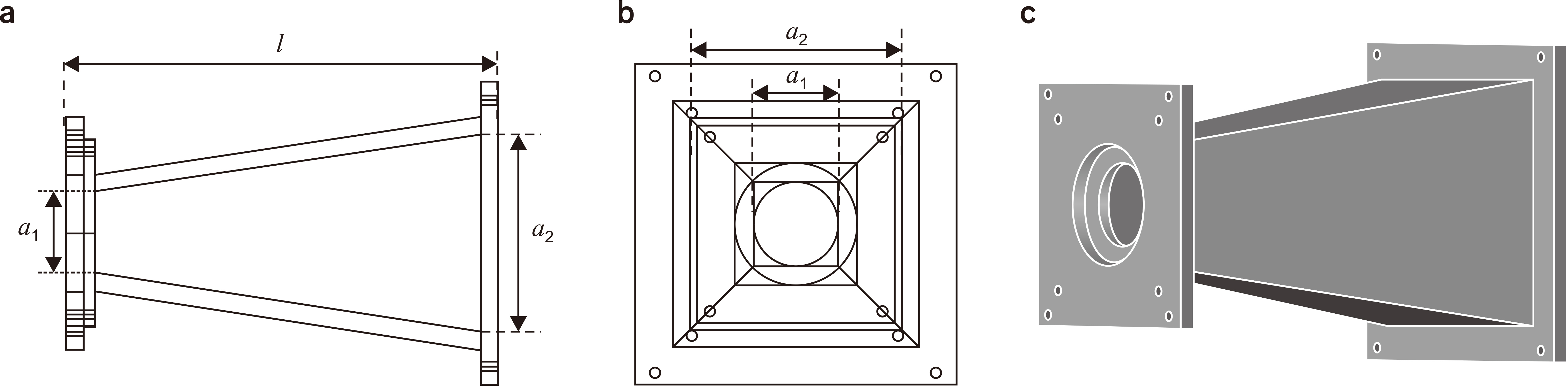
Fig. 1 Design of irradiation device. (a) Side view, (b) front view, (c) 3D view. a1, scattering foil diameter; a2, collimated field size; l, length.

Fig. 2 Calculated and measured percentage depth doses for the determination of incident electron beam energy.
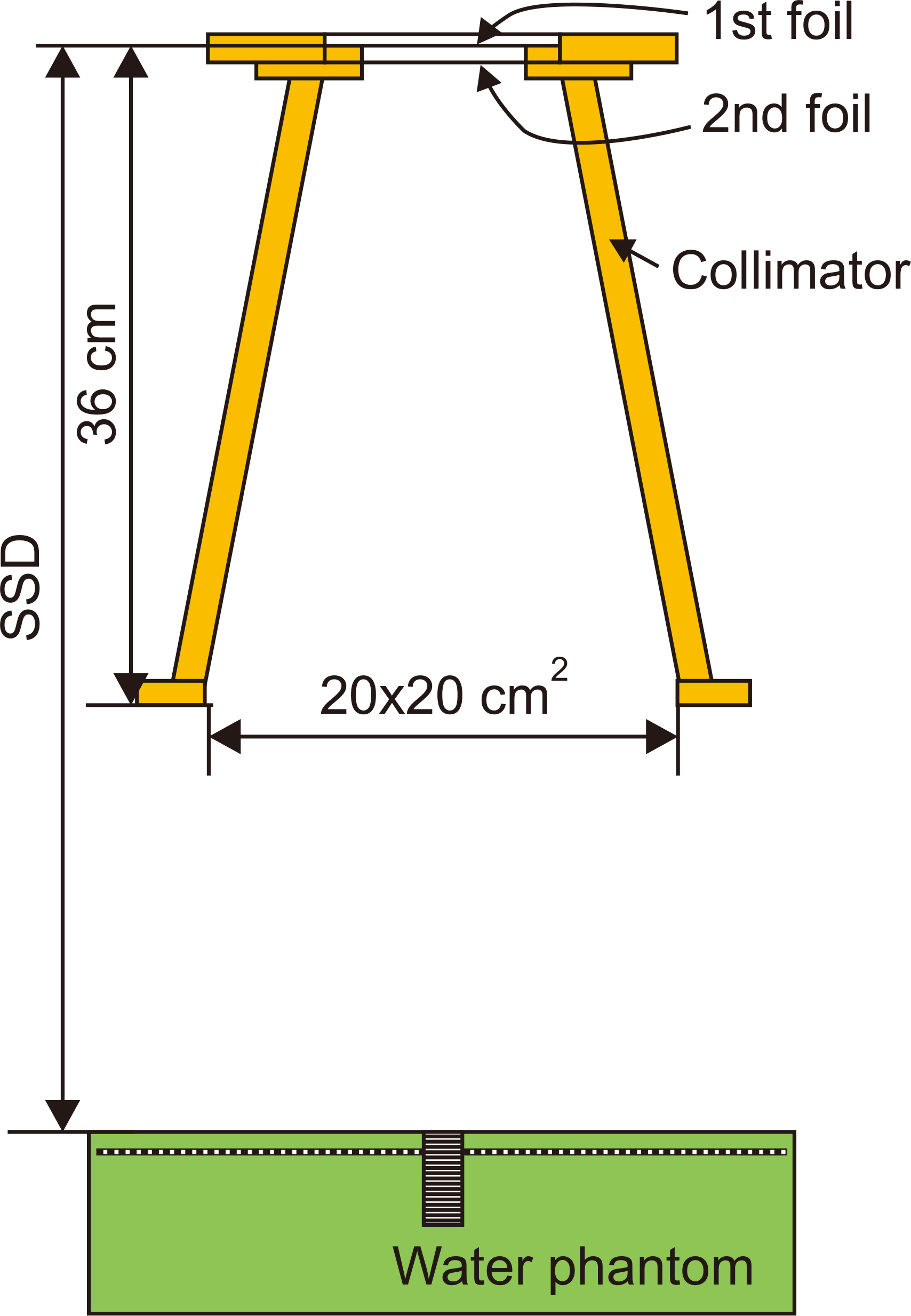
Fig. 3 Monte Carlo calculation geometry. SSD, source to surface distance.
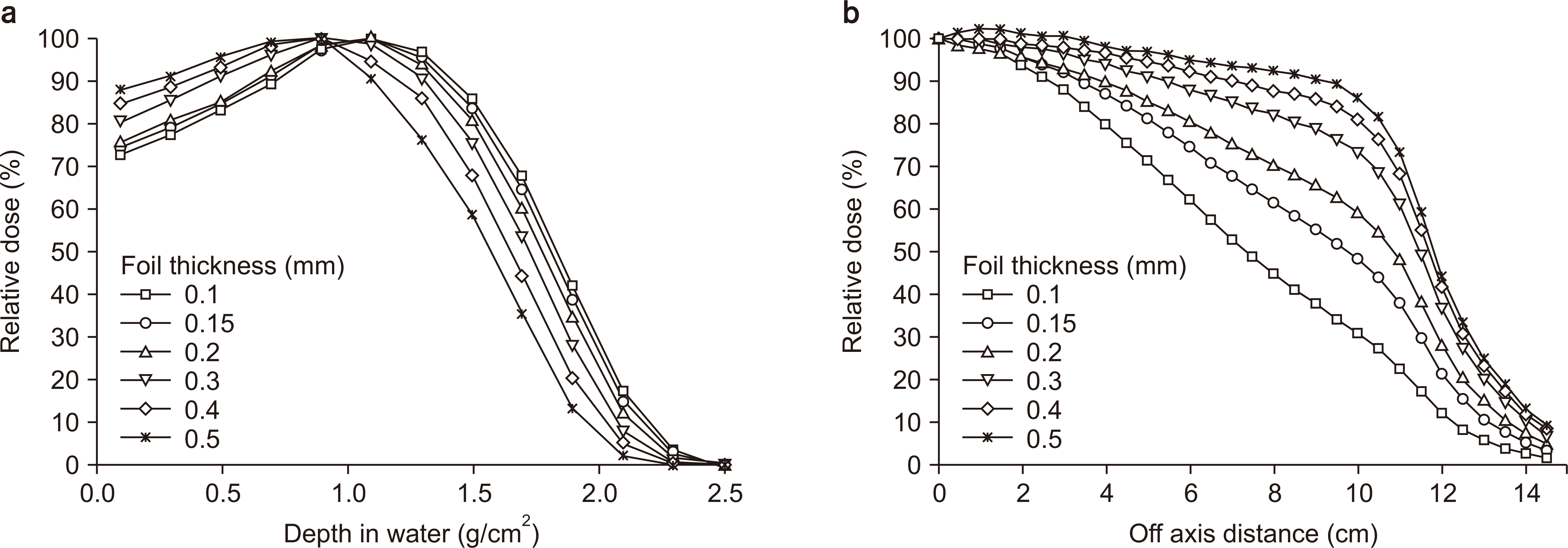
Fig. 4 Calculated percentage depth doses (a) and beam profiles (b) for foil thickness.

Fig. 5 (a) Mean energy at the phantom surface and (b) field diameters (90% and 95% maximum) for foil thickness.
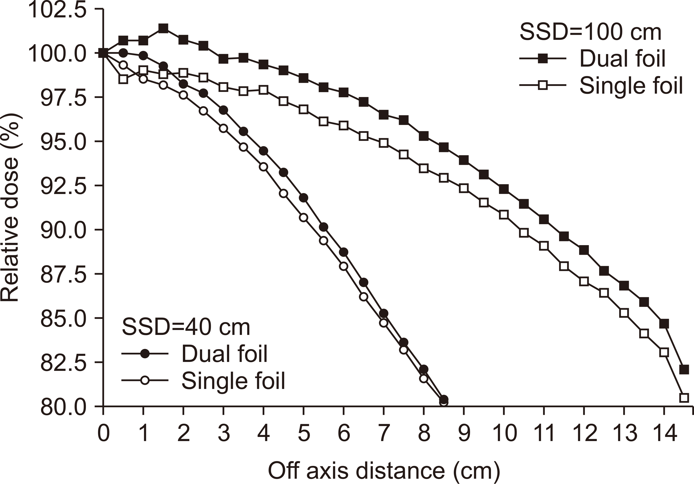
Fig. 6 Calculated beam profiles for single and dual foils. SSD, source to surface distance.

Fig. 7 Experimental setup using DIRAMS LINAC. (a) Reference dosimetry in water using the ionization chamber, (b) film dosimetry. LINAC, linear accelerator; DIRAMS, Dongnam Institute of Radiological and Medical Sciences.

Fig. 8 Measured percentage depth dose for electron irradiator using dual foil system.

Fig. 9 Measured relative dose and linear fit with logarithmic axis at the source to surface distance from 40 to 100 cm.
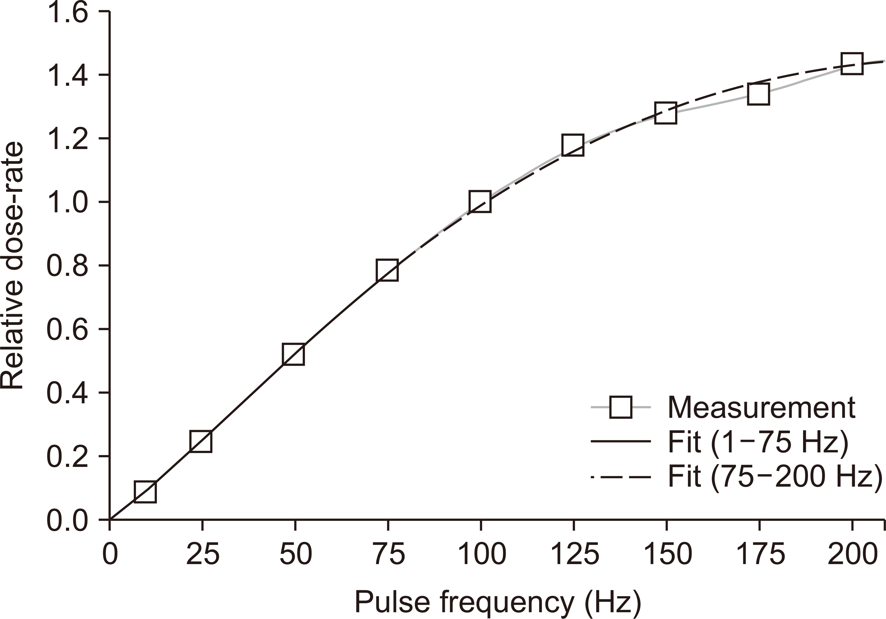
Fig. 10 Relative dose rate as a function of pulse frequency in DIRAMS LINAC (Dongnam Institute of Radiological and Medical Sciences linear accelerator) and a fit line separated into two functions.
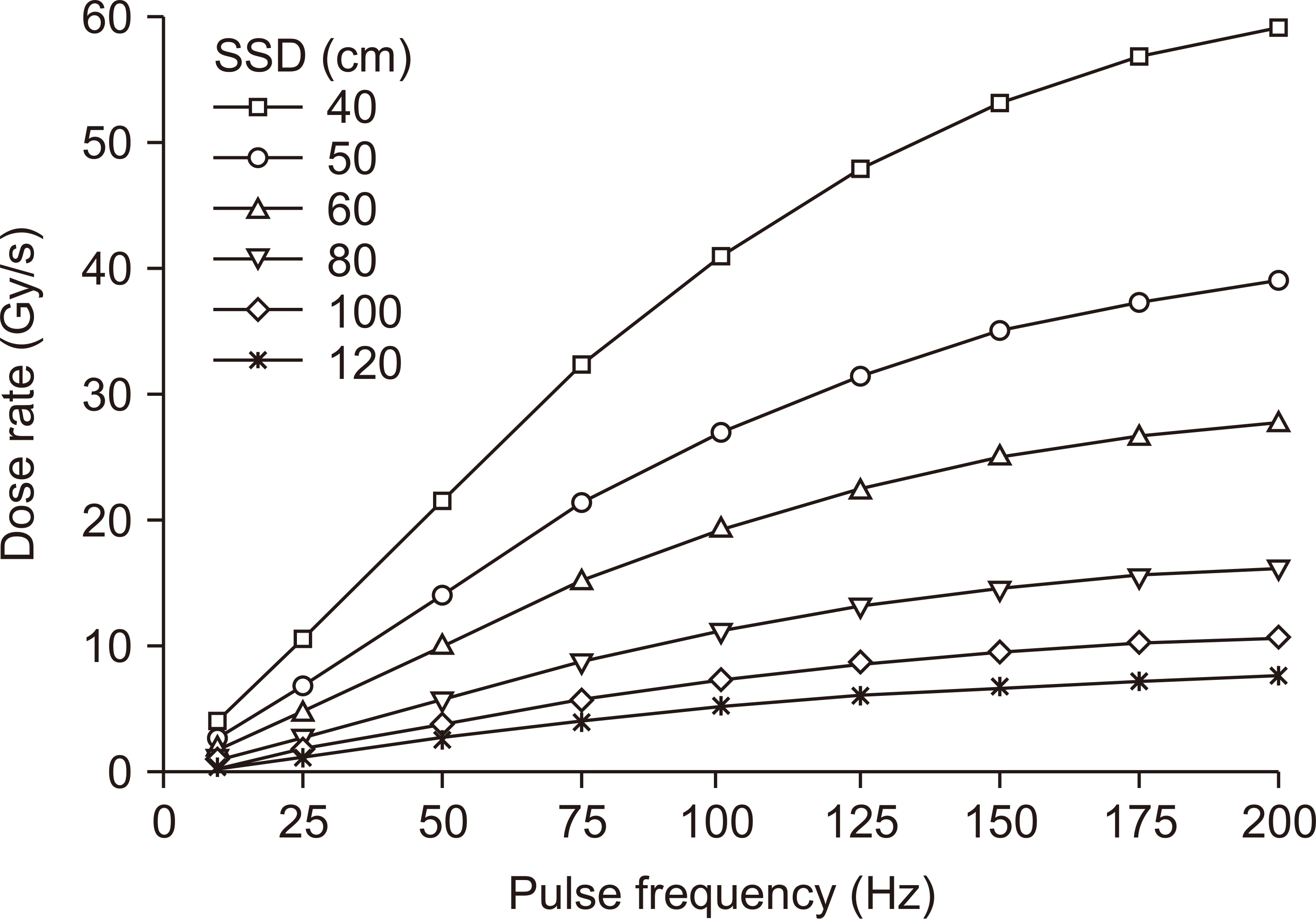
Fig. 11 Determined dose rate (Gy/s) at the depth of dose maximum in water for the electron irradiator system in DIRAMS (Dongnam Institute of Radiological and Medical Sciences). SSD, source to surface distance.
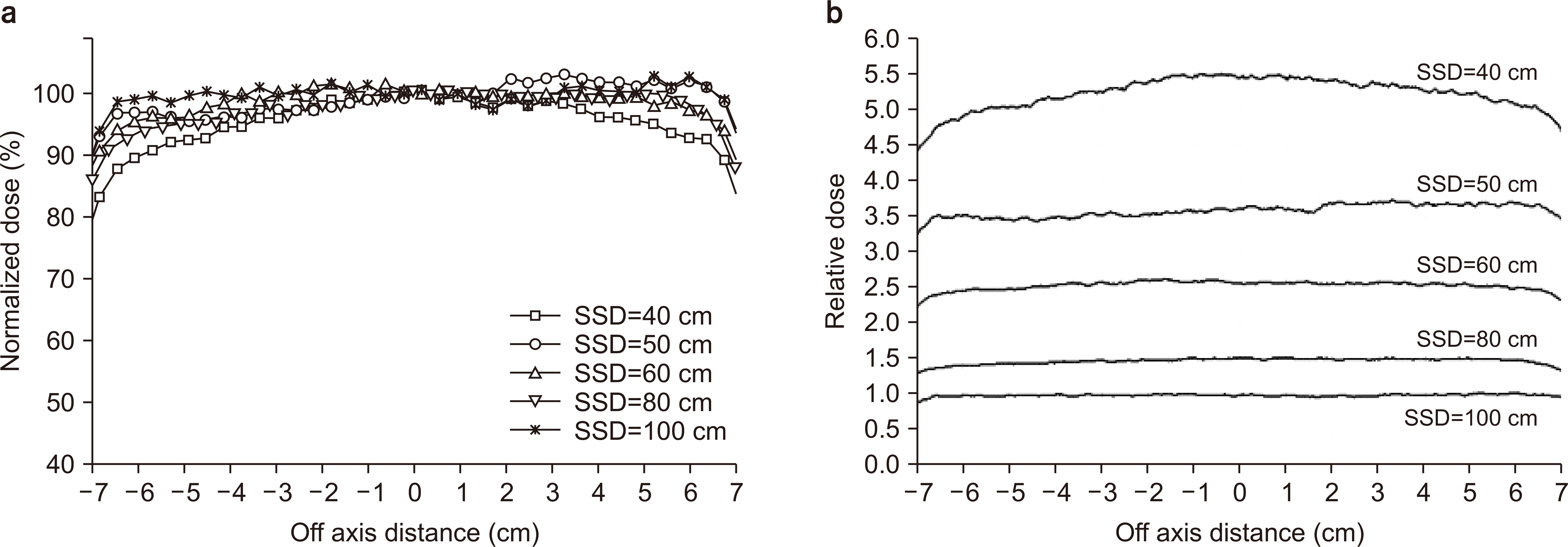
Fig. 12 Measured beam profiles at the SSDs of 40, 50, 60, 80, 100 cm. (a) Normalized dose, (b) relative dose. SSD, source to surface distance.
Cited by 1 articles
-
High-Dose-Rate Electron-Beam Dosimetry Using an Advanced Markus Chamber with Improved Ion-Recombination Corrections
Dong Hyeok Jeong, Manwoo Lee, Heuijin Lim, Sang Koo Kang, Kyoung Won Jang
Prog Med Phys. 2020;31(4):145-152. doi: 10.14316/pmp.2020.31.4.145.
Reference
-
References
1. Lim H, Jeong DH, Lee MW, Lee MJ, Shin SW, Yi J. Design of a radiotherapy machine using the 6 MeV C-band standing-wave accelerator. Paper presented at: International Particle Accelerator Conference (IPAC) 2016. 2016 May 10-16; Busan, Korea. p. 1921–1923.2. Lim H, Jeong DH, Lee MW, Lee MJ, Shin SW, Yi J. Control system of the C-band standing-wave accelerator for the medical application. Paper presented at: International Particle Accelerator Conference (IPAC) 2016. 2016 May 12-16; Busan, Korea. p. 4104–4106.3. Lim H, Jo W, Lee DE, Lee M, Kim SH, Shin SW, et al. Status of the DIRAMS C-band standing-wave accelerator for a radiotherapy machine. Paper presented at: 9th Asia Forum for Accelerators and Detectors; 2018. 2018 Jan 28-31; Daejeon, Korea. p. 28–31.4. Kang SK, Kim SH, Kim HC, Lee KH, Lee SJ, Lee DE, et al. Dosimetry system for medical and biological applications of the electron linear accelerator. Paper presented at: The 23rd International Conference on Accelerators and Beam Utilization (ICABU2019). 2019 Nov 13-15; Daejeon, Korea. p. 153.5. Jang KW, Lee M, Lim H, Kang SK, Lee SJ, Kim SH, et al. 2020; Monte Carlo simulation of an electron irradiation device for medical application of an electron linear accelerator. J Korean Phys Soc. 76:588–591. DOI: 10.3938/jkps.76.588.
Article6. Schüler E, Trovati S, King G, Lartey F, Rafat M, Villegas M, et al. 2017; Experimental platform for ultra-high dose rate FLASH irradiation of small animals using a clinical linear accelerator. Int J Radiat Oncol Biol Phys. 97:195–203. DOI: 10.1016/j.ijrobp.2016.09.018. PMID: 27816362.
Article7. International Atomic Energy Agency. 2004. In : IAEA-TECDOC-1386: Emerging applications of radiation processing; 2003 Apr 28-30; Vienna, Austria. International Atomic Energy Agency;Vienna:8. Petersson K, Jaccard M, Germond JF, Buchillier T, Bochud F, Bourhis J, et al. 2017; High dose-per-pulse electron beam dosimetry - a model to correct for the ion recombination in the Advanced Markus ionization chamber. Med Phys. 44:1157–1167. DOI: 10.1002/mp.12111. PMID: 28094853.
Article9. Lim H, Lee M, Kim MY, Yi J, Lee M, Kang SK, et al. 2016; Measurement of energy parameters for electron gun heater currents and output dose rate for electron beams from a prototype linac. Prog Med Phys. 27:25–30. DOI: 10.14316/pmp.2016.27.1.25.
Article10. Lim H, Jeong DH, Lee M, Lee M, Yi J, Yang K, et al. 2016; Solid-state pulse modulator using Marx generator for a medical linac electron-gun. J Instrum. 11:DOI: 10.1088/1748-0221/11/04/P04003.
Article11. Los Alamos National Laboratory. 2002. The manual of MCNP (Monte Carlo N-particle code system) V2.4.0. Los Alamos National Laboratory;Los Alamos:12. IAEA TRS-398. 2006. Absorbed dose determination in external beam radiotherapy: an international code of practice for dosimetry based on standards of absorbed dose to water (v.12). International Atomic Energy Agency;Vienna: 75–90.13. Ding GX, Rogers DWO. 1995. Energy spectra, angular spread and dose distributions of electron beams from various accelerators used in radiotherapy. PIRS-0439. National Research Council of Canada;Ottawa:14. Patil BJ, Bhoraskar VN, Dhole SD, Chavan ST, Pethe SN, Krishnan R. Optimization of dual scattering foil for 6 to 20 MeV electron beam radiotherapy. Paper presented at: 2011 Particle Accelerator Conference. 2011 March 28-April 1; New York, USA. p. 2157–2159.15. Khan FM. 2003. The physics of radiation therapy. 3rd ed. In : Wilkins; Lippincott Williams & Wilkins;Philadelphia: p. 315–317.16. 2016. Standard calibration procedure of gamma irradiation system. Korea Association of Standards & Testing Organizations;Seoul: KASTO 16-80107-102.
- Full Text Links
-
- Actions
-
Cited
- CITED
-
- Close
- Share
-
- Similar articles
-
- Initial Dosimetry of a Prototype Ultra-High Dose Rate Electron-Beam Irradiator for FLASH RT Preclinical Studies
- High Energy Electron Dosimetry by Alanine/ESR Spectroscopy
- Calculation of Energy Spectra for Electron Beam of Medical Linear Accelerator Using GEANT4
- Measurement of Electron Beam Output for the Prototype Compact Linac
- Dose Characteristics of Stereotactic Radiosurgery in High Energy Linear Accelerator Photon Beam
