Morphologic variant of follicular lymphoma reminiscent of hyaline-vascular Castleman disease
- Affiliations
-
- 1Department of Pathology, Seoul National University Hospital, Seoul, Korea
- 2Department of Pathology, Seoul National University College of Medicine, Seoul, Korea
- KMID: 2501661
- DOI: http://doi.org/10.4132/jptm.2019.12.17
Abstract
- Follicular lymphoma (FL) with hyaline-vascular Castleman disease (FL-HVCD)-like features is a rare morphologic variant, with fewer than 20 cases in the literature. Herein, we report a case of FL-HVCD in a 37-year-old female who presented with isolated neck lymph node enlargement. The excised lymph node showed features reminiscent of HVCD, including regressed germinal centers (GCs) surrounded by onion skin-like mantle zones, lollipop lesions composed of hyalinized blood vessels penetrating into regressed GCs, and hyalinized interfollicular stroma. In addition, focal areas of abnormally conglomerated GCs composed of homogeneous, small centrocytes with strong BCL2, CD10, and BCL6 expression were observed, indicating partial involvement of the FL. Several other lymphoid follicles showed features of in situ follicular neoplasia. Based on the observations, a diagnosis of FL-HVCD was made. Although FLHVCD is very rare, the possibility of this variant should be considered in cases resembling CD. Identification of abnormal, neoplastic follicles and ancillary immunostaining are helpful for proper diagnosis.
Keyword
Figure
-
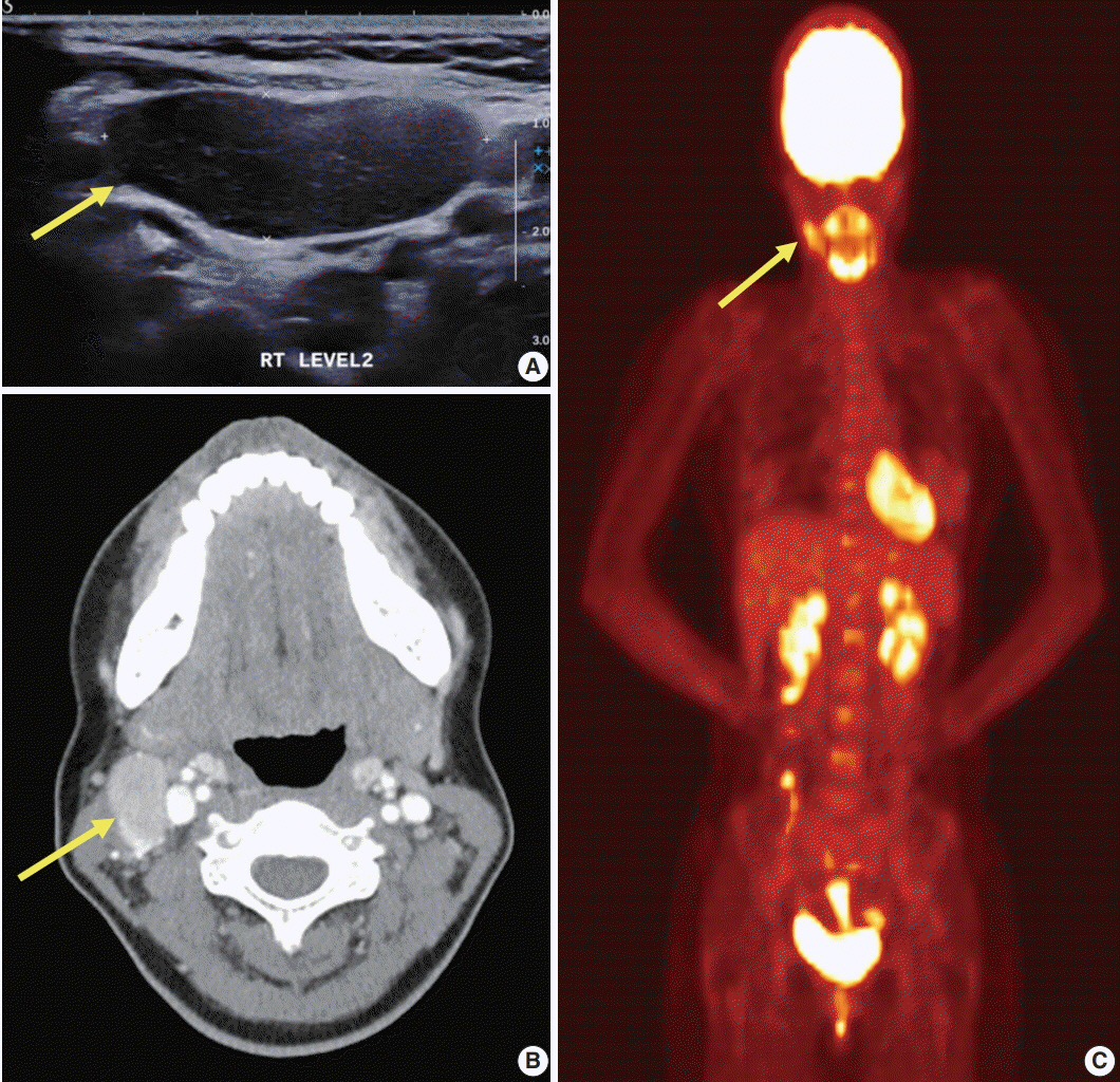
Fig. 1. Imaging work-up of the patient at presentation. (A) Neck ultrasonography shows a hypoechoic mass (arrow). (B) Neck computed tomography shows a well-enhanced ovoid mass at level II area (arrow). (C) 18F-positron emission tomography scan shows hypermetabolic lesion in the right neck (arrow) and no other hypermetabolic lesions.

Fig. 2. Excised lymph node reminiscent of hyaline-vascular Castleman disease (HVCD). Vaguely nodular pattern is noted on low power (A), where regressed lymphoid follicles (B) with expanded onion skin-like mantle zones (C) are observed. (D) Hyalinized blood vessels within follicles are frequently noted. (E) Immunostaining shows the presence of BCL2-positive, BCL6-positive, and CD10-positive cells within the regressed follicles.
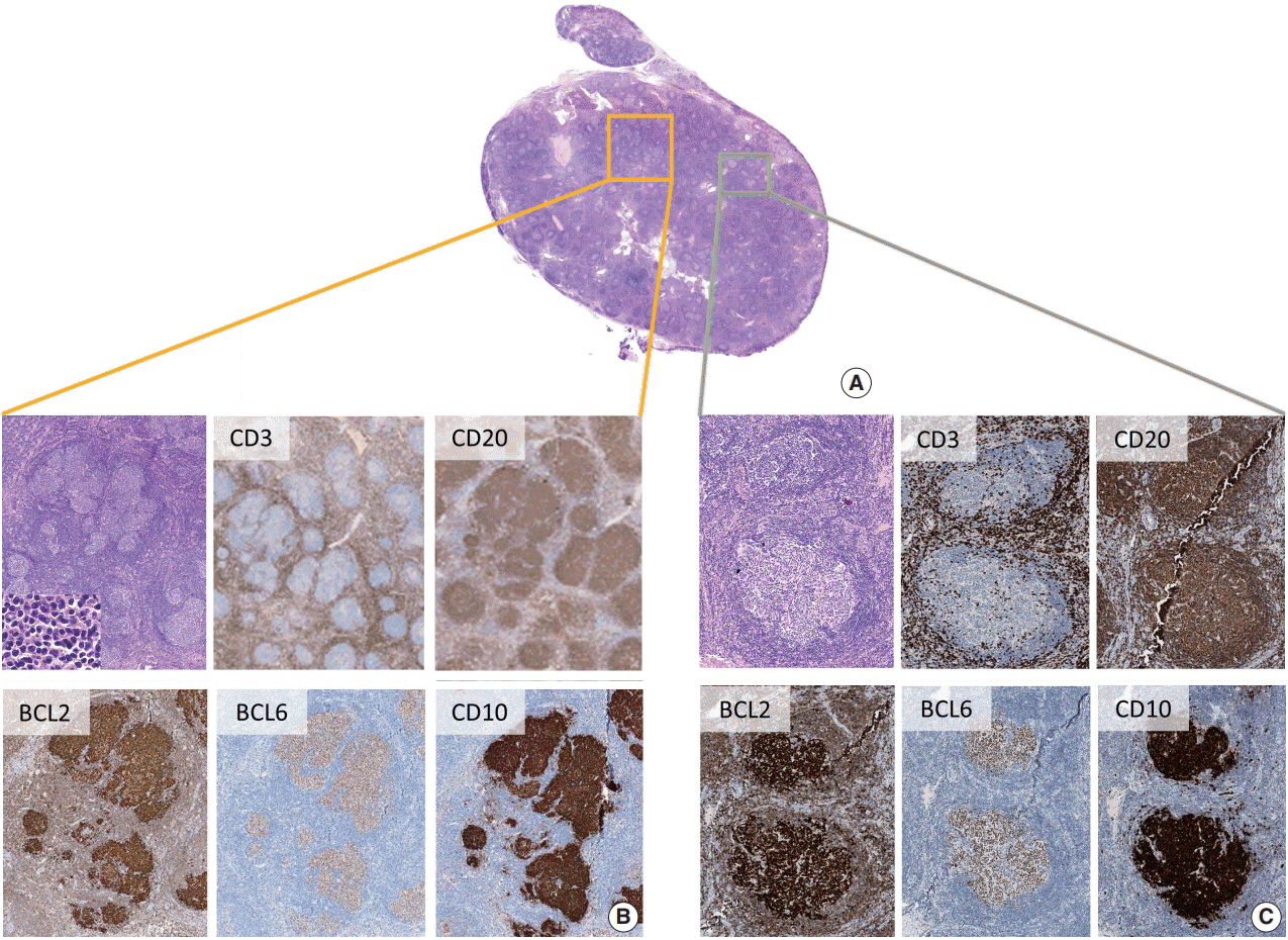
Fig. 3. Features of follicular lymphoma. Upon careful examination, abnormally conglomerated follicles are identified (A, B), and presence of BCL2-positive, BCL6-positive, and CD10-positive neoplastic B-cells is noted (B), confirming follicular lymphoma. (C) In addition, cells having an aberrant BCL2-positive, BCL6-positive, and CD10-positive immunophenotype within otherwise normal-looking follicles are observed, indicating involvement of in situ follicular neoplasia (ISFN).
Reference
-
1. Harris NL, Swerdlow SH, Jaffe ES, et al. Follicular lymphoma. In : Swerdlow SH, Campo E, Harris NL, editors. WHO classification of tumours of haematopoietic and lymphoid tissues. 4th ed. Lyon: IARC Press;2008. p. 220–6.2. Jung HR, Huh J, Ko YH, et al. Classification of malignant lymphoma subtypes in Korean patients: a report of the 4th nationwide study. J Hematopathol. 2019; 12:173–81.
Article3. Goodlad JR, Batstone PJ, Hamilton D, Hollowood K. Follicular lymphoma with marginal zone differentiation: cytogenetic findings in support of a high-risk variant of follicular lymphoma. Histopathology. 2003; 42:292–8.
Article4. Goates JJ, Kamel OW, LeBrun DP, Benharroch D, Dorfman RF. Floral variant of follicular lymphoma. Immunological and molecular studies support a neoplastic process. Am J Surg Pathol. 1994; 18:37–47.5. Kojima M, Matsumoto M, Miyazawa Y, Shimizu K, Itoh H, Masawa N. Follicular lymphoma with prominent sclerosis (“sclerosing variant of follicular lymphoma”) exhibiting a mesenteric bulky mass resembling inflammatory pseudotumor: report of three cases. Pathol Oncol Res. 2007; 13:74–7.
Article6. Kim JM, Ko YH, Lee SS, et al. WHO classification of malignant lymphomas in Korea: report of the third nationwide study. Korean J Pathol. 2011; 45:254–60.
Article7. Warnke RA, Weiss LM, Chan JK, Cleary ML, Dorfman RF. Tumors of the lymph nodes and spleen. In : Rosai J, Sobin LH, editors. Atlans of tumor pathology. 3rd series. Washington, DC: Armed Forces Institute of Pathology;1994. p. 85.8. Nozawa Y, Hirao M, Kamimura K, Hara Y, Abe M. Unusual case of follicular lymphoma with hyaline-vascular follicles. Pathol Int. 2002; 52:794–5.
Article9. Kojima M, Sakurai S, Isoda A, Tsukamoto N, Masawa N, Nakamura N. Follicular lymphoma resembling with hyaline-vascular type of Castleman’s disease: the morphological and immunohistochemical findings of two cases. Cancer Ther. 2009; 7:109–12.10. Siddiqi IN, Brynes RK, Wang E. B-cell lymphoma with hyaline vascular Castleman disease-like features: a clinicopathologic study. Am J Clin Pathol. 2011; 135:901–14.11. Pina-Oviedo S, Wang W, Vicknair E, Manning JT Jr, Medeiros LJ. Follicular lymphoma with hyaline-vascular Castleman disease-like follicles and CD20 positive follicular dendritic cells. Pathology. 2017; 49:544–7.
Article12. Pina-Oviedo S, Miranda RN, Lin P, Manning JT, Medeiros LJ. Follicular lymphoma with hyaline-vascular Castleman-like features: analysis of 6 cases and review of the literature. Hum Pathol. 2017; 68:136–46.
Article13. Kagami Y, Jung J, Choi YS, et al. Establishment of a follicular lymphoma cell line (FLK-1) dependent on follicular dendritic cell-like cell line HK. Leukemia. 2001; 15:148–56.
Article14. Takata K, Sato Y, Nakamura N, et al. Duodenal and nodal follicular lymphomas are distinct: the former lacks activation-induced cytidine deaminase and follicular dendritic cells despite ongoing somatic hypermutations. Mod Pathol. 2009; 22:940–9.
Article15. Chang KC, Wang YC, Hung LY, et al. Monoclonality and cytogenetic abnormalities in hyaline vascular Castleman disease. Mod Pathol. 2014; 27:823–31.
Article16. Freedman A. Follicular lymphoma: 2015 update on diagnosis and management. Am J Hematol. 2015; 90:1171–8.
Article
