Dawn of the Visible Monkey: Segmentation of the Rhesus Monkey for 2D and 3D Applications
- Affiliations
-
- 1Department of Anatomy, Dongguk University School of Medicine, Gyeongju, Korea
- 2Electronics and Telecommunications Research Institute, Daejeon, Korea
- KMID: 2500188
- DOI: http://doi.org/10.3346/jkms.2020.35.e100
Abstract
- Background
To properly utilize the sectioned images in a Visible Monkey dataset, it is essential to segment the images into distinct structures. This segmentation allows the sectioned images to be compiled into two-dimensional or three-dimensional software packages to facilitate anatomy and radiology education, and allows them to be used in experiments involving electromagnetic radiation. The purpose of the present study was to demonstrate the potential of the sectioned images using the segmented images.
Methods
Using sectioned images of a monkey's entire body, 167 structures were segmented using Adobe Photoshop. The segmented images and sectioned images were packaged into the browsing software. Surface models were made from the segmented images using Mimics. Volume models were made from the sectioned images and segmented images using MRIcroGL.
Results
In total, 839 segmented images of 167 structures in the entire body of a monkey were produced at 0.5-mm intervals (pixel size, 0.024 mm; resolution, 8,688 × 5,792; color depth, 24-bit color; BMP format). Using the browsing software, the sectioned images and segmented images were able to be observed continuously and magnified along with the names of the structures. The surface models of PDF file were able to be handled freely using Adobe Reader. In the surface models, the space information of all segmented structures was able to be identified using Sim4Life. On MRIcroGL, the volume model was able to be browsed and sectioned at any angle with real color.
Conclusion
Browsing software, surface models, and volume models are able to be produced based on the segmentation of the sectioned images. These will be helpful for students and researchers studying monkey anatomy and radiology, as well as for biophysicists examining the effects of electromagnetic radiation.
Figure
-
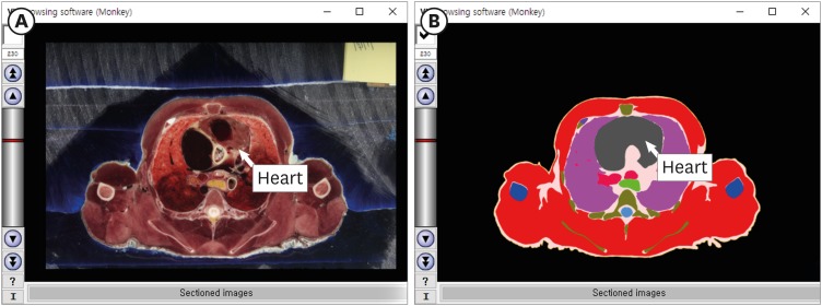
Fig. 1 Browsing software for a rhesus monkey. In the software, (A) the sectioned images and (B) segmented images of the thorax are able to be browsed serially. The name of the structure appears when a mouse pointer is placed over the image.
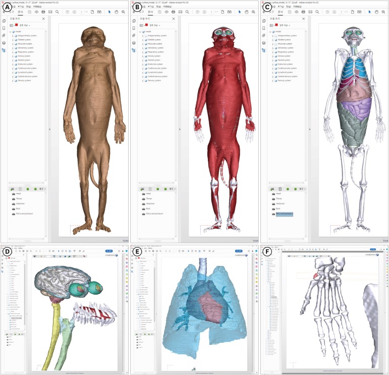
Fig. 2 Surface models of a rhesus monkey in Adobe Reader. The surface models of (A) the skin, (B) muscle, and (C) bones and organs are able to be displayed. (D, E, F) After the surface models are expanded, detailed shapes of segmented structures can be observed. (F) In front of the scaphoid, a sesamoid bone (red color) is visible.
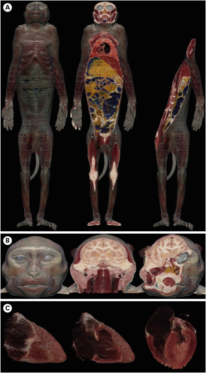
Fig. 3 Volume models of a rhesus monkey on MRIcroGL. The volume models of (A) the whole body, (B) head, and (C) heart can be dissected at any angle (from left to right), similar to the dissection of a real cadaver.
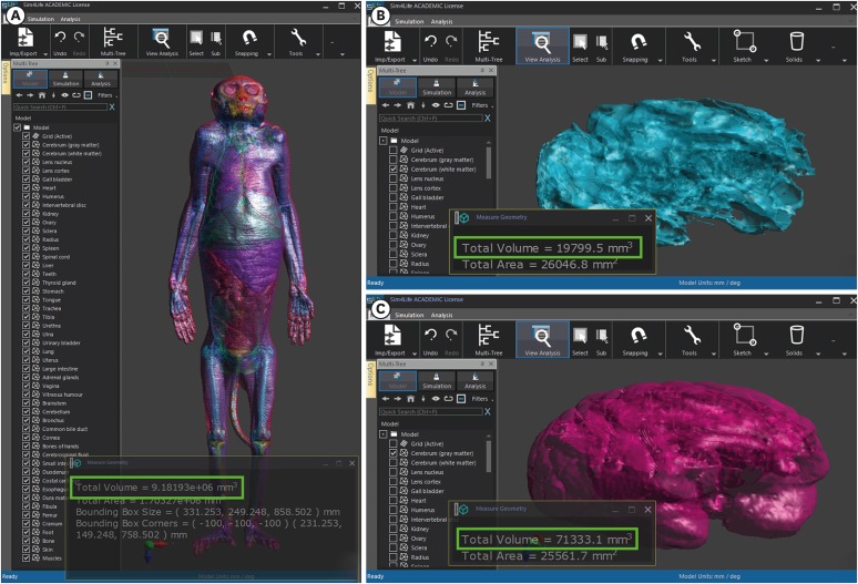
Fig. 4 Voxel models from STL files on Sim4Life software. (A) Voxel volumes (green box) of 167 structures in a whole monkey body, (B) white matter of the cerebrum, and (C) gray matter of the cerebrum are able to be measured.
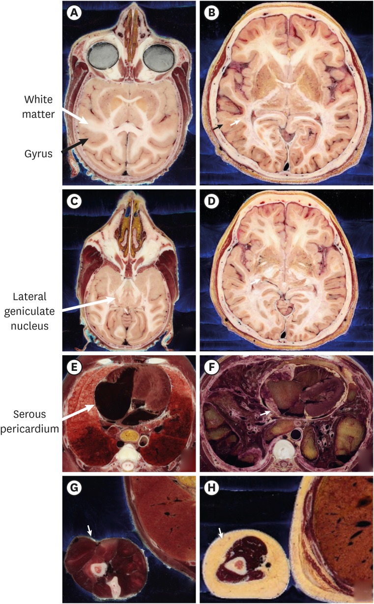
Fig. 5 Comparison of structures between a monkey and a human. In the head, (A) cerebral gyri of the monkey are simpler than (B) those of the human (black arrow). (A) The gray and white matter of the monkey are also simpler than (B) those of the human (white arrow). (A) The eyeball, optic nerve, (C) lateral geniculate nucleus (arrow), and hippocampus of the monkey are larger than (D) that of the human. (E) Because there is minimal fat in the monkey body, the rim of the heart and visceral layer of serous pericardium (arrow) of monkey are very clear, compared with (F) those of the human. (G) In the monkey, there is little fat (arrow) between skin and muscle, as well as between organs; (H) the human exhibits greater volumes of fat in both areas.
Reference
-
1. Thiebaut de Schotten M, Dell'Acqua F, Valabregue R, Catani M. Monkey to human comparative anatomy of the frontal lobe association tracts. Cortex. 2012; 48(1):82–96. PMID: 22088488.
Article2. Olsen RG, Ballinger MB, David TD, Lotz WG. Rewarming of the hypothermic rhesus monkey with electromagnetic radiation. Bioelectromagnetics. 1987; 8(2):183–193. PMID: 3619952.
Article3. Aversi-Ferreira TA, Aversi-Ferreira RA, Bretas RV, Nishimaru H, Nishijo H. Comparative anatomy of the arm muscles of the Japanese monkey (Macaca fuscata) with some comments on locomotor mechanics and behavior. J Med Primatol. 2016; 45(4):165–179. PMID: 27297259.4. Decramer T, Swinnen S, van Loon J, Janssen P, Theys T. White matter tract anatomy in the rhesus monkey: a fiber dissection study. Brain Struct Funct. 2018; 223(8):3681–3688. PMID: 30022250.
Article5. Besle J, Mougin O, Sánchez-Panchuelo RM, Lanting C, Gowland P, Bowtell R, et al. Is human auditory cortex organization compatible with the monkey model? Contrary evidence from ultra-high-field functional and structural MRI. Cereb Cortex. 2019; 29(1):410–428. PMID: 30357410.
Article6. Schilling KG, Gao Y, Christian M, Janve V, Stepniewska I, Landman BA, et al. A web-based atlas combining MRI and histology of the squirrel monkey brain. Neuroinformatics. 2019; 17(1):131–145. PMID: 30006920.
Article7. Gao Y, Chen W, Zhang X. Investigating the influence of spatial constraints on ultimate receive coil performance for monkey brain MRI at 7T. IEEE Trans Med Imaging. 2018; 37(7):1723–1732. PMID: 29969422.8. Park JS, Shin HK, Shin BS, Kwon K. True-color face peeled images with botulinum toxin injection sites and anatomic landmarks. Int J Morphol. 2019; 37(3):1016–1022.
Article9. Lee SB, Chung BS, Chung MS, Youn C, Park JS. Browsing software of the head sectioned images for the android mobile device. Int J Morphol. 2017; 35(4):1377–1382.
Article10. Kwon K, Chung MS, Park JS, Shin BS, Chung BS. Improved software to browse the serial medical images for learning. J Korean Med Sci. 2017; 32(7):1195–1201. PMID: 28581279.
Article11. Kwon K, Shin HK, Shin BS, Park JS. Serially peeled images of the curved surface of the face based on cross-sectional images for use in plastic surgery. J Plast Reconstr Aesthet Surg. 2016; 69(5):727–729.
Article12. Park HS, Chung MS, Shin DS, Jung YW, Park JS. Whole courses of the oculomotor, trochlear, and abducens nerves, identified in sectioned images and surface models. Anat Rec (Hoboken). 2015; 298(2):436–443. PMID: 25212480.
Article13. Chung BS, Jeon CY, Huh JW, Jeong KJ, Har D, Kwack KS, et al. Rise of the Visible Monkey: sectioned images of rhesus monkey. J Korean Med Sci. 2019; 34(8):e66. PMID: 30833883.
Article14. Park JS, Chung MS, Hwang SB, Lee YS, Har DH. Technical report on semiautomatic segmentation using the Adobe Photoshop. J Digit Imaging. 2005; 18(4):333–343. PMID: 16003588.
Article15. Shin DS, Jang HG, Park JS, Park HS, Lee S, Chung MS. Accessible and informative sectioned images and surface models of a cadaver head. J Craniofac Surg. 2012; 23(4):1176–1180. PMID: 22801119.
Article16. Park JS, Chung MS, Chi JG, Park HS, Shin DS. Segmentation of cerebral gyri in the sectioned images by referring to volume model. J Korean Med Sci. 2010; 25(12):1710–1715. PMID: 21165283.
Article17. Yeom YS, Jeong JH, Kim CH, Han MC, Ham BK, Cho KW, et al. HDRK-Woman: whole-body voxel model based on high-resolution color slice images of Korean adult female cadaver. Phys Med Biol. 2014; 59(14):3969–3984. PMID: 24971755.
Article18. Park J, Chung B, Chung M. Digital anatomy using the surface models in portable document format file for self-learning and evaluation. Digit Media. 2017; 3(3):133–137.19. Chung BS, Park JS. Real-color volume models made from real-color sectioned images of Visible Korean. J Korean Med Sci. 2019; 34(10):e86. PMID: 30886552.
Article20. Gabriel C, Gabriel S, Corthout E. The dielectric properties of biological tissues: I. Literature survey. Phys Med Biol. 1996; 41(11):2231–2249. PMID: 8938024.
Article21. Christ A, Kainz W, Hahn EG, Honegger K, Zefferer M, Neufeld E, et al. The Virtual Family--development of surface-based anatomical models of two adults and two children for dosimetric simulations. Phys Med Biol. 2010; 55(2):N23–38. PMID: 20019402.
Article22. Bragg EM, Fairless EA, Liu S, Briggs F. Morphology of visual sector thalamic reticular neurons in the macaque monkey suggests retinotopically specialized, parallel stream-mixed input to the lateral geniculate nucleus. J Comp Neurol. 2017; 525(5):1273–1290. PMID: 27778378.
Article23. Courchesne E, Chisum HJ, Townsend J, Cowles A, Covington J, Egaas B, et al. Normal brain development and aging: quantitative analysis at in vivo MR imaging in healthy volunteers. Radiology. 2000; 216(3):672–682. PMID: 10966694.
Article24. Lee AK, Park JS, Hong SE, Taki M, Wake K, Wiart J, et al. Brain SAR of average male Korean child to adult models for mobile phone exposure assessment. Phys Med Biol. 2019; 64(4):045004. PMID: 30719982.
Article25. Choi C, Nguyen TT, Yeom YS, Lee H, Han H, Shin B, et al. Mesh-type reference Korean phantoms (MRKPs) for adult male and female for use in radiation protection dosimetry. Phys Med Biol. 2019; 64(8):085020. PMID: 30818284.
Article26. Neufeld E, Lloyd B, Schneider B, Kainz W, Kuster N. Functionalized anatomical models for computational life sciences. Front Physiol. 2018; 9:1594. PMID: 30505279.
Article27. Park JS, Jung YW, Choi HD, Lee AK. VK-phantom male with 583 structures and female with 459 structures, based on the sectioned images of a male and a female, for computational dosimetry. J Radiat Res (Tokyo). 2018; 59(3):338–380. PMID: 29659988.
Article28. Werner H, Lopes J, Ribeiro G, Raposo AB, Trajano E, Araujo Júnior E. Three-dimensional virtual traveling navigation and three-dimensional printing models of a normal fetal heart using ultrasonography data. Prenat Diagn. 2019; 39(3):175–177. PMID: 30635939.
Article29. Tofte JN, Westerlind BO, Martin KD, Guetschow BL, Uribe-Echevarria B, Rungprai C, et al. Knee, Shoulder, and Fundamentals of Arthroscopic Surgery Training: Validation of a Virtual Arthroscopy Simulator. Arthroscopy. 2017; 33(3):641–646.e3. PMID: 27989355.
Article
- Full Text Links
-
- Actions
-
Cited
- CITED
-
- Close
- Share
-
- Similar articles
-
- Erratum: Correction of Classification: Rise of the Visible Monkey: Sectioned Images of Rhesus Monkey
- Rise of the Visible Monkey: Sectioned Images of Rhesus Monkey
- Suppurative bite wound by repetitive aggression of dominance hierarchy during group housing in rhesus monkeys
- Reference values of hematological and biochemical parameters in young-adult cynomolgus monkey (Macaca fascicularis) and rhesus monkey (Macaca mulatta) anesthetized with ketamine hydrochloride
- Fixation Task Training in Macaque Monkey and Factors Affecting Fixation
