Short-term impact of microimplant-assisted rapid palatal expansion on the nasal soft tissues in adults: A three-dimensional stereophotogrammetry study
- Affiliations
-
- 1Department of Orthodontics, Dankook University College of Dentistry, Cheonan, Korea. jwlee-1945@daum.net
- KMID: 2471868
- DOI: http://doi.org/10.4041/kjod.2020.50.2.75
Abstract
OBJECTIVE
The aim of this study was to evaluate changes in the nasal soft tissues, including movements of landmarks, changes in linear distances, and volumetric changes, using three-dimensional (3D) stereophotogrammetry after microimplant-assisted rapid palatal expansion (MARPE) in adult patients.
METHODS
Facial data were scanned using a white light scanner before and after MARPE in 30 patients. In total, 7 mm of expansion was achieved over a 4-week expansion period. We determined 10 soft tissue landmarks using reverse engineering software and measured 3D vector changes at those points. In addition, we calculated the distances between points to determine changes in the width of the nasal soft tissues. The volumetric change in the nose was also measured.
RESULTS
All landmarks except pronasale and subnasale showed statistically significant movement on the x-axis. Pronasale, subnasale, alar right, and alar left showed significant movement on the y-axis, while all landmarks except subnasale showed significant movement on the z-axis. The alar base width, alar width, and alar curvature width increased by 1.214, 0.932, and 0.987 mm, respectively. The average volumetric change was 993.33 mm³, and the amount of increase relative to the average initial volume was 2.96%.
CONCLUSIONS
The majority of soft tissue landmarks around the nasal region show significant positional changes after MARPE in adults. The nose tends to widen and move forward and downward. The post-treatment nasal volume may also exhibit a significant increase relative to the initial volume. Clinicians should thoroughly explain the anticipated changes to patients before MARPE initiation.
Keyword
MeSH Terms
Figure
-
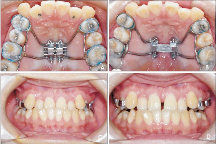
Figure 1 Microimplant-assisted rapid palatal expansion. A, The microimplant-assisted rapid palatal expansion (MARPE) device (MSE-12; Biomaterials, Seoul, Korea). B, After activation of the MARPE appliance. C, Maxillary transverse deficiency before MARPE. D, Diastema after completion of MARPE.
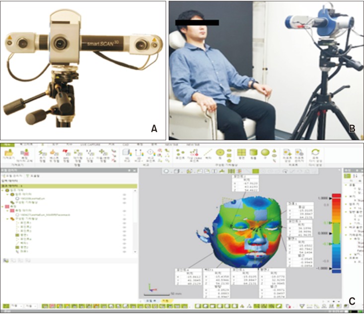
Figure 2 Scanning procedure used before and after microimplant-assisted rapid palatal expansion. A, White light scanner (Smart-Scan 3D; Breuckmann, Braunschweig, Germany). B, Scanning position. C, Reverse engineering software (Geomagic Control X 2017; 3D Systems, Seoul, Korea).
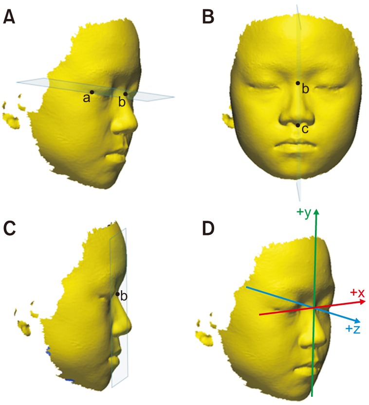
Figure 3 Establishment of reference planes for measurement of nasal soft tissue changes using stereophotogrammetry after microimplant-assisted rapid palatal expansion. A, The horizontal reference plane is defined as the plane passing through the soft tissue nasion (point b) and exocanthion (point a) on both sides. B, The plane perpendicular to the horizontal reference plane and passing through the soft tissue nasion and subnasale (point c) is established as the sagittal reference plane. C, The coronal reference plane is designed such that it is perpendicular to the two existing planes and passed through the soft tissue nasion. D, New coordinate system.
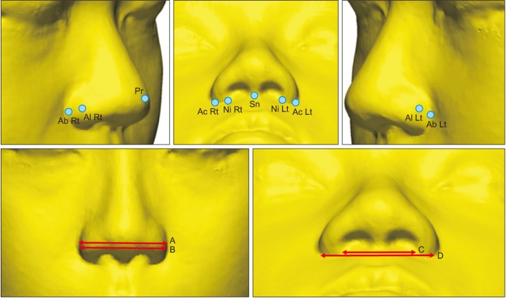
Figure 4 Locations and definitions of nasal soft tissue landmarks for measurement of nasal soft tissue changes using stereophotogrammetry after microimplant-assisted rapid palatal expansion.A, Alar width (Al Rt to Al Lt); B, alar base width (Ab Rt to Ab Lt); C, inferior width of the nostrils (Ni Rt to Ni Lt); D, alar curvature width (Ac Rt to Ac Lt).See Table 2 for definition of each landmark.
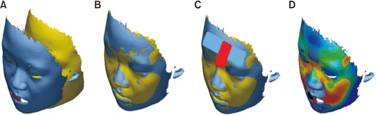
Figure 5 Superimposition and measurements for evaluation of nasal soft tissue changes using stereophotogrammetry after microimplant-assisted rapid palatal expansion (MARPE). A, Facial scan data before and after MARPE. B, Initial registration. C, Best fit alignment. Additional alignment based on specific regions is achieved (forehead, intercanthal region, and dorsum of the nose). D, Shell to shell three-dimensional deviation maps.
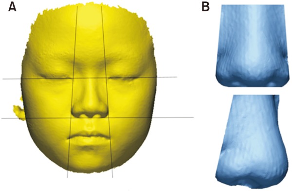
Figure 6 Measurement of changes in the nose volume using stereophotogrammetry after microimplant-assisted rapid palatal expansion. A, Planes outlining the nasal region. B, Cropped nasal area.
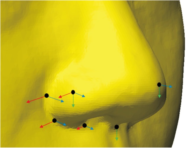
Figure 7 Displacement of landmarks measured by stereophotogrammetry after microimplant-assisted rapid palatal expansion. The length of the arrows indicates the amount of change (red: x-axis, green: y-axis, blue: z-axis).
Cited by 3 articles
-
Midfacial soft tissue changes after maxillary expansion using micro-implant-supported maxillary skeletal expanders in young adults: A retrospective study
Hieu Nguyen, Jeong Won Shin, Hai-Van Giap, Ki Beom Kim, Hwa Sung Chae, Young Ho Kim, Hae Won Choi
Korean J Orthod. 2021;51(3):145-156. doi: 10.4041/kjod.2021.51.3.145.Effectiveness of miniscrew assisted rapid palatal expansion using cone beam computed tomography: A systematic review and meta-analysis
Patchaya Siddhisaributr, Kornkanok Khlongwanitchakul, Niwat Anuwongnukroh, Somchai Manopatanakul, Nita Viwattanatipa
Korean J Orthod. 2022;52(3):182-200. doi: 10.4041/kjod21.256.Skeletal and dentoalveolar effects of different types of microimplant-assisted rapid palatal expansion
Hyeong-Yoon Choi, Sang-Min Lee, Jin-Woo Lee, Dong-Hwa Chung, Mo-Hyeon Lee
Korean J Orthod. 2023;53(4):241-253. doi: 10.4041/kjod23.036.
Reference
-
1. da Silva Filho OG, Santamaria M Jr, Capelozza Filho L. Epidemiology of posterior crossbite in the primary dentition. J Clin Pediatr Dent. 2007; 32:73–78. PMID: 18274476.
Article2. Egermark-Eriksson I, Carlsson GE, Magnusson T, Thilander B. A longitudinal study on malocclusion in relation to signs and symptoms of cranio-mandibular disorders in children and adolescents. Eur J Orthod. 1990; 12:399–407. PMID: 2086260.
Article3. Brunelle JA, Bhat M, Lipton JA. Prevalence and distribution of selected occlusal characteristics in the US population, 1988-1991. J Dent Res. 1996; 75 Spec No:706–713. PMID: 8594094.
Article4. Persson M, Thilander B. Palatal suture closure in man from 15 to 35 years of age. Am J Orthod. 1977; 72:42–52. PMID: 267435.
Article5. Melsen B, Melsen F. The postnatal development of the palatomaxillary region studied on human autopsy material. Am J Orthod. 1982; 82:329–342. PMID: 6961805.
Article6. Takeuchi M, Tanaka E, Nonoyama D, Aoyama J, Tanne K. An adult case of skeletal open bite with a severely narrowed maxillary dental arch. Angle Orthod. 2002; 72:362–370. PMID: 12169037.7. Parr JA, Garetto LP, Wohlford ME, Arbuckle GR, Roberts WE. Sutural expansion using rigidly integrated endosseous implants: an experimental study in rabbits. Angle Orthod. 1997; 67:283–290. PMID: 9267577.8. Lee KJ, Park YC, Park JY, Hwang WS. Miniscrew-assisted nonsurgical palatal expansion before orthognathic surgery for a patient with severe mandibular prognathism. Am J Orthod Dentofacial Orthop. 2010; 137:830–839. PMID: 20685540.
Article9. Wilmes B, Nienkemper M, Drescher D. Application and effectiveness of a mini-implant- and tooth-borne rapid palatal expansion device: the hybrid hyrax. World J Orthod. 2010; 11:323–330. PMID: 21490997.10. Harzer W, Reusser L, Hansen L, Richter R, Nagel T, Tausche E. Minimally invasive rapid palatal expansion with an implant-supported hyrax screw. Biomed Tech (Berl). 2010; 55:39–45. PMID: 20128744.11. Deeb W, Hansen L, Hotan T, Hietschold V, Harzer W, Tausche E. Changes in nasal volume after surgically assisted bone-borne rapid maxillary expansion. Am J Orthod Dentofacial Orthop. 2010; 137:782–789. PMID: 20685533.
Article12. Garib DG, Navarro R, Francischone CE, Oltramari PV. Rapid maxillary expansion using palatal implants. J Clin Orthod. 2008; 42:665–671. PMID: 19075382.13. Park JJ, Park YC, Lee KJ, Cha JY, Tahk JH, Choi YJ. Skeletal and dentoalveolar changes after miniscrew-assisted rapid palatal expansion in young adults: a cone-beam computed tomography study. Korean J Orthod. 2017; 47:77–86. PMID: 28337417.
Article14. MacGinnis M, Chu H, Youssef G, Wu KW, Machado AW, Moon W. The effects of micro-implant assisted rapid palatal expansion (MARPE) on the nasomaxillary complex--a finite element method (FEM) analysis. Prog Orthod. 2014; 15:52. PMID: 25242527.
Article15. Islam R, Kitahara T, Naher L, Hara A, Nakasima A. Lip morphological changes in orthodontic treatment. Class II division 1: malocclusion and normal occlusion at rest and on smiling. Angle Orthod. 2009; 79:256–264. PMID: 19216599.16. Ngan P, Hägg U, Yiu C, Merwin D, Wei SH. Soft tissue and dentoskeletal profile changes associated with maxillary expansion and protraction headgear treatment. Am J Orthod Dentofacial Orthop. 1996; 109:38–49. PMID: 8540481.
Article17. Filho HN, Gonçales ES, Berrentin-Felix G, de Souza César U, Achĵa GL. Evaluation of the facial soft tissues following surgically assisted maxillary expansion associated with the simple V-Y suture. Int J Adult Orthodon Orthognath Surg. 2002; 17:89–97. PMID: 12099321.18. Magnusson A, Bjerklin K, Kim H, Nilsson P, Marcusson A. Three-dimensional computed tomographic analysis of changes to the external features of the nose after surgically assisted rapid maxillary expansion and orthodontic treatment: a prospective longitudinal study. Am J Orthod Dentofacial Orthop. 2013; 144:404–413. PMID: 23992813.19. Hajeer MY, Millett DT, Ayoub AF, Siebert JP. Applications of 3D imaging in orthodontics: part I. J Orthod. 2004; 31:62–70. PMID: 15071154.20. Kau CH, Richmond S, Incrapera A, English J, Xia JJ. Three-dimensional surface acquisition systems for the study of facial morphology and their application to maxillofacial surgery. Int J Med Robot. 2007; 3:97–110. PMID: 17619242.
Article21. van Loon B, Maal TJ, Plooij JM, Ingels KJ, Borstlap WA, Kuijpers-Jagtman AM, et al. 3D Stereophotogrammetric assessment of pre- and postoperative volumetric changes in the cleft lip and palate nose. Int J Oral Maxillofac Surg. 2010; 39:534–540. PMID: 20427150.
Article22. van Loon B, van Heerbeek N, Maal TJ, Borstlap WA, Ingels KJ, Schols JG, et al. Postoperative volume increase of facial soft tissue after percutaneous versus endonasal osteotomy technique in rhinoplasty using 3D stereophotogrammetry. Rhinology. 2011; 49:121–126. PMID: 21468387.
Article23. Oliveira De Felippe NL, Da Silveira AC, Viana G, Kusnoto B, Smith B, Evans CA. Relationship between rapid maxillary expansion and nasal cavity size and airway resistance: short- and long-term effects. Am J Orthod Dentofacial Orthop. 2008; 134:370–382. PMID: 18774083.
Article24. Johnson BM, McNamara JA, Bandeen RL, Baccetti T. Changes in soft tissue nasal widths associated with rapid maxillary expansion in prepubertal and postpubertal subjects. Angle Orthod. 2010; 80:995–1001. PMID: 20677946.
Article25. Aynechi N, Larson BE, Leon-Salazar V, Beiraghi S. Accuracy and precision of a 3D anthropometric facial analysis with and without landmark labeling before image acquisition. Angle Orthod. 2011; 81:245–252. PMID: 21208076.
Article26. de Menezes M, Rosati R, Ferrario VF, Sforza C. Accuracy and reproducibility of a 3-dimensional stereophotogrammetric imaging system. J Oral Maxillofac Surg. 2010; 68:2129–2135. PMID: 20646812.
Article27. Dai F, Yu J, Chen G, Xu T, Jiang R. Changes in buccal facial depth of female patients after extraction and nonextraction orthodontic treatments: a preliminary study. Korean J Orthod. 2018; 48:172–181. PMID: 29732303.
Article28. Eidson L, Cevidanes LH, de Paula LK, Hershey HG, Welch G, Rossouw PE. Three-dimensional evaluation of changes in lip position from before to after orthodontic appliance removal. Am J Orthod Dentofacial Orthop. 2012; 142:410–418. PMID: 22920709.
Article29. Day CJ, Robert T. Three-dimensional assessment of the facial soft tissue changes that occur postoperatively in orthognathic patients. World J Orthod. 2006; 7:15–26. PMID: 16548302.30. Lima SM Jr, de Moraes M, Asprino L. Photoelastic analysis of stress distribution of surgically assisted rapid maxillary expansion with and without separation of the pterygomaxillary suture. J Oral Maxillofac Surg. 2011; 69:1771–1775. PMID: 21292367.31. Corbridge JK, Campbell PM, Taylor R, Ceen RF, Buschang PH. Transverse dentoalveolar changes after slow maxillary expansion. Am J Orthod Dentofacial Orthop. 2011; 140:317–325. PMID: 21889076.
Article32. Sarver DM, Johnston MW. Skeletal changes in vertical and anterior displacement of the maxilla with bonded rapid palatal expansion appliances. Am J Orthod Dentofacial Orthop. 1989; 95:462–466. PMID: 2658544.
Article33. Seong EH, Choi SH, Kim HJ, Yu HS, Park YC, Lee KJ. Evaluation of the effects of miniscrew incorporation in palatal expanders for young adults using finite element analysis. Korean J Orthod. 2018; 48:81–89. PMID: 29564217.
Article34. Zong C, Tang B, Hua F, He H, Ngan P. Skeletal and dentoalveolar changes in the transverse dimension using microimplant-assisted rapid palatal expansion (MARPE) appliances. Semin Orthod. 2019; 25:46–59.
Article35. Abedini S, Elkenawy I, Kim E, Moon W. Three-dimensional soft tissue analysis of the face following micro-implant-supported maxillary skeletal expansion. Prog Orthod. 2018; 19:46. PMID: 30450504.
Article36. Nada RM, van Loon B, Schols JG, Maal TJ, de Koning MJ, Mostafa YA, et al. Volumetric changes of the nose and nasal airway 2 years after tooth-borne and bone-borne surgically assisted rapid maxillary expansion. Eur J Oral Sci. 2013; 121:450–456. PMID: 24028593.37. Kim SJ, Baik HS, Hwang CJ, Yu HS. Diagnosis and evaluation of skeletal Class III patients with facial asymmetry for orthognathic surgery using three-dimensional computed tomography. Semin Orthod. 2015; 21:274–282.
Article38. Bazargani F, Feldmann I, Bondemark L. Three-dimensional analysis of effects of rapid maxillary expansion on facial sutures and bones. Angle Orthod. 2013; 83:1074–1082. PMID: 23745976.
- Full Text Links
-
- Actions
-
Cited
- CITED
-
- Close
- Share
-
- Similar articles
-
- A tomographic study on oro-nasal dimensional changes following rapid palatal expansion
- Skeletal and dentoalveolar effects of different types of microimplant-assisted rapid palatal expansion
- A posteroanterior cephalometric study on the change of maxilla by rapid palatal expansion
- Endotracheal tube damage during surgically assisted rapid palatal expansion surgery; a case report
- Effectiveness of miniscrew assisted rapid palatal expansion using cone beam computed tomography: A systematic review and meta-analysis
