Imaging Sci Dent.
2020 Mar;50(1):65-71. 10.5624/isd.2020.50.1.65.
Three types of ossifying fibroma: A report of 4 cases with an analysis of CBCT features
- Affiliations
-
- 1Department of Pediatric Dentistry, College of Dentistry, Chosun University, Gwangju, Korea.
- 2Department of Oral and Maxillofacial Radiology, College of Dentistry, Chosun University, Gwangju, Korea. hidds@chosun.ac.kr
- KMID: 2471847
- DOI: http://doi.org/10.5624/isd.2020.50.1.65
Abstract
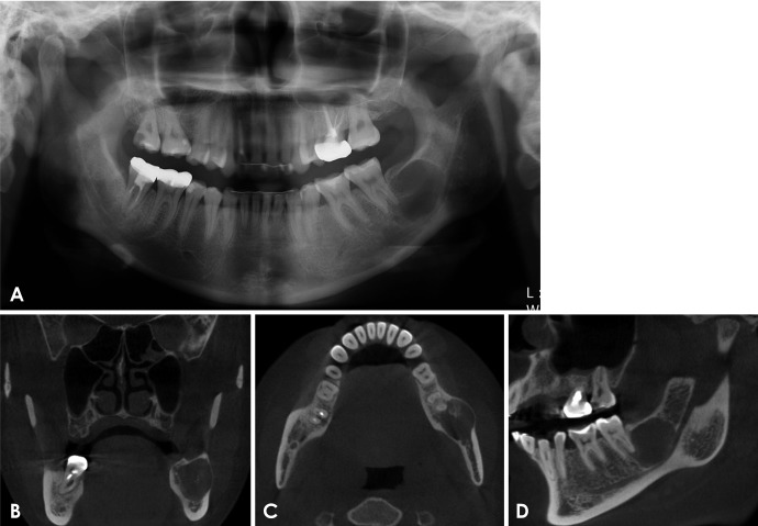
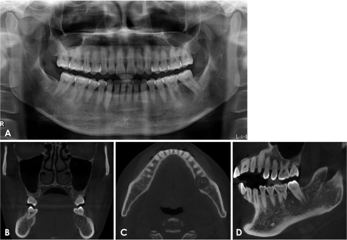
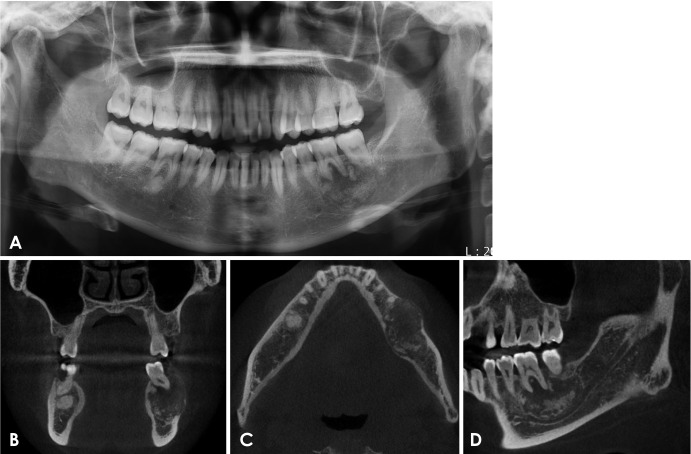
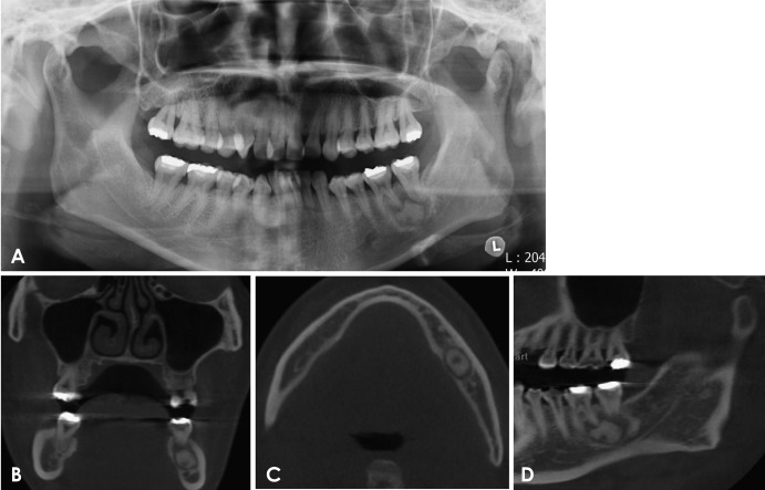
- Ossifying fibroma is a slow-growing benign neoplasm that occurs most often in the jaws, especially the mandible. The tumor is composed of bone that develops within fibrous connective tissue. Some ossifying fibromas consist of cementum-like calcifications, while others contain only bony material; however, a mixture of these calcification types is commonly seen in a single lesion. Of the craniofacial bones, the mandible is the most commonly involved site, with the lesion typically inferior to the premolars and molars. Ossifying fibroma of the jaw shows a female predominance. Some reports of ossifying fibroma have been published in the literature; however, this report continues the research on this topic by detailing 3 types of ossifying fibroma findings on panoramic radiographs and cone-beam computed tomographic images of 4 patients. The radiographs of the presented cases could help clinicians understand the variations in the radiographic appearance of this lesion.
MeSH Terms
Figure
Reference
-
1. Koenig LJ, Tamimi DF, Petrikowski CG, Perschbacher SE. Diagnostic imaging: oral and maxillofacial. 2nd ed. Philadelphia, PA: Elsevier;2017.2. Hamner JE 3rd, Scofield HH, Cornyn J. Benign fibro-osseous jaw lesions of periodontal membrane origin. An analysis of 249 cases. Cancer. 1968; 26:861–878.
Article3. Waldron CA, Giansanti JS. Benign fibro-osseous lesions of the jaws: a clinical-radiologic-histologic review of sixty-five cases. II. Benign fibro-osseous lesions of periodontal ligament origin. Oral Surg Oral Med Oral Pathol. 1973; 35:340–350. PMID: 4510606.4. Eversole LR, Merrell PW, Strub D. Radiographic characteristics of central ossifying fibroma. Oral Surg Oral Med Oral Pathol. 1985; 59:522–527. PMID: 3859811.
Article5. Waldron CA. Fibro-osseous lesions of the jaws. J Oral Maxillofac Surg. 1993; 51:828–835. PMID: 8336219.
Article6. Chang CC, Hung HY, Chang JY, Yu CH, Wang YP, Liu BY, et al. Central ossifying fibroma: a clinicopathologic study of 28 cases. J Formos Med Assoc. 2008; 107:288–294. PMID: 18445542.
Article7. MacDonald-Jankowski DS. Fibro-osseous lesions of the face and jaws. Clin Radiol. 2004; 59:11–25. PMID: 14697371.
Article8. Vegas Bustamante E, Gargallo Albiol J, Berini Aytés L, Gay Escoda C. Benign fibro-osseous lesions of the maxillas: analysis of 11 cases. Med Oral Patol Oral Cir Bucal. 2008; 13:E653–E656. PMID: 18830175.9. Cheng C, Takahashi H, Yao K, Nakayama M, Makoshi T, Nagai H, et al. Cemento-ossifying fibroma of maxillary and sphenoid sinuses: case report and literature review. Acta Otolaryngol Suppl. 2002; (547):118–122. PMID: 12212586.
Article10. Pindborg JJ, Kramer IR. Histological typing of odontogenic tumours, jaw cysts, and allied lesions. World Health Organization. International histological classification of tumours. Geneva: World Health Organization;1971. p. 31–34.11. Kramer IR, Pindborg JJ, Shear M. Neoplasm and other lesions related to bone. Histologic typing of odontogenic tumors. 2nd ed. Heidelberg: Springer-Verlag;1992. p. 28–31.12. Reichart PA, Philipsen HP, Sciubba JJ. The new classification of head and neck tumours (WHO) - any changes? Oral Oncol. 2006; 42:757–758. PMID: 16679047.
Article13. Liu Y, Wang H, You M, Yang Z, Miao J, Shimizutani K, et al. Ossifying fibromas of the jaw bone: 20 cases. Dentomaxillofac Radiol. 2010; 39:57–63. PMID: 20089746.
Article14. Mallya SM, Lam EW. White and Pharoah's oral radiology; principles and interpretation. 8th ed. St. Louis: Elsevier;2019. p. 445–446.


