Imaging Sci Dent.
2020 Mar;50(1):15-22. 10.5624/isd.2020.50.1.15.
Accuracy and reliability of 2-dimensional photography versus 3-dimensional soft tissue imaging
- Affiliations
-
- 1OMFS IMPATH Research Group, Department of Imaging and Pathology, Faculty of Medicine, KU Leuven, Leuven, Belgium. sohaib.shujaat@kuleuven.be
- 2Department of Oral and Maxillofacial Surgery, University Hospitals Leuven, Leuven, Belgium.
- 3Scientific Institute of Public Health, Department of Quality of Medical Laboratories, Brussels, Belgium.
- 4Department of Dental Medicine, Karolinska Institute, Stockholm, Sweden.
- KMID: 2471841
- DOI: http://doi.org/10.5624/isd.2020.50.1.15
Abstract
- PURPOSE
This study was conducted to objectively and subjectively compare the accuracy and reliability of 2-dimensional (2D) photography and 3-dimensional (3D) soft tissue imaging.
MATERIALS AND METHODS
Facial images of 50 volunteers (25 males, 25 females) were captured with a Nikon D800 2D camera (Nikon Corporation, Tokyo, Japan), 3D stereophotogrammetry (SPG), and laser scanning (LS). All subjects were imaged in a relaxed, closed-mouth position with a normal smile. The 2D images were then exported to Mirror® Software (Canfield Scientific, Inc, NJ, USA) and the 3D images into Proplan CMF® software (version 2.1, Materialise HQ, Leuven, Belgium) for further evaluation. For an objective evaluation, 2 observers identified soft tissue landmarks and performed linear measurements on subjects' faces (direct measurements) and both linear and angular measurements on all images (indirect measurements). For a qualitative analysis, 10 dental observers and an expert in facial imaging (subjective gold standard) completed a questionnaire regarding facial characteristics. The reliability of the quantitative data was evaluated using intraclass correlation coefficients, whereas the Fleiss kappa was calculated for qualitative data.
RESULTS
Linear and angular measurements carried out on 2D and 3D images showed excellent inter-observer and intra-observer reliability. The 2D photographs displayed the highest combined total error for linear measurements. SPG performed better than LS, with borderline significance (P=0.052). The qualitative assessment showed no significant differences among the 2D and 3D imaging modalities.
CONCLUSION
SPG was found to a reliable and accurate tool for the morphological evaluation of soft tissue in comparison to 2D imaging and laser scanning.
MeSH Terms
Figure
-
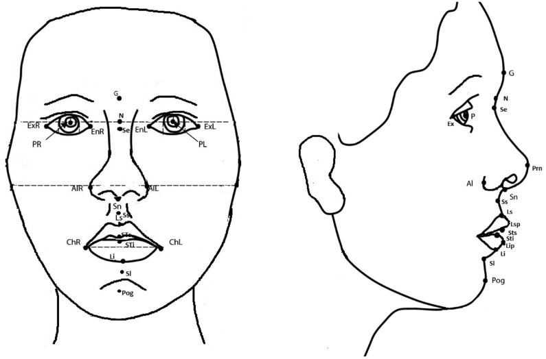
Fig. 1 Facial soft tissue landmarks.
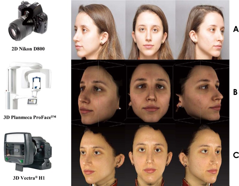
Fig. 2 Image acquisition. A. Two-dimensional camera. B. Three-dimensional laser scanner. C. Three-dimensional stereophotogrammetric device.
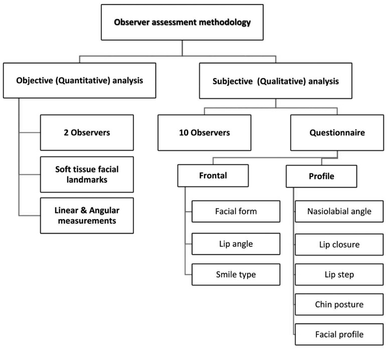
Fig. 3 Flowchart of the observers' analyses.
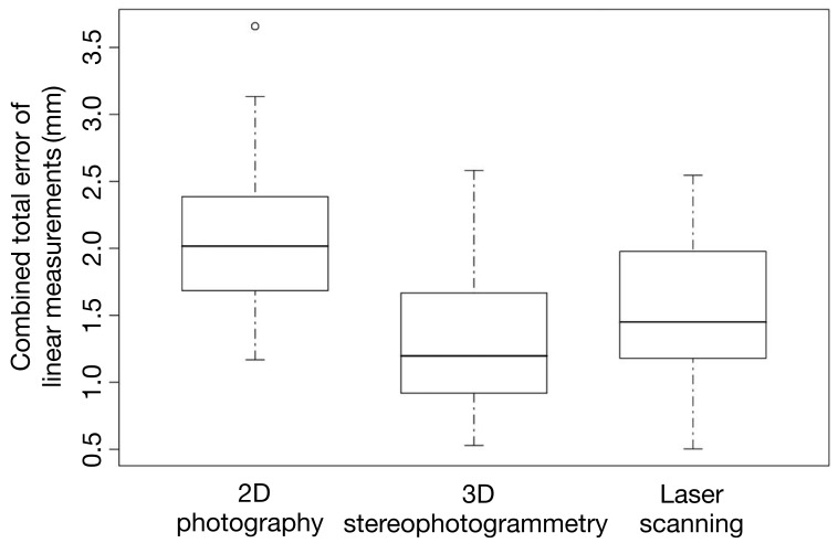
Fig. 4 Combined total error of linear measurements compared to direct anthropometry.
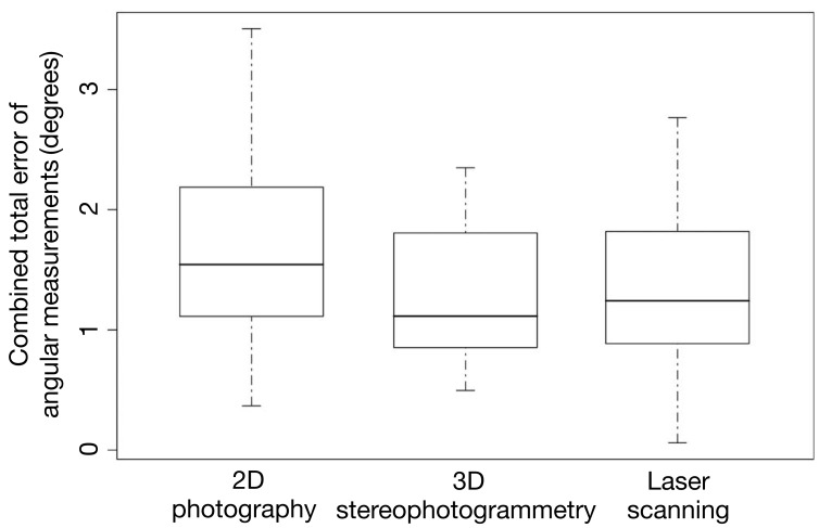
Fig. 5 Combined total error of angular measurements amongst the indirect imaging modalities.
Reference
-
1. Germec-Cakan D, Canter HI, Nur B, Arun T. Comparison of facial soft tissue measurements on three-dimensional images and models obtained with different methods. J Craniofac Surg. 2010; 21:1393–1399. PMID: 20856027.
Article2. Weinberg SM, Scott NM, Neiswanger K, Brandon CA, Marazita ML. Digital three-dimensional photogrammetry: evaluation of anthropometric precision and accuracy using a Genex 3D camera system. Cleft Palate Craniofac J. 2004; 41:507–518. PMID: 15352857.
Article3. Dindaroğlu F, Kutlu P, Duran GS, Görgülü S, Aslan E. Accuracy and reliability of 3D stereophotogrammetry: a comparison to direct anthropometry and 2D photogrammetry. Angle Orthod. 2016; 86:487–494. PMID: 26267357.
Article4. Andrade LM, Rodrigues da Silva AM, Magri LV, Rodrigues da Silva MA. Repeatability study of angular and linear measurements on facial morphology analysis by means of stereophotogrammetry. J Craniofac Surg. 2017; 28:1107–1111. PMID: 28212123.
Article5. Su S, Sinha S, Gabriel V. Evaluating accuracy and reliability of active stereophotogrammetry using MAVIS III Wound Camera for three-dimensional assessment of hypertrophic scars. Burns. 2017; 43:1263–1270. PMID: 28363664.
Article6. Farkas LG, Bryson W, Klotz J. Is photogrammetry of the face reliable? Plast Reconstr Surg. 1980; 66:346–355. PMID: 7422721.
Article7. Aung SC, Ngim RC, Lee ST. Evaluation of the laser scanner as a surface measuring tool and its accuracy compared with direct facial anthropometric measurements. Br J Plast Surg. 1995; 48:551–558. PMID: 8548155.
Article8. Kusnoto B, Evans CA. Reliability of a 3D surface laser scanner for orthodontic applications. Am J Orthod Dentofacial Orthop. 2002; 122:342–348. PMID: 12411877.
Article9. Da Silveira AC, Martinez O, Da Silveira D, Daw JL Jr, Cohen M. Three-dimensional technology for documentation and record keeping for patients with facial clefts. Clin Plast Surg. 2004; 31:141–148. PMID: 15145659.
Article10. Ceinos R, Tardivo D, Bertrand MF, Lupi-Pegurier L. Inter- and intra-operator reliability of facial and dental measurements using 3D-stereophotogrammetry. J Esthet Restor Dent. 2016; 28:178–189. PMID: 26887926.
Article11. Baysal A, Sahan AO, Ozturk MA, Uysal T. Reproducibility and reliability of three-dimensional soft tissue landmark identification using three-dimensional stereophotogrammetry. Angle Orthod. 2016; 86:1004–1009. PMID: 27023408.
Article12. Lam WY, Hsung RT, Choi WW, Luk HW, Cheng LY, Pow EH. A clinical technique for virtual articulator mounting with natural head position by using calibrated stereophotogrammetry. J Prosthet Dent. 2018; 119:902–908. PMID: 28969919.
Article13. Hassan B, Giménez Gonzáles B, Tahmaseb A, Jacobs R, Bornstein MM. Three-dimensional facial scanning technology: applications and future trends. Forum Implantol. 2014; 10:77–86.14. Fink M, Medelnik J, Strobel K, Hirschfelder U, Hofmann E. Metric precision via soft-tissue landmarks in three-dimensional structured-light scans of human faces. J Orofac Orthop. 2014; 75:133–143. PMID: 24577017.
Article15. Kusnoto B, Evans CA. Reliability of a 3D surface laser scanner for orthodontic applications. Am J Orthod Dentofacial Orthop. 2002; 122:342–348. PMID: 12411877.
Article16. Lincoln KP, Sun AY, Prihoda TJ, Sutton AJ. Comparative accuracy of facial models fabricated using traditional and 3D imaging techniques. J Prosthodont. 2016; 25:207–215. PMID: 26381058.
Article17. Ramieri GA, Spada MC, Nasi A, Tavolaccini A, Vezzetti E, Tornincasa S, et al. Reconstruction of facial morphology from laser scanned data. Part I: reliability of the technique. Dentomaxillofac Radiol. 2006; 35:158–164. PMID: 16618848.
Article18. Weinberg SM, Naidoo S, Govier DP, Martin RA, Kane AA, Marazita ML. Anthropometric precision and accuracy of digital three-dimensional photogrammetry: comparing the Genex and 3dMD imaging systems with one another and with direct anthropometry. J Craniofac Surg. 2006; 17:477–483. PMID: 16770184.19. Naini FB, Akram S, Kepinska J, Garagiola U, McDonald F, Wertheim D. Validation of a new three-dimensional imaging system using comparative craniofacial anthropometry. Maxillofac Plast Reconstr Surg. 2017; 39:23. PMID: 28894726.
Article20. Borman H, Ozgür F. A simple instrument to define the Frankfurt horizontal plane for soft-tissue measurements of the face. Plast Reconstr Surg. 1998; 102:580–581. PMID: 9703107.
Article21. Canfiled [Internet]. VECTRA H1 user guide. Parsippany: Canfield;cited 2019 Apr 23. Available from: http://canfieldupgrade.com/assets/media/VECTRA-H1-User-Guide.pdf.22. Cicchetti DV. Guidelines, criteria, and rules of thumb for evaluating normed and standardized assessment instruments in psychology. Psychol Assess. 1994; 6:284–290.
Article23. Landis JR, Koch GG. The measurement of observer agreement for categorical data. Biometrics. 1977; 33:159–174. PMID: 843571.
Article24. Zogheib T, Jacobs R, Bornstein MM, Agbaje JO, Anumendem D, Klazen Y, et al. Comparison of 3D scanning versus 2D photography for the identification of facial soft-tissue landmarks. Open Dent J. 2018; 12:61–71. PMID: 29492171.
Article25. Metzler P, Sun Y, Zemann W, Bartella A, Lehner M, Obwegeser JA, et al. Validity of the 3D VECTRA photogrammetric surface imaging system for cranio-maxillofacial anthropometric measurements. Oral Maxillofac Surg. 2014; 18:297–304. PMID: 23559195.
Article26. de Menezes M, Rosati R, Ferrario VF, Sforza C. Accuracy and reproducibility of a 3-dimensional stereophotogrammetric imaging system. J Oral Maxillofac Surg. 2010; 68:2129–2135. PMID: 20646812.
Article27. Storms AS, Vansant L, Shaheen E, Coucke W, de Llano-Pérula MC, Jacobs R, et al. Three-dimensional aesthetic assessment of class II patients before and after orthognathic surgery and its association with quantitative surgical changes. Int J Oral Maxillofac Surg. 2017; 46:1664–1671. PMID: 28751183.
Article28. Stebel A, Desmedt D, Bronkhorst E, Kuijpers MA, Fudalej PS. Rating nasolabial appearance on three-dimensional images in cleft lip and palate: a comparison with standard photographs. Eur J Orthod. 2016; 38:197–201. PMID: 25900054.
Article29. Hajeer MY, Ayoub AF, Millett DT, Bock M, Siebert JP. Three-dimensional imaging in orthognathic surgery: the clinical application of a new method. Int J Adult Orthodon Orthognath Surg. 2002; 17:318–330. PMID: 12593004.
- Full Text Links
-
- Actions
-
Cited
- CITED
-
- Close
- Share
-
- Similar articles
-
- Usefulness of Two-dimensioanl CT & Three-dimensional CT in Blow-out Fracture
- Diagnostic Accuracy and Usefulness of Three Dimensional Image of Helical CT in Maxillofacial Fractures
- A proposal of landmarks for craniofacial analysis using three-dimensional CT imaging
- The Vectra M3 3-dimensional digital stereophotogrammetry system: A reliable technique for detecting chin asymmetry
- Linear accuracy of cone-beam computed tomography and a 3-dimensional facial scanning system: An anthropomorphic phantom study
