Uterine arteriovenous malformation with repeated vaginal bleeding after dilatation and curettage
- Affiliations
-
- 1Department of Obstetrics and Gynecology, Chosun University Hospital, Chosun University College of Medicine, Gwangju, Korea. themoon6pence@hanmail.net
- KMID: 2471688
- DOI: http://doi.org/10.5468/ogs.2019.62.2.142
Abstract
- Uterine arteriovenous vascular malformation (UAVM) is a disease that causes excessive bleeding. The symptoms do not subside without proper treatment and this can lead to life-threatening situations. The correct diagnosis of UAVM can be complicated if the patient's uterus did not completely discharge everything during abortion (in broader terms, retaining remnants of the products of conception). In this case, Doppler ultrasonography and computed tomography angiography with 3-dimensional rendering were used to analyze the cause of bleeding and provide proper treatment of this patient. Then, uterine artery embolization, dilatation, and curettage were performed safely and successfully. The patient no longer had symptoms of vaginal spotting during the planned follow up care. UAVM is uncommon; however, if reproductive-age women show repeated abnormal vaginal bleeding after dilatation and curettage, a diagnosis of UAVM must be considered based on the medical history and examination.
MeSH Terms
Figure
-
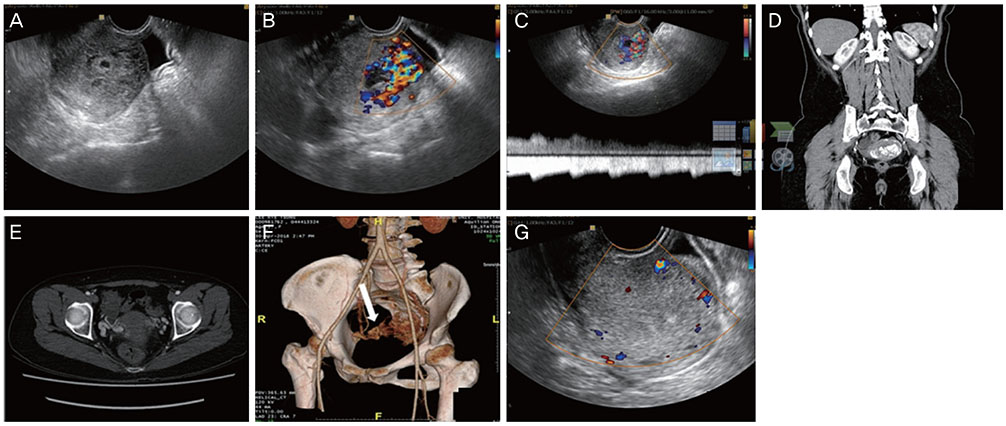
Fig. 1 (A) Transvaginal US shows heterogenous endometrium with an internal echolucent structure. (B) Color Doppler US images reveal multidirectional flow within the posterior wall of the endometrium. (C) Spectral flow Doppler shows high velocity flow pattern (PSV >60 cm/s). (D, E) CT scan shows prominent vascularity within thickened posterior uterine fundus and high enhanced lesion. (F, white arrow) CT angiography with 3D rendering shows the hypervascular tangles of tortuous vessel. (G) After 2 months UAE, follow-up transvaginal US demonstrated complete resolution of UAVM and no vascularity within the lesion. PSV, peak systolic velocity; CT, computed tomography; 3D, 3-dimensional; UAE, uterine artery embolization; UAVM, uterine arteriovenous vascular malformation.
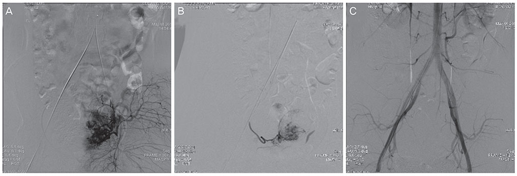
Fig. 2 (A) Left uterine artery. (B) Right uterine artery. Angiography shows UAVM with an early contrast vein filling. (C) Embolization of the AVM was completed. UAVM, uterine arteriovenous vascular malformation; AVM, arteriovenous vascular malformation
Reference
-
1. Grivell RM, Reid KM, Mellor A. Uterine arteriovenous malformations: a review of the current literature. Obstet Gynecol Surv. 2005; 60:761–767.
Article2. Ahn HY, Park IY, Lee G, Kim SJ, Shin JC. Uterine arteriovenous malformation. Arch Gynecol Obstet. 2005; 271:172–175.
Article3. Oguz Y, Gonca Eldem F, Cil B, Sanhal C, Gençosmanoğlu-Türkmen G, Aykan Y, et al. Uterine arteriovenous malformation. Gynecol Obstet Reprod Med. 2018.
Article4. Polat P, Suma S, Kantarcý M, Alper F, Levent A. Color Doppler US in the evaluation of uterine vascular abnormalities. Radiographics. 2002; 22:47–53.
Article5. Kim SM, Ahn HY, Choi MJ, Kang YD, Park JW, Park CH, et al. Uterine arteriovenous malformation with positive serum beta-human chorionic gonadotropin: embolization of both uterine arteries and extra-uterine feeding arteries. Obstet Gynecol Sci. 2016; 59:554–558.
Article6. Müngen E. Vascular abnormalities of the uterus: have we recently over-diagnosed them? Ultrasound Obstet Gynecol. 2003; 21:529–531.
Article7. Rosa e Silva JC, de Aguiar FM, de Sa Rosa e Silva AC, Candido dos Reis FJ, Poli Neto OB, Nogueira AA. Conservative management of large uterine arteriovenous malformation: case report. Pol J Radiol. 2015; 19:202–205.
Article8. Singh N, Tripathi R, Mala YM, Tyagi S, Tyagi S, Singh C. Varied presentation of uterine arteriovenous malformations and their management by uterine artery embolisation. J Obstet Gynaecol. 2014; 34:104–106.
Article9. Delotte J, Chevallier P, Benoit B, Castillon JM, Bongain A. Pregnancy after embolization therapy for uterine arteriovenous malformation. Fertil Steril. 2006; 85:228.
Article10. Hashim H, Nawawi O. Uterine arteriovenous malformation. Malays J Med Sci. 2013; 20:76–80.11. Nonaka T, Yahata T, Kashima K, Tanaka K. Resolution of uterine arteriovenous malformation and successful pregnancy after treatment with a gonadotropin-releasing hormone agonist. Obstet Gynecol. 2011; 117:452–455.
Article
