Korean J Radiol.
2019 Dec;20(12):1646-1652. 10.3348/kjr.2019.0262.
Scoring System to Stratify Malignancy Risks for Mammographic Microcalcifications Based on Breast Imaging Reporting and Data System 5th Edition Descriptors
- Affiliations
-
- 1Department of Radiology, Gangnam Severance Hospital, Yonsei University College of Medicine, Seoul, Korea. jhyouk@yuhs.ac
- 2Department of Radiology, Chonbuk National University Medical School and Hospital, Institute of Medical Science, Research Institute of Clinical Medicine, Jeonju, Korea.
- KMID: 2471677
- DOI: http://doi.org/10.3348/kjr.2019.0262
Abstract
OBJECTIVE
To develop a scoring system stratifying the malignancy risk of mammographic microcalcifications using the 5th edition of the Breast Imaging Reporting and Data System (BI-RADS).
MATERIALS AND METHODS
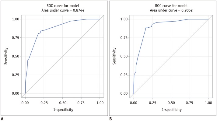
One hundred ninety-four lesions with microcalcifications for which surgical excision was performed were independently reviewed by two radiologists according to the 5th edition of BI-RADS. Each category's positive predictive value (PPV) was calculated and a scoring system was developed using multivariate logistic regression. The scores for benign and malignant lesions or BI-RADS categories were compared using an independent t test or by ANOVA. The area under the receiver operating characteristic curve (AUROC) was assessed to determine the discriminatory ability of the scoring system. Our scoring system was validated using an external dataset.
RESULTS
After excision, 69 lesions were malignant (36%). The PPV of BI-RADS descriptors and categories for calcification showed significant differences. Using the developed scoring system, mean scores for benign and malignant lesions or BI-RADS categories were significantly different (p < 0.001). The AUROC of our scoring system was 0.874 (95% confidence interval, 0.840-0.909) and the PPV of each BI-RADS category determined by the scoring system was as follows: category 3 (0%), 4A (6.8%), 4B (19.0%), 4C (68.2%), and 5 (100%). The validation set showed an AUROC of 0.905 and PPVs of 0%, 8.3%, 11.9%, 68.3%, and 94.7% for categories 3, 4A, 4B, 4C, and 5, respectively.
CONCLUSION
A scoring system based on BI-RADS morphology and distribution descriptors could be used to stratify the malignancy risk of mammographic microcalcifications.
Keyword
MeSH Terms
Figure
Reference
-
1. Rao AA, Feneis J, Lalonde C, Ojeda-Fournier H. A pictorial review of changes in the BI-RADS fifth edition. Radiographics. 2016; 36:623–639. PMID: 27082663.
Article2. Kim J, Kim EK, Kim MJ, Moon HJ, Yoon JH. “Category 4A” microcalcifications: how should this subcategory be applied to microcalcifications seen on mammography? Acta Radiol. 2018; 59:147–153. PMID: 28490180.
Article3. Kim SY, Kim HY, Kim EK, Kim MJ, Moon HJ, Yoon JH. Evaluation of malignancy risk stratification of microcalcifications detected on mammography: a study based on the 5th edition of BI-RADS. Ann Surg Oncol. 2015; 22:2895–2901. PMID: 25608770.
Article4. Sickles EA, D'Orsi CJ, Bassett LW, Appleton CM, Berg WA, Burnside ES, et al. ACR BI-RADS®; mammography. In : Sickles EA, editor. ACR BI-RADS® atlas, breast imaging reporting and data system. Reston, VA: American College of Radiology;2013.5. DeLong ER, DeLong DM, Clarke-Pearson DL. Comparing the areas under two or more correlated receiver operating characteristic curves: a nonparametric approach. Biometrics. 1988; 44:837–845. PMID: 3203132.
Article6. Iwase M, Tsunoda H, Nakayama K, Morishita E, Hayashi N, Suzuki K, et al. Overcalling low-risk findings: grouped amorphous calcifications found at screening mammography associated with minimal cancer risk. Breast Cancer. 2017; 24:579–584. PMID: 27873170.
Article7. Elezaby M, Li G, Bhargavan-Chatfield M, Burnside ES, DeMartini WB. ACR BI-RADS assessment category 4 subdivisions in diagnostic mammography: utilization and outcomes in the national mammography database. Radiology. 2018; 287:416–422. PMID: 29315061.8. Narayan AK, Keating DM, Morris EA, Mango VL. Calling all calcifications: a retrospective case control study. Clin Imaging. 2019; 53:151–154. PMID: 30340079.
Article9. Oligane HC, Berg WA, Bandos AI, Chen SS, Sohrabi S, Anello M, et al. Grouped amorphous calcifications at mammography: frequently atypical but rarely associated with aggressive malignancy. Radiology. 2018; 288:671–679. PMID: 29916773.10. Grimm LJ, Johnson DY, Johnson KS, Baker JA, Soo MS, Hwang ES, et al. Suspicious breast calcifications undergoing stereotactic biopsy in women ages 70 and over: breast cancer incidence by BI-RADS descriptors. Eur Radiol. 2017; 27:2275–2281. PMID: 27752832.
Article11. Bent CK, Bassett LW, D'Orsi CJ, Sayre JW. The positive predictive value of BI-RADS microcalcification descriptors and final assessment categories. AJR Am J Roentgenol. 2010; 194:1378–1383. PMID: 20410428.
Article12. Burnside ES, Ochsner JE, Fowler KJ, Fine JP, Salkowski LR, Rubin DL, et al. Use of microcalcification descriptors in BI-RADS 4th edition to stratify risk of malignancy. Radiology. 2007; 242:388–395. PMID: 17255409.
Article
- Full Text Links
- Actions
-
Cited
- CITED
-
- Close
- Share
- Similar articles
-
- Breast Imaging Reporting and Data System (BI-RADS): Advantages and Limitations
- Scoring System to Predict Malignancy for MRI-Detected Lesions in Breast Cancer Patients: Diagnostic Performance and Effect on Second-Look Ultrasonography
- Thyroid Imaging Reporting and Data System (TIRADS)
- Clinical Utility of MicroPure US Imaging for Breast Microcalcifications
- The Biologic Singificance of Mammographic Calcification


