Biliary cystadenoma in an endemic zone of hydatid cyst: A rare surgical surprise
- Affiliations
-
- 1Department of General Surgery, Post Graduate Institute of Medical Education and Research, Chandigarh, India. kamanlil@yahoo.com
- 2Department of Histopathology, Post Graduate Institute of Medical Education and Research, Chandigarh, India.
- KMID: 2471195
- DOI: http://doi.org/10.14701/ahbps.2020.24.1.85
Abstract
- The advancement of radiological investigations has led to the early and incidental detection of hepatic cystic lesions. These are most commonly the simple cysts but can be malignant as well. Despite the recent advances, these lesions still pose a diagnostic as well as therapeutic challenge. The biliary cystadenomas and carcinomas form around 5% of all the malignant cystic lesions of liver. These lesions are hardly diagnosed preoperatively and are usually a histopathological surprise. They warrant a surgical excision. Herewith, the authors are describing a case of cystic hepatic neoplasm initially misdiagnosed as hydatid cyst of liver and discovered to be a vascular cystic lesion intraoperatively. This patient underwent resection of the lesion and was discovered to harbour biliary cystadenoma on histopathological specimen.
Figure
-
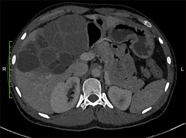
Fig. 1 Multiloculated hypodense lesion (9 to 50 HU) with with multiple thick septae seen in segment IV A, IV B and V. The lesion is abutting the anterior abdominal wall anteriorly, medially it is abutting the psoas muscle and IVC and displacing few small bowel loops. It is also abutting the antropyloric region of stomach and duodenum and also displacing head of pancreas.
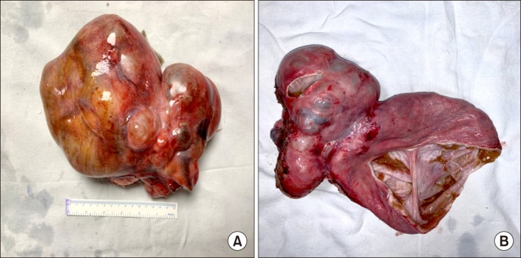
Fig. 2 (A) Resected gross specimen of ~18×16×9 cm with bosselated surface containing multiple cysts. (B) Cut section of whole specimen containing multiple cystic lesions. Luminal aspect of the cyst was smooth and contains mucinous fluid.
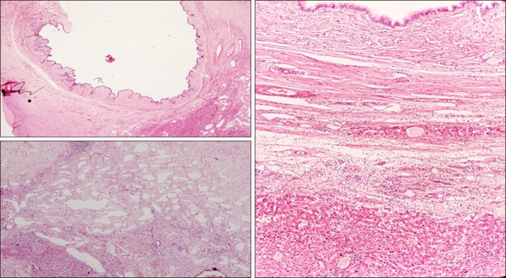
Fig. 3 Histopathology report: multiloculated cyst contains variable size of cystic spaces lined by mucinous epithelium, at places it is replaced by macrophages sheets. It contains fibrous septae, fibrocollagenous tissue and moderate degree of lymphocytes rich chronic inflammation with lymphoid aggregates. Many proliferating vascular and lymphatic channels are also seen. There was no evidence of ovarian stroma. No neoplastic cells were seen. Overall features of biliary cyst adenoma without ovarian stroma.
Reference
-
1. Lantinga MA, Gevers TJ, Drenth JP. Evaluation of hepatic cystic lesions. World J Gastroenterol. 2013; 19:3543–3554. PMID: 23801855.
Article2. Choi BY, Nguyen MH. The diagnosis and management of benign hepatic tumors. J Clin Gastroenterol. 2005; 39:401–412. PMID: 15815209.
Article3. Bacher H, Cerwenka H, Werkgartner G, El-Shabrawi A, Höss G, Preidler K, et al. Primary biliary cystadenocarcinoma perforating the duodenum and left intrahepatic biliary tree--mimicking a hydatid cyst. Liver. 1999; 19:39–41. PMID: 9928764.4. Del Poggio P, Buonocore M. Cystic tumors of the liver: a practical approach. World J Gastroenterol. 2008; 14:3616–3620. PMID: 18595127.5. Frick MP, Feinberg SB. Biliary cystadenoma. AJR Am J Roentgenol. 1982; 139:393–395. PMID: 6979903.
Article6. Simo KA, Mckillop IH, Ahrens WA, Martinie JB, Iannitti DA, Sindram D. Invasive biliary mucinous cystic neoplasm: a review. HPB (Oxford). 2012; 14:725–740. PMID: 23043661.
Article7. Williams DM, Vitellas KM, Sheafor D. Biliary cystadenocarcinoma: seven year follow-up and the role of MRI and MRCP. Magn Reson Imaging. 2001; 19:1203–1208. PMID: 11755730.
Article8. Korobkin M, Stephens DH, Lee JK, Stanley RJ, Fishman EK, Francis IR, et al. Biliary cystadenoma and cystadenocarcinoma: CT and sonographic findings. AJR Am J Roentgenol. 1989; 153:507–511. PMID: 2669463.
Article9. Scherer K, Gupta N, Caine WP, Panda M. Differential diagnosis and management of a recurrent hepatic cyst: a case report and review of literature. J Gen Intern Med. 2009; 24:1161–1165. PMID: 19633897.
Article10. Koffron A, Rao S, Ferrario M, Abecassis M. Intrahepatic biliary cystadenoma: role of cyst fluid analysis and surgical management in the laparoscopic era. Surgery. 2004; 136:926–936. PMID: 15467680.
Article11. van Roekel V, Marx WJ, Baskin W, Greenlaw RL. Cystadenoma of the liver. J Clin Gastroenterol. 1982; 4:167–172. PMID: 7086109.
Article12. Manouras A, Markogiannakis H, Lagoudianakis E, Katergiannakis V. Biliary cystadenoma with mesenchymal stroma: report of a case and review of the literature. World J Gastroenterol. 2006; 12:6062–6069. PMID: 17009411.
Article13. Erdogan D, Busch OR, Rauws EA, van Delden OM, Gouma DJ, van-Gulik TM. Obstructive jaundice due to hepatobiliary cystadenoma or cystadenocarcinoma. World J Gastroenterol. 2006; 12:5735–5738. PMID: 17007033.
Article14. Läuffer JM, Baer HU, Maurer CA, Stoupis C, Zimmerman A, Büchler MW. Biliary cystadenocarcinoma of the liver: the need for complete resection. Eur J Cancer. 1998; 34:1845–1851. PMID: 10023304.
Article15. Gupta A, Pattnaik B, Das A, Kaman L. Von Meyenburg complex and complete ductal plate malformation along with Klatskin tumour: a rare association. BMJ Case Rep. 2016; 2016:bcr2016215220. DOI: 10.1136/bcr-2016-215220.
Article
- Full Text Links
-
- Actions
-
Cited
- CITED
-
- Close
- Share
-
- Similar articles
-
- A Case of a Choledochal Cyst with a Mucinous Cystadenoma of the Pancreas
- A Clinical Case Report of Hydatid Cyst of Liver
- Rare variant of type V choledochal cyst masquerading as a biliary cystadenoma
- Laparoscopic Resection of Adrenal Cyst Mistaken as Intrahepatic Biliary Cystadenoma
- Rupture of Right Hepatic Duct into Hydatid Cyst
