Preoperative imaging of the inferior alveolar nerve canal by cone-beam computed tomography and 1-year neurosensory recovery following mandibular setback through bilateral sagittal split ramus osteotomy: a randomized clinical trial
- Affiliations
-
- 1Department of Oral and Maxillofacial Surgery and Implant Research Center, Islamic Azad University, Tehran Dental Branch, Tehran, Iran. hamidmahaseni@gmail.com
- 2Department of Dental Anatomy, Dental School, Islamic Azad University, Tehran, Iran.
- 3Craniomaxillofacial Research Center, Islamic Azad University, Tehran Dental Branch, Tehran, Iran.
- KMID: 2471111
- DOI: http://doi.org/10.5125/jkaoms.2020.46.1.41
Abstract
OBJECTIVES
One of the most common complications of bilateral sagittal split ramus osteotomy (BSSRO) is neurosensory impairment of the inferior alveolar nerve (IAN). Accurate preoperative determination of the position of the IAN canal within the mandible using cone-beam computed tomography (CBCT) is recommended to prevent IAN dysfunction during BSSRO and facilitate neurosensory improvement after BSSRO.
MATERIALS AND METHODS
This randomized clinical trial consisted of 86 surgical sites in 43 patients (30 females and 13 males), including 21 cases (42 sides) and 22 controls (44 sides). Panoramic and lateral cephalographs were obtained from all patients. In the experimental group, CBCT was also performed from both sides of the ramus and mandibular body. Neurosensory function of the IAN was subjectively assessed using a 5-point scale preoperatively and 7 days, 1 month, 3 months, 6 months, and 12 months post-surgery. Data were analyzed using Fisher's test, Spearman's test, t-test, linear mixed-model regression, and repeated-measures ANCOVA (α=0.05, 0.01).
RESULTS
Mean sensory scores in the control group were 1.57, 2.61, 3.34, 3.73, and 4.20 over one year and were 1.69, 3.00, 3.60, 4.19, and 4.48 in the CBCT group. Significant effects were detected for CBCT intervention (P=0.002) and jaw side (P=0.003) but not for age (P=0.617) or displacement extent (P=0.122).
CONCLUSION
Preoperative use of CBCT may help surgeons to practice more conservative surgery. Neurosensory deficits might heal faster on the right side.
MeSH Terms
Figure
-

Fig. 1 A. Schematic view of the mandibular body. a: distance between inferior alveolar nerve (IAN) canal and buccal cortex of the mandible, b: thickness of the buccal cortex, c: distance between IAN canal in inferior border. B. Measurements of cone-beam computed tomography. C. Distance between the mandibular foramen (at the center of the red circle) and sigmoid notch. The mandibular foramen was located on images as the starting point of the mandibular canal.
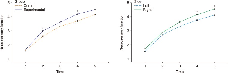
Fig. 2 Marginal means of neurosensory function in the control/experimental groups (left panel) and jaw sides (right panel), estimated at an age of 25.7 years and at a displacement extent of 4.3 mm. The time points 1 to 5 indicate 7 days, 1 month, 3 months, 6 months, and 12 months after surgery, respectively. Significant comparisons (*P<0.01) between control versus experimental groups (left panel) and between left versus right sides (right panel).
Reference
-
1. Antony PG, Sebastian A, Varghese KG, Sobhana CR, Mohan S, Soumithran CS, et al. Neurosensory evaluation of inferior alveolar nerve after bilateral sagittal split ramus osteotomy of mandible. J Oral Biol Craniofac Res. 2017; 7:81–88. PMID: 28706780.
Article2. Aizenbud D, Ciceu C, Hazan-Molina H, Abu-El-Naaj I. Relationship between inferior alveolar nerve imaging and neurosensory impairment following bilateral sagittal split osteotomy in skeletal class III cases with mandibular prognathism. Int J Oral Maxillofac Surg. 2012; 41:461–468. PMID: 22115977.
Article3. Tamás F. Position of the mandibular canal. Int J Oral Maxillofac Surg. 1987; 16:65–69. PMID: 3104497.
Article4. Ylikontiola L, Kinnunen J, Oikarinen K. Factors affecting neurosensory disturbance after mandibular bilateral sagittal split osteotomy. J Oral Maxillofac Surg. 2000; 58:1234–1239. PMID: 11078134.
Article5. Brusati R, Fiamminghi L, Sesenna E, Gazzotti A. Functional disturbances of the inferior alveolar nerve after sagittal osteotomy of the mandibular ramus: operating technique for prevention. J Maxillofac Surg. 1981; 9:123–125. PMID: 6943243.
Article6. Ylikontiola L, Moberg K, Huumonen S, Soikkonen K, Oikarinen K. Comparison of three radiographic methods used to locate the mandibular canal in the buccolingual direction before bilateral sagittal split osteotomy. Oral Surg Oral Med Oral Pathol Oral Radiol Endod. 2002; 93:736–742. PMID: 12142882.
Article7. Westermark A, Bystedt H, von Konow L. Inferior alveolar nerve function after mandibular osteotomies. Br J Oral Maxillofac Surg. 1998; 36:425–428. PMID: 9881783.
Article8. Hasani A, Ahmadi Moshtaghin F, Roohi P, Rakhshan V. Diagnostic value of cone beam computed tomography and panoramic radiography in predicting mandibular nerve exposure during third molar surgery. Int J Oral Maxillofac Surg. 2017; 46:230–235. PMID: 27810140.
Article9. Neves FS, Souza TC, Almeida SM, Haiter-Neto F, Freitas DQ, Bóscolo FN. Correlation of panoramic radiography and cone beam CT findings in the assessment of the relationship between impacted mandibular third molars and the mandibular canal. Dentomaxillofac Radiol. 2012; 41:553–557. PMID: 22282507.
Article10. Arora A, Patil BA, Sodhi A. Validity of the vertical tube-shift method in determining the relationship between the mandibular third molar roots and the inferior alveolar nerve canal. J Korean Assoc Oral Maxillofac Surg. 2015; 41:66–73. PMID: 25922817.
Article11. Szalma J, Lempel E, Jeges S, Szabó G, Olasz L. The prognostic value of panoramic radiography of inferior alveolar nerve damage after mandibular third molar removal: retrospective study of 400 cases. Oral Surg Oral Med Oral Pathol Oral Radiol Endod. 2010; 109:294–302. PMID: 19846324.
Article12. Kositbowornchai S, Densiri-aksorn W, Piumthanaroj P. Ability of two radiographic methods to identify the closeness between the mandibular third molar root and the inferior alveolar canal: a pilot study. Dentomaxillofac Radiol. 2010; 39:79–84. PMID: 20100918.
Article13. Tantanapornkul W, Okochi K, Bhakdinaronk A, Ohbayashi N, Kurabayashi T. Correlation of darkening of impacted mandibular third molar root on digital panoramic images with cone beam computed tomography findings. Dentomaxillofac Radiol. 2009; 38:11–16. PMID: 19114418.
Article14. Nakamori K, Tomihara K, Noguchi M. Clinical significance of computed tomography assessment for third molar surgery. World J Radiol. 2014; 6:417–423. PMID: 25071882.
Article15. Obwegeser HL. Orthognathic surgery and a tale of how three procedures came to be: a letter to the next generations of surgeons. Clin Plast Surg. 2007; 34:331–355. PMID: 17692696.
Article16. Westermark A, Bystedt H, von Konow L. Patients' evaluation of the final result of sagittal split osteotomy: is it influenced by impaired sensitivity of the lower lip and chin. Int J Adult Orthodon Orthognath Surg. 1999; 14:135–139. PMID: 10686836.17. Yamamoto R, Nakamura A, Ohno K, Michi KI. Relationship of the mandibular canal to the lateral cortex of the mandibular ramus as a factor in the development of neurosensory disturbance after bilateral sagittal split osteotomy. J Oral Maxillofac Surg. 2002; 60:490–495. PMID: 11988921.
Article18. Rajchel J, Ellis E 3rd, Fonseca RJ. The anatomical location of the mandibular canal: its relationship to the sagittal ramus osteotomy. Int J Adult Orthodon Orthognath Surg. 1986; 1:37–47. PMID: 3457874.19. Yoshioka I, Tanaka T, Khanal A, Habu M, Kito S, Kodama M, et al. Relationship between inferior alveolar nerve canal position at mandibular second molar in patients with prognathism and possible occurrence of neurosensory disturbance after sagittal split ramus osteotomy. J Oral Maxillofac Surg. 2010; 68:3022–3027. PMID: 20739116.
Article20. De Vos W, Casselman J, Swennen GR. Cone-beam computerized tomography (CBCT) imaging of the oral and maxillofacial region: a systematic review of the literature. Int J Oral Maxillofac Surg. 2009; 38:609–625. PMID: 19464146.
Article21. Hashiba Y, Ueki K, Marukawa K, Shimada M, Yoshida K, Shimizu C, et al. A comparison of lower lip hypoesthesia measured by trigeminal somatosensory-evoked potential between different types of mandibular osteotomies and fixation. Oral Surg Oral Med Oral Pathol Oral Radiol Endod. 2007; 104:177–185. PMID: 17448708.
Article22. Anderson LC, Kosinski TF, Mentag PJ. A review of the intraosseous course of the nerves of the mandible. J Oral Implantol. 1991; 17:394–403. PMID: 1813647.23. Gowgiel JM. The position and course of the mandibular canal. J Oral Implantol. 1992; 18:383–385. PMID: 1298823.24. Ozturk A, Potluri A, Vieira AR. Position and course of the mandibular canal in skulls. Oral Surg Oral Med Oral Pathol Oral Radiol. 2012; 113:453–458. PMID: 22676925.
Article25. Colella G, Cannavale R, Vicidomini A, Lanza A. Neurosensory disturbance of the inferior alveolar nerve after bilateral sagittal split osteotomy: a systematic review. J Oral Maxillofac Surg. 2007; 65:1707–1715. PMID: 17719387.
Article26. Al-Bishri A, Barghash Z, Rosenquist J, Sunzel B. Neurosensory disturbance after sagittal split and intraoral vertical ramus osteotomy: as reported in questionnaires and patients' records. Int J Oral Maxillofac Surg. 2005; 34:247–251. PMID: 15741031.
Article27. Cunningham LL, Tiner BD, Clark GM, Bays RA, Keeling SD, Rugh JD. A comparison of questionnaire versus monofilament assessment of neurosensory deficit. J Oral Maxillofac Surg. 1996; 54:454–459. PMID: 8600262.
Article28. Chen N, Neal CE, Lingenbrink P, Bloomquist D, Kiyak HA. Neurosensory changes following orthognathic surgery. Int J Adult Orthodon Orthognath Surg. 1999; 14:259–267. PMID: 10895640.29. Bell WH. Modern practice in orthognathic and reconstructive surgery. Philadelphia: Saunders;1992.30. Manor Y, Blinder D, Taicher S. Sequence of treatment in mandibular prognathism patients. Cranio. 2006; 24:95–97. PMID: 16711270.
Article31. Romeo U, Del Vecchio A, Palaia G, Tenore G, Visca P, Maggiore C. Bone damage induced by different cutting instruments--an in vitro study. Braz Dent J. 2009; 20:162–168. PMID: 19738951.
- Full Text Links
-
- Actions
-
Cited
- CITED
-
- Close
- Share
-
- Similar articles
-
- Anatomical position of the mandibular canal in relation to the buccal cortical bone: relevance to sagittal split osteotomy
- A Study on Bone-Contact to Inter-Segmental Length Ratio of Rigid Fixation Screws used in Bssro for Mandibular Setback
- Neurosensory Recovery Of The Inferior Alveolar Nerve After Bilateral Sagittal Split Osteotomy
- Cone Beam Computed Tomography Analysis of Mandibular Anatomical Variation in a Patient with Facial Asymmetry
- Study On The Relationship Of The Inferior Alveolar Nerve Position Between Buccal And Lingual Side Using Ct And Orthpantomogram
