Radiomorphometric analysis of edentulous posterior mandibular ridges in the first molar region: a cone-beam computed tomography study
- Affiliations
-
- 1Department of Oral and Maxillofacial Radiology, Faculty of Dentistry, Necmettin Erbakan University, Konya, Turkey. gul_dent@hotmail.com
- KMID: 2470828
- DOI: http://doi.org/10.5051/jpis.2020.50.1.28
Abstract
- PURPOSE
The aim of our study was to determine the prevalence and degree of lingual concavities in the first molar region of the mandible to reduce the risk of perforating the lingual cortical bone during dental implant insertion.
METHODS
A total of 163 suitable cross-sectional cone-beam computed tomography images of edentulous mandibular first molar regions were evaluated. The mandibular morphology was classified as a U-configuration (undercut), a P-configuration (parallel), or a C-configuration (convex), depending on the shape of the alveolar ridge. The characteristics of lingual concavities, including their depth, angle, vertical location, and additional parameters, were measured.
RESULTS
Lingual undercuts had a prevalence of 32.5% in the first molar region. The mean concavity angle was 63.34°±8.26°, and the mean linear concavity depth (LCD) was 3.03±0.99 mm. The mean vertical distances of point P from the alveolar crest (Vc) and from the inferior mandibular border were 9.39±3.39 and 16.25±2.44, respectively. Men displayed a larger vertical height from the alveolar crest to 2 mm coronal to the inferior alveolar nerve (Vcb) and a wider LCD than women (P<0.05). Negative correlations were found between age and buccolingual width at 2 mm apical to the alveolar crest, between age and Vcb, between age and Vc, and between age and LCD (P<0.05).
CONCLUSION
The prevalence of lingual concavities was 32.5% in this study. Age and gender had statistically significant effects on the lingual morphology. The risk of lingual perforation was higher in young men than in the other groups analyzed.
MeSH Terms
Figure
-
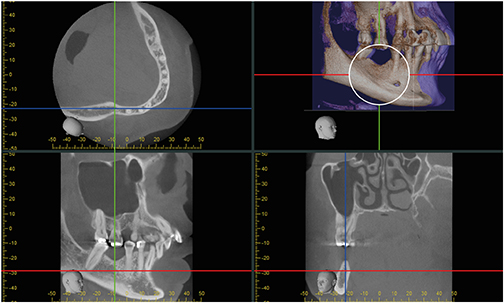
Figure 1 Selection of a cross-sectional slice taken from a sagittal image.
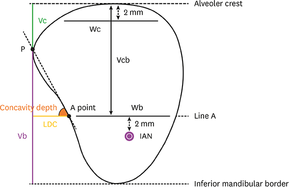
Figure 2 Schematic illustration of measurements taken from a cross-sectional slice [11]. Wc: buccolingual widths at 2 mm apical to the alveolar crest, Wb: buccolingual widths at the level of line A, Vcb: vertical distance from the alveolar ridge to line A, LCD: linear concavity depth, Vc: vertical distances between point P and the alveolar crest, Vb: vertical distances between point P and the inferior mandibular border.
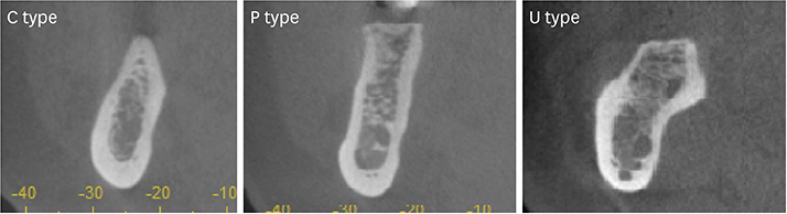
Figure 3 Cross-sectional views of the first molar region, showing the 3 types. C: convex, P: parallel, U: undercut.
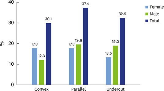
Figure 4 Distribution of ridge types according to gender (%).
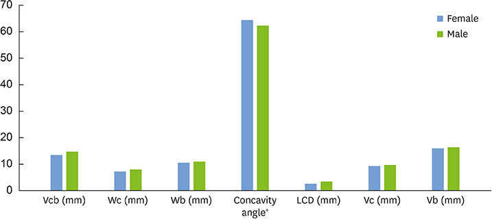
Figure 5 Measurements according to sex. Vcb: vertical distance from the alveolar ridge to line A, Wc: buccolingual widths at 2 mm apical to the alveolar crest, Wb: buccolingual widths at the level of line A, LCD: linear concavity depth, Vc: vertical distances between point P and the alveolar crest, Vb: vertical distances between point P and the inferior mandibular border.
Reference
-
1. Rajput BS, Merita S, Parihar AS, Vyas T, Kaur P, Chansoria S. Assessment of lingual concavities in submandibular fossa region in patients requiring dental implants-A cone beam computed tomography study. J Contemp Dent Pract. 2018; 19:1329–1333.2. Panjnoush M, Eil N, Kheirandish Y, Mofidi N, Shamshiri AR. Evaluation of the concavity depth and inclination in jaws using CBCT. Caspian J Dent Res. 2016; 5:17–23.3. Kalpidis CD, Konstantinidis AB. Critical hemorrhage in the floor of the mouth during implant placement in the first mandibular premolar position: a case report. Implant Dent. 2005; 14:117–124.
Article4. de Souza LA, Souza Picorelli Assis NM, Ribeiro RA, Pires Carvalho AC, Devito KL. Assessment of mandibular posterior regional landmarks using cone-beam computed tomography in dental implant surgery. Ann Anat. 2016; 205:53–59.
Article5. Tyndall DA, Price JB, Tetradis S, Ganz SD, Hildebolt C, Scarfe WC, et al. Position statement of the American Academy of Oral and Maxillofacial Radiology on selection criteria for the use of radiology in dental implantology with emphasis on cone beam computed tomography. Oral Surg Oral Med Oral Pathol Oral Radiol. 2012; 113:817–826.
Article6. Zohrabian VM, Sonick M, Hwang D, Abrahams JJ. Dental Implants. Semin Ultrasound CT MR. 2015; 36:415–426.
Article7. Aranyarachkul P, Caruso J, Gantes B, Schulz E, Riggs M, Dus I, et al. Bone density assessments of dental implant sites: 2. Quantitative cone-beam computerized tomography. Int J Oral Maxillofac Implants. 2005; 20:416–424.8. Naitoh M, Hirukawa A, Katsumata A, Ariji E. Evaluation of voxel values in mandibular cancellous bone: relationship between cone-beam computed tomography and multislice helical computed tomography. Clin Oral Implants Res. 2009; 20:503–506.
Article9. Sammartino G, Marenzi G, Citarella R, Ciccarelli R, Wang HL. Analysis of the occlusal stress transmitted to the inferior alveolar nerve by an osseointegrated threaded fixture. J Periodontol. 2008; 79:1735–1744.
Article10. Chiapasco M, Abati S, Romeo E, Vogel G. Clinical outcome of autogenous bone blocks or guided bone regeneration with e-PTFE membranes for the reconstruction of narrow edentulous ridges. Clin Oral Implants Res. 1999; 10:278–288.
Article11. Chan HL, Brooks SL, Fu JH, Yeh CY, Rudek I, Wang HL. Cross-sectional analysis of the mandibular lingual concavity using cone beam computed tomography. Clin Oral Implants Res. 2011; 22:201–206.
Article12. Leong DJ, Chan HL, Yeh CY, Takarakis N, Fu JH, Wang HL. Risk of lingual plate perforation during implant placement in the posterior mandible: a human cadaver study. Implant Dent. 2011; 20:360–363.
Article13. Dubois L, de Lange J, Baas E, Van Ingen J. Excessive bleeding in the floor of the mouth after endosseus implant placement: a report of two cases. Int J Oral Maxillofac Surg. 2010; 39:412–415.
Article14. Loukas M, Kinsella CR Jr, Kapos T, Tubbs RS, Ramachandra S. Anatomical variation in arterial supply of the mandible with special regard to implant placement. Int J Oral Maxillofac Surg. 2008; 37:367–371.
Article15. Tomljenovic B, Herrmann S, Filippi A, Kühl S. Life-threatening hemorrhage associated with dental implant surgery: a review of the literature. Clin Oral Implants Res. 2016; 27:1079–1084.
Article16. Flanagan D. Important arterial supply of the mandible, control of an arterial hemorrhage, and report of a hemorrhagic incident. J Oral Implantol. 2003; 29:165–173.
Article17. Huang RY, Cochran DL, Cheng WC, Lin MH, Fan WH, Sung CE, et al. Risk of lingual plate perforation for virtual immediate implant placement in the posterior mandible: a computer simulation study. J Am Dent Assoc. 2015; 146:735–742.
Article18. Froum S, Casanova L, Byrne S, Cho SC. Risk assessment before extraction for immediate implant placement in the posterior mandible: a computerized tomographic scan study. J Periodontol. 2011; 82:395–402.
Article19. Herranz-Aparicio J, Marques J, Almendros-Marqués N, Gay-Escoda C. Retrospective study of the bone morphology in the posterior mandibular region. Evaluation of the prevalence and the degree of lingual concavity and their possible complications. Med Oral Patol Oral Cir Bucal. 2016; 21:e731–6.
Article20. Nickenig HJ, Wichmann M, Eitner S, Zöller JE, Kreppel M. Lingual concavities in the mandible: a morphological study using cross-sectional analysis determined by CBCT. J Craniomaxillofac Surg. 2015; 43:254–259.
Article21. Kamburoğlu K, Acar B, Yüksel S, Paksoy CS. CBCT quantitative evaluation of mandibular lingual concavities in dental implant patients. Surg Radiol Anat. 2015; 37:1209–1215.
Article22. Quirynen M, Mraiwa N, van Steenberghe D, Jacobs R. Morphology and dimensions of the mandibular jaw bone in the interforaminal region in patients requiring implants in the distal areas. Clin Oral Implants Res. 2003; 14:280–285.
Article23. Aranha Watanabe PC, Moreira Lopes De Faria L, Mardegan Issa JP, Caldeira Monteiro SA, Tiossi R. Morphodigital evaluation of the trabecular bone pattern in the mandible using digitized panoramic and periapical radiographs. Minerva Stomatol. 2009; 58:73–80.24. Ou CY, Chen CJ, Lin CP, Huang KC, Tseng CC. Morphologic analysis of mandibular lingual concavity by computed tomography image in Taiwan population. J Taiwan Periodontol. 2012; 17:191–197.25. Parnia F, Fard EM, Mahboub F, Hafezeqoran A, Gavgani FE. Tomographic volume evaluation of submandibular fossa in patients requiring dental implants. Oral Surg Oral Med Oral Pathol Oral Radiol Endod. 2010; 109:e32–6.
Article26. Braut V, Bornstein MM, Lauber R, Buser D. Bone dimensions in the posterior mandible: a retrospective radiographic study using cone beam computed tomography. Part 1--analysis of dentate sites. Int J Periodontics Restorative Dent. 2012; 32:175–184.27. Salemi F, Shokri A, Forouzandeh M, Karami M, Khalili Z. Mandibular lingual concavity: a cross-sectional analysis using cone beam computed tomography. J Clin Diagn Res. 2018; 12:ZC37–41.
Article28. Borahan MO, Dönmez FG, Ulay G, Sadıkoğlu AN, Pekiner FN. Assesment of submandibular fossa depth using cone beam computed tomography. Yeditepe J Dent. 2018; 14:51–56.
Article29. Uchida Y, Goto M, Danjo A, Yamashita Y, Kuraoka A. Anatomic measurement of the depth and location of the sublingual fossa. Int J Oral Maxillofac Surg. 2012; 41:1571–1576.
Article
- Full Text Links
-
- Actions
-
Cited
- CITED
-
- Close
- Share
-
- Similar articles
-
- Cone beam computed tomography findings of ectopic mandibular third molar in the mandibular condyle: report of a case
- Radiographic evaluation of the course and visibility of the mandibular canal
- Computed tomographic analysis of maxillary sinus anatomy relevant to sinus lift procedures in edentulous ridges in Taiwanese patients
- Stafne bone cavity and cone-beam computed tomography: a report of two cases
- Assessment of the relationship between the mandibular third molar and the mandibular canal using panoramic radiograph and cone beam computed tomography
