J Periodontal Implant Sci.
2019 Aug;49(4):237-247. 10.5051/jpis.2019.49.4.237.
Computed tomographic analysis of maxillary sinus anatomy relevant to sinus lift procedures in edentulous ridges in Taiwanese patients
- Affiliations
-
- 1Department of Periodontics, Chang Gung Memorial Hospital, Chang Gung University College of Medicine, Kaohsiung, Taiwan.
- 2Department of Orthodontics, Chang Gung Memorial Hospital, Chang Gung University College of Medicine, Kaohsiung, Taiwan.
- 3Department of Dentistry, Chang Gung Memorial Hospital, Chang Gung University College of Medicine, Kaohsiung, Taiwan. dentwu@hotmail.com
- KMID: 2455801
- DOI: http://doi.org/10.5051/jpis.2019.49.4.237
Abstract
- PURPOSE
To analyze the maxillary sinus anatomy over edentulous ridges in the bilateral posterior maxillary area in Taiwanese patients using cone-beam computed tomography (CBCT).
METHODS
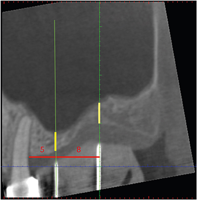
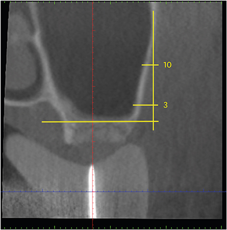
In total, 101 anatomical sites from 61 patients, including 32 premolar and 69 molar regions, were analyzed using CBCT. Measurements were made of the width and height of edentulous ridges, the thickness of the lateral wall of the maxillary sinus, and the presence of a sinus septum and the posterior superior alveolar artery (PSAA). A statistical analysis of the measurements was performed, and correlations among the measurements were assessed.
RESULTS
The average ridge width was 10.26±3.16 mm, with a significantly greater ridge width in the second molar region than in the premolar region. The mean residual ridge height was 8.55±4.09 mm, and ridge height showed an opposite trend from ridge width for the premolar and molar regions. A sinus septum was present at 5.9% of the sites, and the PSAA was observed in 24.5%. The average thickness of the lateral wall of the maxillary sinus was 2.08±0.94 mm, with no significant difference between the tooth position and lateral wall thickness.
CONCLUSIONS
This study presents the anatomical features of the maxillary sinus, which should be considered in sinus lift procedures for implant placement, in the Taiwanese population. The use of CBCT is recommended to avoid intraoperative complications.
Keyword
MeSH Terms
Figure
Reference
-
1. Shanbhag S, Karnik P, Shirke P, Shanbhag V. Cone-beam computed tomographic analysis of sinus membrane thickness, ostium patency, and residual ridge heights in the posterior maxilla: implications for sinus floor elevation. Clin Oral Implants Res. 2014; 25:755–760.
Article2. Kang SJ, Shin SI, Herr Y, Kwon YH, Kim GT, Chung JH. Anatomical structures in the maxillary sinus related to lateral sinus elevation: a cone beam computed tomographic analysis. Clin Oral Implants Res. 2013; 24:Suppl A100. 75–81.
Article3. Rosano G, Taschieri S, Gaudy JF, Weinstein T, Del Fabbro M. Maxillary sinus vascular anatomy and its relation to sinus lift surgery. Clin Oral Implants Res. 2011; 22:711–715.
Article4. Neugebauer J, Ritter L, Mischkowski RA, Dreiseidler T, Scherer P, Ketterle M, et al. Evaluation of maxillary sinus anatomy by cone-beam CT prior to sinus floor elevation. Int J Oral Maxillofac Implants. 2010; 25:258–265.5. Acharya A, Hao J, Mattheos N, Chau A, Shirke P, Lang NP. Residual ridge dimensions at edentulous maxillary first molar sites and periodontal bone loss among two ethnic cohorts seeking tooth replacement. Clin Oral Implants Res. 2014; 25:1386–1394.
Article6. Ball R, Shu C, Xi P, Rioux M, Luximon Y, Molenbroek J. A comparison between Chinese and Caucasian head shapes. Appl Ergon. 2010; 41:832–839.
Article7. Gu Y, McNamara JA Jr, Sigler LM, Baccetti T. Comparison of craniofacial characteristics of typical Chinese and Caucasian young adults. Eur J Orthod. 2011; 33:205–211.
Article8. Nunes LS, Bornstein MM, Sendi P, Buser D. Anatomical characteristics and dimensions of edentulous sites in the posterior maxillae of patients referred for implant therapy. Int J Periodontics Restorative Dent. 2013; 33:337–345.
Article9. Sahrmann P, Naenni N, Jung RE, Held U, Truninger T, Hämmerle CH, et al. Success of 6-mm implants with single-tooth restorations: a 3-year randomized controlled clinical trial. J Dent Res. 2016; 95:623–628.
Article10. Mertens C, Meyer-Bäumer A, Kappel H, Hoffmann J, Steveling HG. Use of 8-mm and 9-mm implants in atrophic alveolar ridges: 10-year results. Int J Oral Maxillofac Implants. 2012; 27:1501–1508.11. Deporter D, Pharoah M, Yeh S, Todescan R, Atenafu EG. Performance of titanium alloy sintered porous-surfaced (SPS) implants supporting mandibular overdentures during a 20-year prospective study. Clin Oral Implants Res. 2014; 25:e189–e195.
Article12. Thoma DS, Cha JK, Jung UW. Treatment concepts for the posterior maxilla and mandible: short implants versus long implants in augmented bone. J Periodontal Implant Sci. 2017; 47:2–12.
Article13. Atieh MA, Zadeh H, Stanford CM, Cooper LF. Survival of short dental implants for treatment of posterior partial edentulism: a systematic review. Int J Oral Maxillofac Implants. 2012; 27:1323–1331.14. Wang HL, Katranji A. ABC sinus augmentation classification. Int J Periodontics Restorative Dent. 2008; 28:383–389.15. Tatum H Jr. Maxillary and sinus implant reconstructions. Dent Clin North Am. 1986; 30:207–229.16. Tatum OH Jr, Lebowitz MS, Tatum CA, Borgner RA. Sinus augmentation. Rationale, development, long-term results. N Y State Dent J. 1993; 59:43–48.17. Boyne PJ, James RA. Grafting of the maxillary sinus floor with autogenous marrow and bone. J Oral Surg. 1980; 38:613–616.18. Pjetursson BE, Rast C, Brägger U, Schmidlin K, Zwahlen M, Lang NP. Maxillary sinus floor elevation using the (transalveolar) osteotome technique with or without grafting material. Part I: Implant survival and patients' perception. Clin Oral Implants Res. 2009; 20:667–676.
Article19. Kim MJ, Jung UW, Kim CS, Kim KD, Choi SH, Kim CK, et al. Maxillary sinus septa: prevalence, height, location, and morphology. A reformatted computed tomography scan analysis. J Periodontol. 2006; 77:903–908.
Article20. Krennmair G, Ulm CW, Lugmayr H, Solar P. The incidence, location, and height of maxillary sinus septa in the edentulous and dentate maxilla. J Oral Maxillofac Surg. 1999; 57:667–671.
Article21. Maestre-Ferrín L, Galán-Gil S, Rubio-Serrano M, Peñarrocha-Diago M, Peñarrocha-Oltra D. Maxillary sinus septa: a systematic review. Med Oral Patol Oral Cir Bucal. 2010; 15:e383–e386.22. Pommer B, Ulm C, Lorenzoni M, Palmer R, Watzek G, Zechner W. Prevalence, location and morphology of maxillary sinus septa: systematic review and meta-analysis. J Clin Periodontol. 2012; 39:769–773.
Article23. Solar P, Geyerhofer U, Traxler H, Windisch A, Ulm C, Watzek G. Blood supply to the maxillary sinus relevant to sinus floor elevation procedures. Clin Oral Implants Res. 1999; 10:34–44.
Article24. Güncü GN, Yildirim YD, Wang HL, Tözüm TF. Location of posterior superior alveolar artery and evaluation of maxillary sinus anatomy with computerized tomography: a clinical study. Clin Oral Implants Res. 2011; 22:1164–1167.
Article25. Zijderveld SA, van den Bergh JP, Schulten EA, ten Bruggenkate CM. Anatomical and surgical findings and complications in 100 consecutive maxillary sinus floor elevation procedures. J Oral Maxillofac Surg. 2008; 66:1426–1438.
Article26. Hur MS, Kim JK, Hu KS, Bae HE, Park HS, Kim HJ. Clinical implications of the topography and distribution of the posterior superior alveolar artery. J Craniofac Surg. 2009; 20:551–554.
Article27. Elian N, Wallace S, Cho SC, Jalbout ZN, Froum S. Distribution of the maxillary artery as it relates to sinus floor augmentation. Int J Oral Maxillofac Implants. 2005; 20:784–787.28. Traxler H, Windisch A, Geyerhofer U, Surd R, Solar P, Firbas W. Arterial blood supply of the maxillary sinus. Clin Anat. 1999; 12:417–421.
Article29. Barone A, Santini S, Sbordone L, Crespi R, Covani U. A clinical study of the outcomes and complications associated with maxillary sinus augmentation. Int J Oral Maxillofac Implants. 2006; 21:81–85.30. Kim YK, Hwang JW, Yun PY. Closure of large perforation of sinus membrane using pedicled buccal fat pad graft: a case report. Int J Oral Maxillofac Implants. 2008; 23:1139–1142.31. Maksoud MA. Complications after maxillary sinus augmentation: a case report. Implant Dent. 2001; 10:168–171.
Article32. Wallace SS, Tarnow DP, Froum SJ, Cho SC, Zadeh HH, Stoupel J, et al. Maxillary sinus elevation by lateral window approach: evolution of technology and technique. J Evid Based Dent Pract. 2012; 12:161–171.
Article33. Wallace SS, Mazor Z, Froum SJ, Cho SC, Tarnow DP. Schneiderian membrane perforation rate during sinus elevation using piezosurgery: clinical results of 100 consecutive cases. Int J Periodontics Restorative Dent. 2007; 27:413–419.34. Danesh-Sani SA, Loomer PM, Wallace SS. A comprehensive clinical review of maxillary sinus floor elevation: anatomy, techniques, biomaterials and complications. Br J Oral Maxillofac Surg. 2016; 54:724–730.
Article35. Kaufman E. Maxillary sinus elevation surgery: an overview. J Esthet Restor Dent. 2003; 15:272–283.
Article36. Stern A, Green J. Sinus lift procedures: an overview of current techniques. Dent Clin North Am. 2012; 56:219–233.
Article37. Guerrero JS, Al-Jandan BA. Lateral wall sinus floor elevation for implant placement: revisiting fundamentals and the surgical technique. J Calif Dent Assoc. 2013; 41:185–187.38. Barone A, Santini S, Marconcini S, Giacomelli L, Gherlone E, Covani U. Osteotomy and membrane elevation during the maxillary sinus augmentation procedure. A comparative study: piezoelectric device vs. conventional rotative instruments. Clin Oral Implants Res. 2008; 19:511–515.
Article39. Lambert F, Lecloux G, Rompen E. One-step approach for implant placement and subantral bone regeneration using bovine hydroxyapatite: a 2- to 6-year follow-up study. Int J Oral Maxillofac Implants. 2010; 25:598–606.40. Baldini N, D'Elia C, Bianco A, Goracci C, de Sanctis M, Ferrari M. Lateral approach for sinus floor elevation: large versus small bone window - a split-mouth randomized clinical trial. Clin Oral Implants Res. 2017; 28:974–981.
Article41. Yang SM, Park SI, Kye SB, Shin SY. Computed tomographic assessment of maxillary sinus wall thickness in edentulous patients. J Oral Rehabil. 2012; 39:421–428.
Article42. Neiva RF, Gapski R, Wang HL. Morphometric analysis of implant-related anatomy in Caucasian skulls. J Periodontol. 2004; 75:1061–1067.
Article43. Schropp L, Wenzel A, Kostopoulos L, Karring T. Bone healing and soft tissue contour changes following single-tooth extraction: a clinical and radiographic 12-month prospective study. Int J Periodontics Restorative Dent. 2003; 23:313–323.44. Tan WL, Wong TL, Wong MC, Lang NP. A systematic review of post-extractional alveolar hard and soft tissue dimensional changes in humans. Clin Oral Implants Res. 2012; 23:Suppl 5. 1–21.
Article45. Cho SC, Wallace SS, Froum SJ, Tarnow DP. Influence of anatomy on Schneiderian membrane perforations during sinus elevation surgery: three-dimensional analysis. Pract Proced Aesthet Dent. 2001; 13:160–163.46. Janner SF, Caversaccio MD, Dubach P, Sendi P, Buser D, Bornstein MM. Characteristics and dimensions of the Schneiderian membrane: a radiographic analysis using cone beam computed tomography in patients referred for dental implant surgery in the posterior maxilla. Clin Oral Implants Res. 2011; 22:1446–1453.
Article
- Full Text Links
- Actions
-
Cited
- CITED
-
- Close
- Share
- Similar articles
-
- CLINICO-ANATOMICAL STUDY OF SEPTUM IN THE MAXILLARY SINUS
- Computed tomographic findings of maxillary sinus cancer
- Sinus lifts in the presence of pseudoantral and mucous retention cysts
- Clinical anatomy of the maxillary sinus: application to sinus floor augmentation
- Analysis of location and prevalence of maxillary sinus septa


