Clinical anatomy of the maxillary sinus: application to sinus floor augmentation
- Affiliations
-
- 1Seattle Science Foundation, Seattle, WA, USA. joei@seattlesciencefoundation.org
- 2Dental and Oral Medical Center, Kurume University School of Medicine, Kurume, Japan.
- 3Division of Gross and Clinical Anatomy, Department of Anatomy, Kurume University School of Medicine, Kurume, Japan.
- 4Department of Anatomy, Jagiellonian University Medical College, Krakow, Poland.
- 5Department of Medical Education, Jagiellonian University Medical College, Krakow, Poland.
- 6Department of Anatomical Sciences, St. George's University, St. George's, Grenada, West Indies.
- KMID: 2442324
- DOI: http://doi.org/10.5115/acb.2019.52.1.17
Abstract
- The anatomy of the maxillary sinus, especially its vascular anatomy, and its relationships with the teeth and alveolar processes have been well documented. The development of cone-beam computed tomography has resulted in dentists being more familiar with maxillary sinus floor augmentation procedures. This paper aims to revisit the classic anatomy of the maxillary sinus and review the newly published literature in order to help dentists diagnose in more detail and perform safer surgery of the maxillary sinus.
Keyword
MeSH Terms
Figure
-
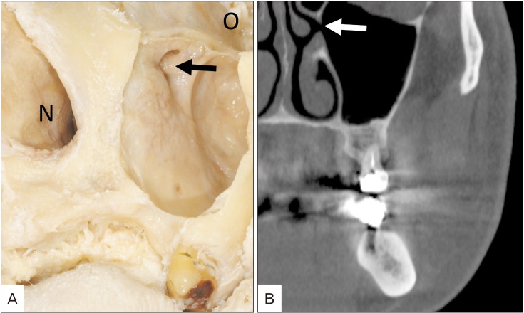
Fig. 1 Ostium on the left maxillary sinus (arrows). (A) Cadaveric dissection (anterolateral view). (B) Computed tomography (coronal image). N, nasal cavity; O, orbit.
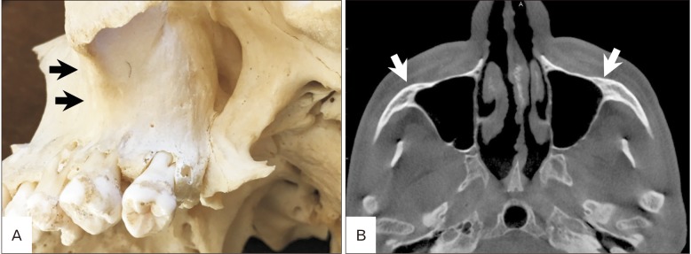
Fig. 2 The maxillary sinus extending into the zygomatic process (arrows). (A) Dry skull (inferolateral view). (B) Computed tomography (axial image).
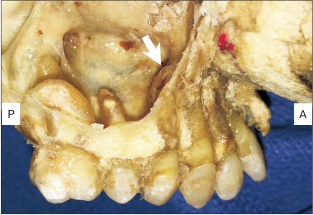
Fig. 3 Relationship between teeth and maxillary sinus (right side). Note the root of the first premolar (arrow) is located most medially.
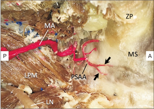
Fig. 4 Course of the right posterior superior alveolar artery. Note the two branches of posterior superior alveolar artery enter the posterior wall of the maxillary sinus (arrows). LN, lingual nerve; LPM, lateral pterygoid muscle; MA, maxillary artery; MS, maxillary sinus; PSAA, posterior superior alveolar artery; ZP, zygomatic process.
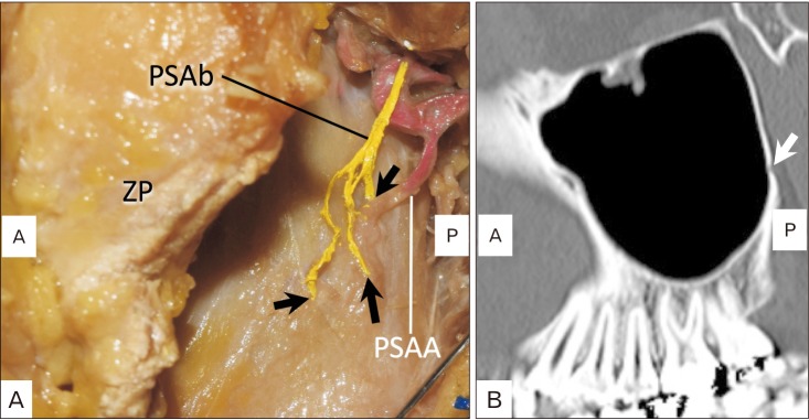
Fig. 5 Course of the left posterior superior a lveolar branch of the maxil lar y ner ve. Note the three branches enter the posterior wall of the maxillary sinus (arrows). (A) Lateral view of the posterior superior alveolar branch of the maxillary nerve (PSAb) in the cadaveric dissection (left side). (B) Computed tomography (sagittal image). A, anterior; P, posterior; PSAA, posterior superior alveolar artery; ZP, zygomatic process.
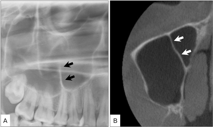
Fig. 6 Septum inside the right maxillary sinus (arrows). (A) Panoramic radiography. (B) Computed tomography (axial image).
Reference
-
1. Mavrodi A, Paraskevas G. Evolution of the paranasal sinuses' anatomy through the ages. Anat Cell Biol. 2013; 46:235–238. PMID: 24386595.2. Nuñez-Castruita A, López-Serna N, Guzmán-López S. Prenatal development of the maxillary sinus: a perspective for paranasal sinus surgery. Otolaryngol Head Neck Surg. 2012; 146:997–1003. PMID: 22267494.3. Standring S. Gray's anatomy: the anatomical basis of clinical practice. 41st ed. London: Elsevier Health Sciences;2015.4. Lang J. Clinical anatomy of the nose, nasal cavity, and paranasal sinuses. New York: Thieme Medical Publishers;1989.5. Duncavage J. The maxillary sinus: medical and surgical management. New York: Thieme Medical Publishers;2011.6. Chanavaz M. Maxillary sinus: anatomy, physiology, surgery, and bone grafting related to implantology: eleven years of surgical experience (1979-1990). J Oral Implantol. 1990; 16:199–209. PMID: 2098563.7. Eberhardt JA, Torabinejad M, Christiansen EL. A computed tomographic study of the distances between the maxillary sinus floor and the apices of the maxillary posterior teeth. Oral Surg Oral Med Oral Pathol. 1992; 73:345–346. PMID: 1545967.8. Kilic C, Kamburoglu K, Yuksel SP, Ozen T. An assessment of the relationship between the maxillary sinus floor and the maxillary posterior teeth root tips using dental cone-beam computerized tomography. Eur J Dent. 2010; 4:462–467. PMID: 20922167.9. Roque-Torres GD, Ramirez-Sotelo LR, Vaz SL, Bóscolo SM, Bóscolo FN. Association between maxillary sinus pathologies and healthy teeth. Braz J Otorhinolaryngol. 2016; 82:33–38. PMID: 26727608.10. Kim MJ, Jung UW, Kim CS, Kim KD, Choi SH, Kim CK, Cho KS. Maxillary sinus septa: prevalence, height, location, and morphology: a reformatted computed tomography scan analysis. J Periodontol. 2006; 77:903–908. PMID: 16671885.11. Krennmair G, Ulm CW, Lugmayr H, Solar P. The incidence, location, and height of maxillary sinus septa in the edentulous and dentate maxilla. J Oral Maxillofac Surg. 1999; 57:667–671. PMID: 10368090.12. Underwood AS. An inquiry into the anatomy and pathology of the maxillary sinus. J Anat Physiol. 1910; 44(Pt 4):354–369. PMID: 17232856.13. Lee WJ, Lee SJ, Kim HS. Analysis of location and prevalence of maxillary sinus septa. J Periodontal Implant Sci. 2010; 40:56–60. PMID: 20498761.14. Maestre-Ferrín L, Carrillo-García C, Galán-Gil S, Peñarrocha-Diago M, Peñarrocha-Diago M. Prevalence, location, and size of maxillary sinus septa: panoramic radiograph versus computed tomography scan. J Oral Maxillofac Surg. 2011; 69:507–511. PMID: 21238847.15. Velásquez-Plata D, Hovey LR, Peach CC, Alder ME. Maxillary sinus septa: a 3-dimensional computerized tomographic scan analysis. Int J Oral Maxillofac Implants. 2002; 17:854–860. PMID: 12507246.16. González-Santana H, Peñarrocha-Diago M, Guarinos-Carbó J, Sorní-Bröker M. A study of the septa in the maxillary sinuses and the subantral alveolar processes in 30 patients. J Oral Implantol. 2007; 33:340–343. PMID: 18240793.17. Bell GW, Joshi BB, Macleod RI. Maxillary sinus disease: diagnosis and treatment. Br Dent J. 2011; 210:113–118. PMID: 21311531.18. Kqiku L, Biblekaj R, Weiglein AH, Kqiku X, Städtler P. Arterial blood architecture of the maxillary sinus in dentate specimens. Croat Med J. 2013; 54:180–184. PMID: 23630145.19. Flanagan D. Arterial supply of maxillary sinus and potential for bleeding complication during lateral approach sinus elevation. Implant Dent. 2005; 14:336–338. PMID: 16361882.20. Lovasova K, Kachlik D, Rozpravkova M, Matusevska M, Ferkova J, Kluchova D. Three-dimensional CAD/CAM imaging of the maxillary sinus in ageing process. Ann Anat. 2018; 218:69–82. PMID: 29627610.21. Gosau M, Rink D, Driemel O, Draenert FG. Maxillary sinus anatomy: a cadaveric study with clinical implications. Anat Rec (Hoboken). 2009; 292:352–354. PMID: 19248167.22. Velasco-Torres M, Padial-Molina M, Avila-Ortiz G, García-Delgado R, O'Valle F, Catena A, Galindo-Moreno P. Maxillary sinus dimensions decrease as age and tooth loss increase. Implant Dent. 2017; 26:288–295. PMID: 28125519.23. Paatero YV. Orthoradial jaw pantomography. Ann Med Intern Fenn Suppl. 1959; 48(Supp 28):222–227. PMID: 13637452.24. Malina-Altzinger J, Damerau G, Grätz KW, Stadlinger PD. Evaluation of the maxillary sinus in panoramic radiography: a comparative study. Int J Implant Dent. 2015; 1:17. PMID: 27747639.25. Bornstein MM, Scarfe WC, Vaughn VM, Jacobs R. Cone beam computed tomography in implant dentistry: a systematic review focusing on guidelines, indications, and radiation dose risks. Int J Oral Maxillofac Implants. 2014; 29(Suppl):55–77. PMID: 24660190.26. Dula K, Benic GI, Bornstein M, Dagassan-Berndt D, Filippi A, Hicklin S, Kissling-Jeger F, Luebbers HT, Sculean A, Sequeira-Byron P, Walter C, Zehnder M. SADMFR guidelines for the use of cone-beam computed tomography/digital volume tomography. Swiss Dent J. 2015; 125:945–953. PMID: 26399521.27. Maestre-Ferrín L, Galán-Gil S, Carrillo-García C, Peñarrocha-Diago M. Radiographic findings in the maxillary sinus: comparison of panoramic radiography with computed tomography. Int J Oral Maxillofac Implants. 2011; 26:341–346. PMID: 21483887.28. Toraman Alkurt M, Peker I, Degerli S, Cebeci AR, Sadik E. Comparison of cone-beam computed tomography and panoramic radiographs in detecting maxillary sinus septa. J Istanb Univ Fac Dent. 2016; 50:8–14. PMID: 28955570.29. Tehranchi M, Taleghani F, Shahab S, Nouri A. Prevalence and location of the posterior superior alveolar artery using cone-beam computed tomography. Imaging Sci Dent. 2017; 47:39–44. PMID: 28361028.30. Chitsazi MT, Shirmohammadi A, Faramarzi M, Esmaieli F, Chitsazi S. Evaluation of the position of the posterior superior alveolar artery in relation to the maxillary sinus using the Cone-Beam computed tomography scans. J Clin Exp Dent. 2017; 9:e394–e399. PMID: 28298981.31. Pandharbale AA, Gadgil RM, Bhoosreddy AR, Kunte VR, Ahire BS, Shinde MR, Joshi SS. Evaluation of the posterior superior alveolar artery using cone beam computed tomography. Pol J Radiol. 2016; 81:606–610. PMID: 28058075.32. Danesh-Sani SA, Movahed A, ElChaar ES, Chong Chan K, Amintavakoli N. Radiographic evaluation of maxillary sinus lateral wall and posterior superior alveolar artery anatomy: a cone-beam computed tomographic study. Clin Implant Dent Relat Res. 2017; 19:151–160. PMID: 27238049.33. Khojastehpour L, Dehbozorgi M, Tabrizi R, Esfandnia S. Evaluating the anatomical location of the posterior superior alveolar artery in cone beam computed tomography images. Int J Oral Maxillofac Surg. 2016; 45:354–358. PMID: 26516028.34. Güncü GN, Yildirim YD, Wang HL, Tözüm TF. Location of posterior superior alveolar artery and evaluation of maxillary sinus anatomy with computerized tomography: a clinical study. Clin Oral Implants Res. 2011; 22:1164–1167. PMID: 21244499.35. Ilgüy D, Ilgüy M, Dolekoglu S, Fisekcioglu E. Evaluation of the posterior superior alveolar artery and the maxillary sinus with CBCT. Braz Oral Res. 2013; 27:431–437. PMID: 24036981.36. Solar P, Geyerhofer U, Traxler H, Windisch A, Ulm C, Watzek G. Blood supply to the maxillary sinus relevant to sinus floor elevation procedures. Clin Oral Implants Res. 1999; 10:34–44. PMID: 10196788.37. Traxler H, Windisch A, Geyerhofer U, Surd R, Solar P, Firbas W. Arterial blood supply of the maxillary sinus. Clin Anat. 1999; 12:417–421. PMID: 10545857.38. Maes JJ, Clement PA. The usefulness of irrigation of the maxillary sinus in children with maxillary sinusitis on the basis of the Water's X-ray. Rhinology. 1987; 25:259–264. PMID: 2448862.39. Eley KA, Watt-Smith SR, Boland P, Potter M, Golding SJ. MRI pre-treatment tumour volume in maxillary complex squamous cell carcinoma treated with surgical resection. J Craniomaxillofac Surg. 2014; 42:119–124. PMID: 23777920.40. Yuan XP, Li CX, Cao Y, Singh S, Zhong R. Inflammatory myofibroblastic tumour of the maxillary sinus: CT and MRI findings. Clin Radiol. 2012; 67:e53–e57. PMID: 22974570.41. Ng SH, Chang TC, Ko SF, Yen PS, Wan YL, Tang LM, Tsai MH. Nasopharyngeal carcinoma: MRI and CT assessment. Neuroradiology. 1997; 39:741–746. PMID: 9351114.42. Asaumi J, Konouchi H, Hisatomi M, Kishi K. Odontogenic myxoma of maxillary sinus: CT and MR-pathologic correlation. Eur J Radiol. 2001; 37:1–4. PMID: 11274831.43. Yasumoto M, Taura S, Shibuya H, Honda M. Primary malignant lymphoma of the maxillary sinus: CT and MRI. Neuroradiology. 2000; 42:285–289. PMID: 10872174.44. Gray CF, Redpath TW, Smith FW, Staff RT, Bainton R. Assessment of the sinus lift operation by magnetic resonance imaging. Br J Oral Maxillofac Surg. 1999; 37:285–289. PMID: 10475650.45. Gray CF, Staff RT, Redpath TW, Needham G, Renny NM. Assessment of maxillary sinus volume for the sinus lift operation by three-dimensional magnetic resonance imaging. Dentomaxillofac Radiol. 2000; 29:154–158. PMID: 10849541.46. Gray CF, Redpath TW, Bainton R, Smith FW. Magnetic resonance imaging assessment of a sinus lift operation using reoxidised cellulose (Surgicel) as graft material. Clin Oral Implants Res. 2001; 12:526–530. PMID: 11564114.47. Senel FC, Duran S, Icten O, Izbudak I, Cizmeci F. Assessment of the sinus lift operation by magnetic resonance imaging. Br J Oral Maxillofac Surg. 2006; 44:511–514. PMID: 16540215.48. Lim D, Parumo R, Chai MB, Shanmuganathan J. Transnasal endoscopy removal of dislodged dental implant: a case report. J Oral Implantol. 2017; 43:228–231. PMID: 27996585.49. Jeong KI, Kim SG, Oh JS, You JS. Implants displaced into the maxillary sinus: a systematic review. Implant Dent. 2016; 25:547–551. PMID: 26974033.50. Matti E, Emanuelli E, Pusateri A, Muniz CC, Pagella F. Transnasal endoscopic removal of dental implants from the maxillary sinus. Int J Oral Maxillofac Implants. 2013; 28:905–910. PMID: 23748326.51. Andreasi Bassi M, Andrisani C, Lico S, Ormanier Z, Barlattani A Jr, Ottria L. Endoscopic management of the schneiderian membrane perforation during transcrestal sinus augmentation: a case report. Oral Implantol (Rome). 2016; 9:157–163. PMID: 28042444.52. Zheng J, Zhang S, Lu E, Yang C, Zhang W, Zhao J. Endoscopic lift of the maxillary sinus floor in Beagles. Br J Oral Maxillofac Surg. 2014; 52:845–849. PMID: 25174319.53. Kunihiro T, Araki Y, Oba T. Minimally invasive endoscopic middle meatal antrostomy for the prevention of maxillary sinusitis in association with dental implantation in the posterior maxilla: a proposal. Fukuoka Igaku Zasshi. 2014; 105:182–189. PMID: 25639025.
- Full Text Links
-
- Actions
-
Cited
- CITED
-
- Close
- Share
-
- Similar articles
-
- A Case of Maxillary Sinusitis after Sinus Floor Augmentation
- Delayed Occurrence of Maxillary Sinusitis after Simultaneous Maxillary Sinus Augmentation and Implant: A Case Report and Literature Review
- Management of Perioperative Pathologic Conditions Involving Maxillary Sinus for Dental Implant Placement and Sinus Augmentation: Report of Case Series and Literature Review
- Sinus floor augmentation at the time of tooth removal
- The use of Autologous Venous Blood for Maxillary Sinus Floor Augmentation in Conjunction with the Sinus Membrane Elevation: An Experimental Study
