Manual Preparation of Donor Lenticule Using Artificial Anterior Chamber for Descemet's Membrane Stripping Endothelial Keratoplasty
- Affiliations
-
- 1Department of Ophthalmology, Chuncheon Sacred Heart Hospital, Hallym University College of Medicine, Chuncheon, Korea.
- 2Department of Ophthalmology, Yeouido St. Mary's Hospital, College of Medicine, The Catholic University of Koea, Seoul, Korea. huanghs@hanmail.net
- KMID: 2470745
- DOI: http://doi.org/10.3341/jkos.2020.61.2.209
Abstract
- PURPOSE
To report a patient with a pseudophakic bullous keratopathy (PBK) who underwent Descemet's membrane stripping endothelial keratoplasty (DSEK) with manual preparation of the donor corneal graft.
CASE SUMMARY
A 61-year-old female presented with visual disturbance in her right eye. Five months prior, she was treated with phacoemulsification and intraocular lens exchange surgery of the right eye, and a very severe corneal edema was revealed by slit-lamp examination. We diagnosed PBK and planned DSEK with manual preparation of a donor corneal graft because of the non-availability of a microkeratome or a femtosecond laser. After making the corneal graft using an artificial anterior chamber, crescent knife and cornea dissector, the keratoplasty proceeded using the graft. Three months after surgery, her graft was well-maintained on the right eye. The patient's visual acuity was 0.3, and the corneal endothelial cell count was 1,844/mm².
CONCLUSIONS
Manual preparation of the donor corneal graft for DSEK is suitable as a second choice treatment method when the availability of surgical devices is limited.
Keyword
MeSH Terms
Figure
-

Figure 1 (A) Slit lamp photography of the right eye on the first visit shows severe corneal stromal edema. Severe descemet's folds are also shown on the right eye. (B) Slit lamp photography of the right eye 5 months after manual prepared Descemet's membrane stripping endothelial keratoplasty. The margins of the centrally adhered posterior corneal disk are seen. Some residual stromal edema is visible in the posterior corneal disk, and central corneal thickness is 571 µm.

Figure 2 Manual preparation procedures. (A) Corneoscleral button on the top of the artificial anterior chamber. (B) Tight fixation for manual dividing. (C) Making 5 mm wide and 80% depth of incision with the crescent knife. (D) Dividing starts from the incision with the crescent knife. (E) Straight cornea dissector proceed the lamellar dividing to the top of the cornea. (F) Curved cornea dissector goes to the counterpart limbus beyond the equator.
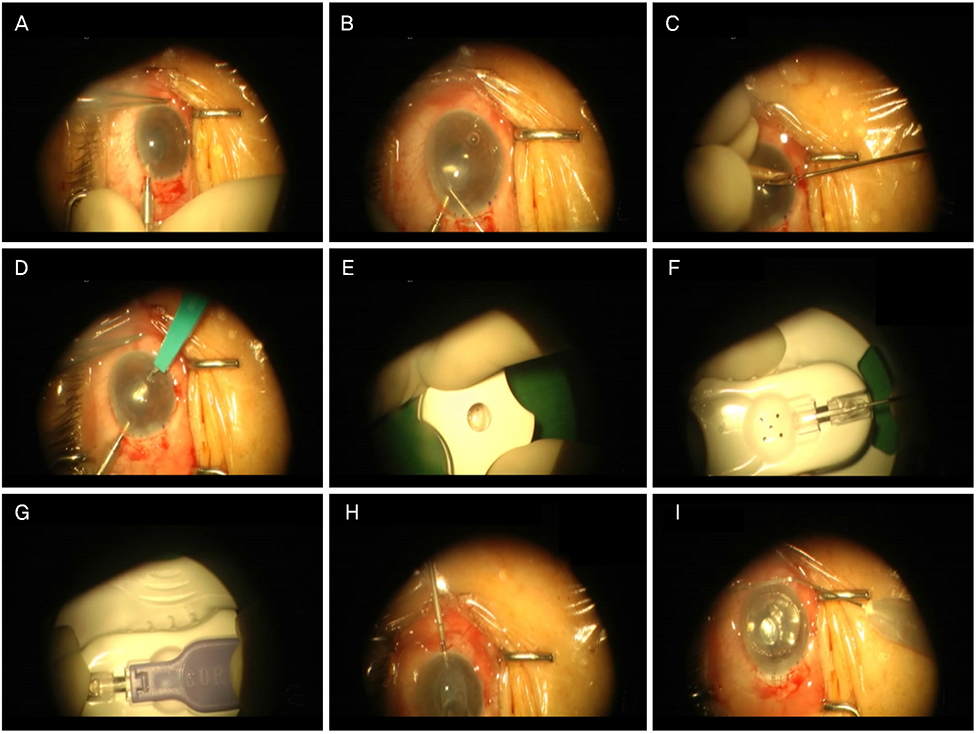
Figure 3 Descemet's membrane stripping endothelial keratoplasty procedures. (A) Making main incision. (B) Scoring and Descemet's membrane stripping. (C) Iridectomy for preventing the pupillary block. (D) Making incision for mciroforceps. (E) Making the graft punch with 7.5 mm trephine. (F) Graft loading on the injector. (G) Graft injection. (H) Graft centering in the anterior chamber. (I) Air injection.
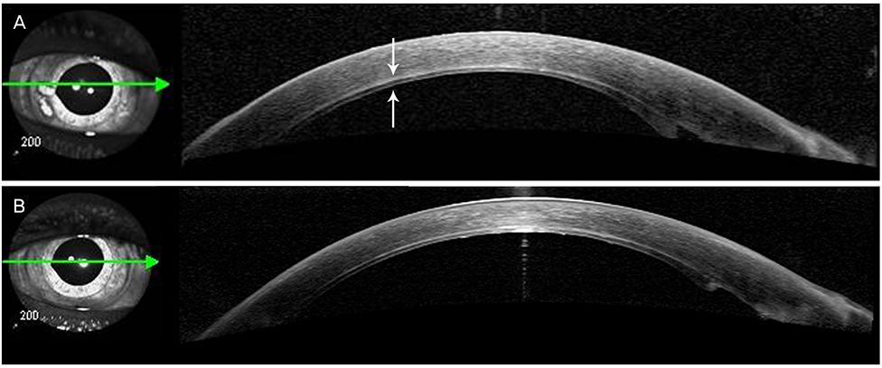
Figure 4 (A) Optical coherence tomography taken 5 days after the Descemet's membrane stripping endothelial keratoplasty with manual preparation on the right eye shows the corneal graft (white arrows). It is well attached to the posterior surface of the recipient cornea. (B) One month after the surgery, we can find the well-maintained donor graft.
Reference
-
1. Melles GR, Eggink FA, Lander F, et al. A surgical technique for posterior lamellar keratoplasty. Cornea. 1998; 17:618–626.
Article2. Price FW Jr, Price MO. Descemet’s stripping with endothelial keratoplasty in 200 eyes: Early challenges and techniques to enhance donor adherence. J Cataract Refract Surg. 2006; 32:411–418.3. Gorovoy MS. Descemet-stripping automated endothelial keratoplasty. Cornea. 2006; 25:886–889.
Article4. Cheng YY, Pels E, Nuijts RM. Femtosecond-laser-assisted Descemet’s stripping endothelial keratoplasty. J Cataract Refract Surg. 2007; 33:152–155.
Article
- Full Text Links
-
- Actions
-
Cited
- CITED
-
- Close
- Share
-
- Similar articles
-
- A Case of Double Descemet's Membrane after Penetrating Keratoplasty Converted from Deep Anterior Lamellar Keratoplasty
- Factors Influencing Corneal Endothelial Cell Density after Descemet Stripping Automated Endothelial Keratoplasty
- Descemet Membrane Endothelial Keratoplasty to Treat Graft Failure after Descemet Stripping Endothelial Keratoplasty
- The Correlations between Donor Endothelial Lenticule Thickness and Visual Prognosis in DSAEK
- Early Result of Femtosecond Laser Assisted Descemet's Membrane Stripping Endothelial Keratoplasty
