J Korean Ophthalmol Soc.
2014 Mar;55(3):449-453. 10.3341/jkos.2014.55.3.449.
A Case of Double Descemet's Membrane after Penetrating Keratoplasty Converted from Deep Anterior Lamellar Keratoplasty
- Affiliations
-
- 1Department of Ophthalmology, Kyungpook National University School of Medicine, Daegu, Korea.
- 2Metro Eye Center, Daegu, Korea. eyedr.lee@gmail.com
- KMID: 2218278
- DOI: http://doi.org/10.3341/jkos.2014.55.3.449
Abstract
- PURPOSE
To report a case of double Descemet's membrane in a patient who had penetrating keratoplasty after rupture of Descemet's membrane during deep anterior lamellar keratoplasty (DALK).
CASE SUMMARY
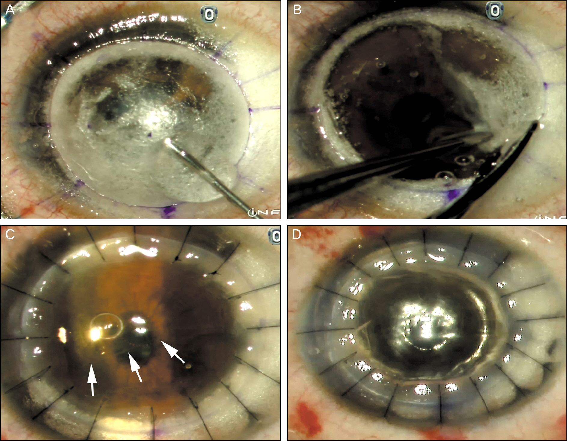
A 24-year-old female had keratoconus in her right eye and underwent DALK for treatment. Descemet's membrane was ruptured while separating the corneal stroma from Descemet's membrane with the big bubble technique. The operation method was changed from DALK to penetrating keratoplasty. Detached Descemet's membrane was observed in the anterior chamber after suturing. Sterile air was injected into the anterior chamber to attach the Descemet's membrane. Five days after the surgery, Descemet's membrane was detached and a second air injection was performed. Corneal edema was improved but Descemet's membrane was re-detached. Double Descemet's membrane was observed by anterior segment optical coherence tomography (OCT). The detached Descemet's membrane originated from the recipient's cornea and not from the donor's cornea. Detached Descemet's membrane was removed successfully. Patient's cornea was clear and best corrected visual acuity was 20/25.
CONCLUSIONS
When penetrating keratoplasty is performed instead of DALK, the surgeon should completely remove the remnant corneal stroma and Descemet's membrane. Remnant Descemet's membrane can be disregarded as it comes from the donor cornea. Unnecessary anterior chamber air injection causes endothelial damage. Anterior segment OCT is a useful tool to identify anatomical structures of transplanted cornea.
Keyword
MeSH Terms
Figure
Reference
-
References
1. Edrington TB, Szczotka LB, Barr JT, et al. Rigid contact lens fitting relationships in keratoconus. Collaborative Longitudinal Evaluation of Keratoconus (CLEK) Study Group. Optom Vis Sci. 1999; 76:692–9.2. Wollensak G, Spoerl E, Seiler T. Riboflavin/ultraviolet-a-induced collagen crosslinking for the treatment of keratoconus. Am J Ophthalmol. 2003; 135:620–7.
Article3. Doh SH, Kim MS. Influence of preoperative corneal thickness to postoperative astigmatism and endothelial cell in keratoconus penetrating keratoplasty. J Korean Ophthalmol Soc. 2005; 46:1978–82.4. Amayem AF, Anwar M. Fluid lamellar keratoplasty in keratoconus. Ophthalmology. 2000; 107:76–9. discussion 80.5. Soong HK, Katz DG, Farjo AA, et al. Central lamellar keratoplasty for optical indications. Cornea. 1999; 18:249–56.
Article6. Archila EA. Deep lamellar keratoplasty dissection of host tissue with intrastromal air injection. Cornea. 1984-1985; 3:217–8.
Article7. Shimazaki J, Shimmura S, Ishioka M, Tsubota K. Randomized clinical trial of deep lamellar keratoplasty vs penetrating keratoplasty. Am J Ophthalmol. 2002; 134:159–65.8. Tsubota K, Kaido M, Monden Y, et al. A new surgical technique for deep lamellar keratoplasty with single running suture adjustment. Am J Ophthalmol. 1998; 126:1–8.
Article9. Anwar M, Teichmann KD. Deep lamellar keratoplasty: surgical techniques for anterior lamellar keratoplasty with and without baring of Descemet's membrane. Cornea. 2002; 21:374–83.10. Fogla R, Padmanabhan P. Results of deep lamellar keratoplasty using the big-bubble technique in patients with keratoconus. Am J Ophthalmol. 2006; 141:254–9.
Article11. van Dooren BT, Mulder PG, Nieuwendaal CP, et al. Endothelial cell density after deep anterior lamellar keratoplasty (Melles technique). Am J Ophthalmol. 2004; 137:397–400.
Article12. Sugita J, Kondo J. Deep lamellar keratoplasty with complete removal of pathological stroma for vision improvement. Br J Ophthalmol. 1997; 81:184–8.
Article13. Fontana L, Parente G, Tassinari G. Clinical outcomes after deep anterior lamellar keratoplasty using the big-bubble technique in patients with keratoconus. Am J Ophthalmol. 2007; 143:117–24.
Article14. Smadja D, Colin J, Krueger RR, et al. Outcomes of deep anterior lamellar keratoplasty for keratoconus: learning curve and advan-tages of the big bubble technique. Cornea. 2012; 31:859–63.15. Bourne WM. Cellular changes in transplanted human corneas. Cornea. 2001; 20:560–9.
Article16. Hong A, Caldwell MC, Kuo AN, Afshari NA. Air bubble-associated endothelial trauma in descemet stripping automated endothelial keratoplasty. Am J Ophthalmol. 2009; 148:256–9.
Article17. Minasian M, Ayliffe W. Fixed dilated pupil following deep lamellar keratoplasty (Urrets-Zavalia syndrome). Br J Ophthalmol. 2002; 86:115–6.
Article18. Maurino V, Allan BD, Stevens JD, Tuft SJ. Fixed dilated pupil (Urrets-Zavalia syndrome) after air/gas injection after deep lamellar keratoplasty for keratoconus. Am J Ophthalmol. 2002; 133:266–8.
Article19. Bhojwani RD, Noble B, Chakrabarty AK, Stewart OG. Sequestered viscoelastic after deep lamellar keratoplasty using viscodissection. Cornea. 2003; 22:371–3.
Article20. Patel N, Mearza A, Rostron CK, Chow J. Corneal ectasia following deep lamellar keratoplasty. Br J Ophthalmol. 2003; 87:799–800.
Article
- Full Text Links
- Actions
-
Cited
- CITED
-
- Close
- Share
- Similar articles
-
- Clinical Results of Deep Anterior Lamellar Keratoplasty
- Three Cases of Urrets-Zavalia Syndrome Following Deep Lamellar Keratoplasty (DLKP)
- Clinical Evaluation of Full-thickness Deep Lamellar Keratoplasty
- A Case of Anterior Synechiolysis with Lamellar Corneal Dissection in Penetrating Keratoplasty
- Descemet Membrane Endothelial Keratoplasty after Penetrating Keratoplasty Graft Failure


