Strategic surgical-combined orthodontic treatment planning of patient with missing incisors on maxilla: a case report
- Affiliations
-
- 1Department of Orthodontics, Institute of Oral Biosciences and School of Dentistry, Chonbuk National University, Jeonju, Republic of Korea. kjgortho@jbnu.ac.kr
- 2Research Institute of Clinical Medicine of Chonbuk National University-Biomedical Research Institute of Chonbuk National University Hospital, Jeonju, Republic of Korea.
- KMID: 2470630
- DOI: http://doi.org/10.14368/jdras.2019.35.4.244
Abstract
- Proper positioning of maxillary incisors is key to success of surgery combined treatment. Establishing surgery plan would be a difficult job if maxillary incisors are lost. Patient who lost all of her maxillary incisors due to accident came for orthodontic treatment. Through careful modification of maxillary archform, pre-surgical orthodontic treatment was conducted with four prosthetic space consolidation. Position of incisors was decided by help of 3D prosthetic set-up, and 1-jaw surgery was planned. After relative short treatment period of 28 months, final prosthesis was done. When alveolar bone loss happens, harmonious prosthesis of upper incisors is difficult. Utilizing mandibular set-back surgery and incisor positioning using 3D set-up could make a better environment for treatment outcome. Strategic pre-surgical orthodontic treatment can allow shorter time and less number of prosthetics.
MeSH Terms
Figure
-
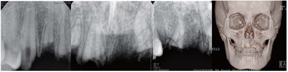
Fig. 1 Radiographs of patient obtained at the time of trauma.
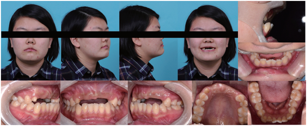
Fig. 2 Extraoral and intraoral photos of patient obtained before orthodontic treatment.
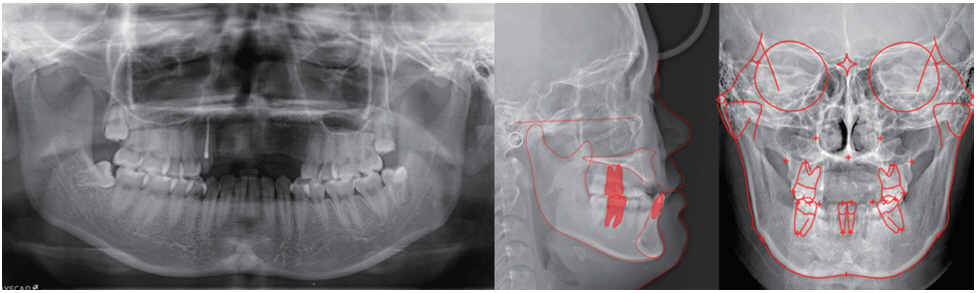
Fig. 3 Cephalometric and panoramic x-rays obtained before orthodontic treatment.
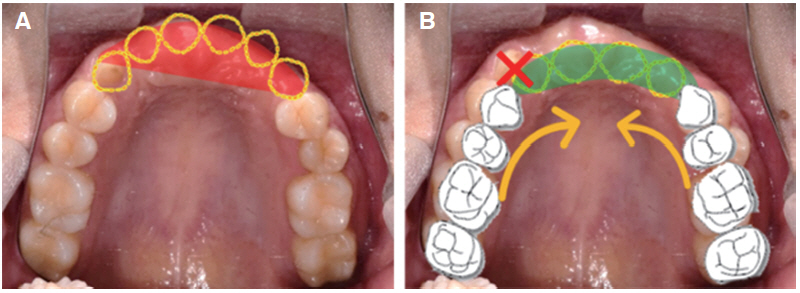
Fig. 4 Pre-surgical orthodontic treatment plan. (A) camouflage treatment with 6 prosthetics, (B) 4 prosthetics with modification of maxillary dentition.

Fig. 5 Pre-surgical orthodontic treatment progress. (A) Photographic image at 4 months, (B) Photographic image at 9 months, (C) Photographic image at 12 months.
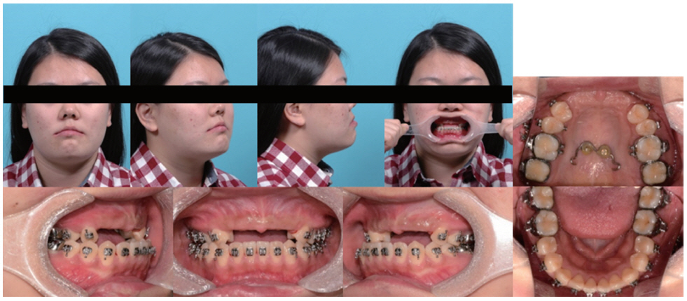
Fig. 6 Extraoral and intraoral photos of patient obtained after 12 months of treatment (pre-surgical assessment).
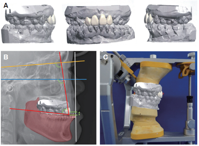
Fig. 7 Model surgery progress. (A) Incisor positioning with 3D program, (B) Paper surgery with positioned incisors, (C) Final occlusion for surgical wafer.

Fig. 8 Post-surgical orthodontic treatment progress. (A) 4 weeks after surgery, (B) 7 months after surgery, (C) Appliance removal with provisional restoration.

Fig. 9 Extraoral and intraoral photos, radiograph data obtained after final prosthesis.

Fig. 10 Superimposition of pre-treatment and treatment result. Red line: pre-treatment, Green line: post-treatment.
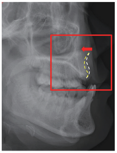
Fig. 11 X-ray superimposition. White line: at-trauma, Yellow line: after 2-years. Resorption of maxillary alveolar bone (Red box) is prominent (Red arrow).
Reference
-
References
1. Proffit WR, Fields HW Jr, Sarver DM. 2012. Contemporary Orthodontics. 5th ed. Elsevier;p. 693–9.2. Graber T, Vanarsdall R, Vig K. 2017. Orthodontics, current principles and technique. 6th ed. Elsevier;p. 784–91.3. Ngan P, Moon W. 2015; Evolution of Class III treatment in orthodontics. Am J Orthod Dentofacial Orthop. 148:22–36. DOI: 10.1016/j.ajodo.2015.04.012. PMID: 26124025.4. Nicodemo D, Pereira MD, Ferreira LM. 2008; Effect of orthognathic surgery for class III correction on quality of life as measured by SF-36. Int J Oral Maxillofac Surg. 37:131–4. DOI: 10.1016/j.ijom.2007.07.024. PMID: 17919889.5. Lin J, Gu Y. 2003; Preliminary investigation of nonsurgical treatment of severe skeletal Class III malocclusion in the permanent dentition. Angle Orthod. 73:401–10. PMID: 12940561.6. Atwood DA. 1963; Postextraction changes in the adult mandible as illustrated by microradiographs of midsagittal sections and serial cephalometric roentgenograms. J Prosthet Dent. 13:810–24. DOI: 10.1016/0022-3913(63)90225-7.7. Atwood DA. 1979; Bone loss of edentulous alveolar ridges. J Periodontol. 50:11–21. DOI: 10.1902/jop.1979.50.4s.11.8. Cohenca N, Stabholz A. 2007; Decoronation - a conservative method to treat ankylosed teeth for preservation of alveolar ridge prior to permanent prosthetic reconstruction: literature review and case presentation. Dent Traumatol. 23:87–94. DOI: 10.1111/j.1600-9657.2006.00454.x. PMID: 17367456.9. Wolford LM, Chemello PD, Hilliard FW. 1993; Occlusal plane alteration in orthognathic surgery. J Oral Maxillofac Surg. 51:730–40. DOI: 10.1016/S0278-2391(10)80410-0. PMID: 8509911.10. Aboul-Hosn Centenero S, Hernandez-Alfaro F. 2012; 3D planning in orthognathic surgery: CAD/CAM surgical splints and prediction of the soft and hard tissues results - our experience in 16 cases. J Craniomaxillofac Surg. 40:162–8. DOI: 10.1016/j.jcms.2011.03.014. PMID: 21458285.11. Hammoudeh JA, Howell LK, Boutros S, Scott MA, Urata MM. 2015; Current status of surgical planning for orthognathic surgery: traditional methods versus 3D surgical planning. Plast Reconstr Surg Glob Open. 3:e307. DOI: 10.1097/GOX.0000000000000184. PMID: 25750846. PMCID: PMC4350313.
- Full Text Links
-
- Actions
-
Cited
- CITED
-
- Close
- Share
-
- Similar articles
-
- The case report of the skeletal Angle's Class II malocclusion with the upper central incisor missing
- An alternative approach for the management of missing lateral incisors through the intentional retention of primary canines
- A Simulation Model for Strategic Planning in a Community Hospital
- A case report of orthodontic treatment of cleft palate accompany teeth congenital missing
- Extensive Adenomatoid Odontogenic Tumor of the Maxilla: A Case Report of Conservative Surgical Excision and Orthodontic Alignment of Impacted Canine
