Right Ventricular Analysis Using Real-time Three-dimensional Echocardiography for Preload Dependency
- Affiliations
-
- 1Division of Cardiology, Department of Internal Medicine, College of Medicine, The Catholic University of Korea, Seoul, Korea. chanseok@catholic.ac.kr
- KMID: 2468379
- DOI: http://doi.org/10.4250/jcvi.2019.0079
Abstract
- BACKGROUND
The importance of the right ventricle (RV) has been increasingly recognized, and accurate RV measurement has become necessary. However, assessment of the RV with two-dimensional (2D) echocardiography has several limitations. As the development of novel methods for RV measurement continues, we can expect more accordant values related to RV geometry.
METHODS
Fifty-eight subjects who were examined by transthoracic echocardiography (TTE) immediately before and after hemodialysis (HD) were enrolled. Real-time, full-volume, three-dimensional (3D) echocardiographic images were acquired and analyzed using dedicated software. Conventional RV parameters for RV size and function were measured for comparison with pre-HD and post-HD values by both 2D-TTE and 3D-TTE.
RESULTS
3D RV volumes and ejection fractions were significantly decreased after HD. The values of the 3D image-derived RV dimensions, tricuspid annular plane systolic excursion (TAPSE), fractional area change (FAC), and longitudinal strain were also affected by acute preload changes (TAPSE pre-HD: 22.4 ± 4.0 mm, post-HD: 19.0 ± 4.2 mm, p < 0.001; FAC pre-HD: 49.6% ± 5.9%, post-HD: 46.4% ± 5.5%, p < 0.001; septal longitudinal strain pre-HD: -20.1% ± 3.7%, post-HD: -16.8% ± 3.8%, p < 0.001). With the exception of FAC, most 2D RV parameters were well correlated with the 3D values.
CONCLUSIONS
Various parameters representing RV anatomy and function were acquired easily and more accurately from 3D echocardiographic images than from 2D images but were affected by acute preload changes. 3D TTE could be a new modality for assessing RV function and size, but each value from 3D TTE should be interpreted with caution while considering the loading condition of the patients.
Keyword
Figure
-

Figure 1 Example of RV 3D analysis using the TomTec 4D function. (A) Select the RV focused 4-chamber view, including LV apex and MV. (B) Click on the right mouse button on the image and click the “4D RV function”. (C) When the multi-planar images are shown, put a dot on the LV apex, MV, RV apex, tricuspid valve, aortic valve, and anterior and posterior junctions of RV-LV. Then, click ‘beutel revision’. (D) Observe the diastolic RV image, adjust the RV borders, and click the ‘tracking revision’ button. (E) Observe the modified RV data that are shown. (F) Systolic RV data were also acquired using the same process. (G) After this process, RV volume curves were shown. (H) Click the “2D” button on the left side to display the parameters of 2D. LV: left ventricle, MV: mitral valve, RV: right ventricle.
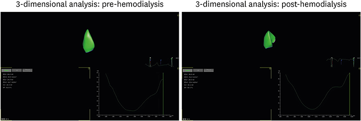
Figure 2 Three-dimensional right ventricular volume changes with hemodialysis.
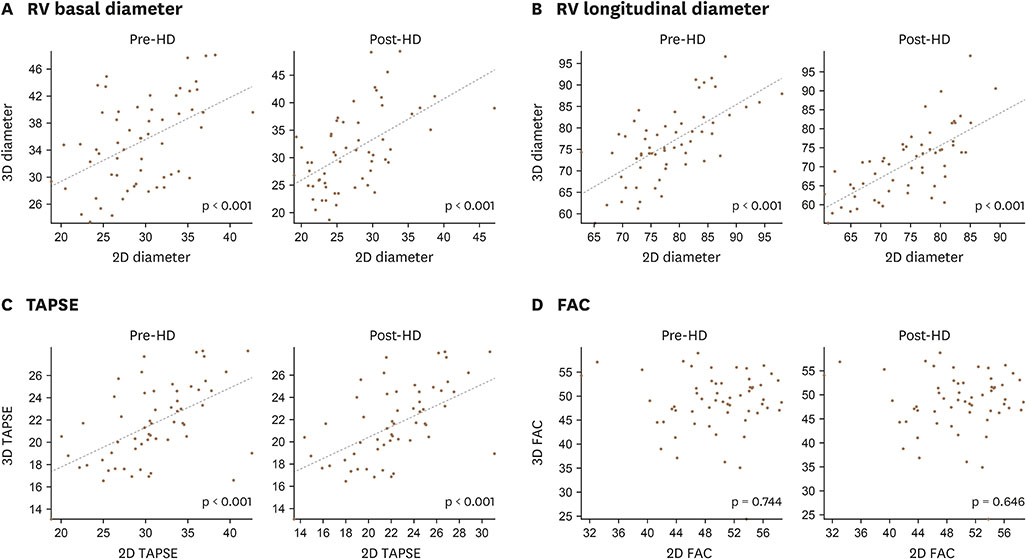
Figure 3 Correlation between 3D and 2D echocardiographic parameters: RV basal diameter (A), RV longitudinal diameter (B), TAPSE (C), FAC (D). FAC: fractional area change, RV: right ventricle, TAPSE: tricuspid annular plane systolic excursion.

Figure 4 Reliability test for the fractional area change and ejection fraction of the right ventricle, Bland-Altman analyses of pre-hemodialysis and post-hemodialysis patients (top: intra-observer, bottom: inter-observer). FAC: fractional area change, HD: hemodialysis, RVEF: right ventricular ejection fraction.
Reference
-
1. Eidem BW, O'Leary PW, Tei C, Seward JB. Usefulness of the myocardial performance index for assessing right ventricular function in congenital heart disease. Am J Cardiol. 2000; 86:654–658.
Article2. Franco V. Right ventricular remodeling in pulmonary hypertension. Heart Fail Clin. 2012; 8:403–412.
Article3. Maffessanti F, Gripari P, Tamborini G, et al. Evaluation of right ventricular systolic function after mitral valve repair: a two-dimensional Doppler, speckle-tracking, and three-dimensional echocardiographic study. J Am Soc Echocardiogr. 2012; 25:701–708.
Article4. Jauhiainen T, Järvinen VM, Hekali PE, Poutanen VP, Penttilä A, Kupari M. MR gradient echo volumetric analysis of human cardiac casts: focus on the right ventricle. J Comput Assist Tomogr. 1998; 22:899–903.5. Pons-Lladó G. Assessment of cardiac function by CMR. Eur Radiol. 2005; 15:Suppl 2. B23–B32.6. Shiota T, McCarthy PM, White RD, et al. Initial clinical experience of real-time three-dimensional echocardiography in patients with ischemic and idiopathic dilated cardiomyopathy. Am J Cardiol. 1999; 84:1068–1073.
Article7. Qin JX, Jones M, Shiota T, et al. Validation of real-time three-dimensional echocardiography for quantifying left ventricular volumes in the presence of a left ventricular aneurysm: in vitro and in vivo studies. J Am Coll Cardiol. 2000; 36:900–907.
Article8. Fijalkowski M, Koprowski A, Gruchala M, et al. Effect of preload reduction by hemodialysis on myocardial ultrasonic characterization, left atrial volume, and Doppler tissue imaging in patients with end-stage renal disease. J Am Soc Echocardiogr. 2006; 19:1359–1364.
Article9. Drighil A, Madias JE, Mathewson JW, et al. Haemodialysis: effects of acute decrease in preload on tissue Doppler imaging indices of systolic and diastolic function of the left and right ventricles. Eur J Echocardiogr. 2008; 9:530–535.
Article10. Nagueh SF, Appleton CP, Gillebert TC, et al. Recommendations for the evaluation of left ventricular diastolic function by echocardiography. Eur J Echocardiogr. 2009; 10:165–193.
Article11. Lang RM, Bierig M, Devereux RB, et al. Recommendations for chamber quantification: a report from the American Society of Echocardiography's Guidelines and Standards Committee and the Chamber Quantification Writing Group, developed in conjunction with the European Association of Echocardiography, a branch of the European Society of Cardiology. J Am Soc Echocardiogr. 2005; 18:1440–1463.
Article12. Tamborini G, Marsan NA, Gripari P, et al. Reference values for right ventricular volumes and ejection fraction with real-time three-dimensional echocardiography: evaluation in a large series of normal subjects. J Am Soc Echocardiogr. 2010; 23:109–115.
Article13. Ghio S, Klersy C, Magrini G, et al. Prognostic relevance of the echocardiographic assessment of right ventricular function in patients with idiopathic pulmonary arterial hypertension. Int J Cardiol. 2010; 140:272–278.
Article14. Forfia PR, Fisher MR, Mathai SC, et al. Tricuspid annular displacement predicts survival in pulmonary hypertension. Am J Respir Crit Care Med. 2006; 174:1034–1041.
Article15. Anavekar NS, Gerson D, Skali H, Kwong RY, Yucel EK, Solomon SD. Two-dimensional assessment of right ventricular function: an echocardiographic-MRI correlative study. Echocardiography. 2007; 24:452–456.
Article16. Anavekar NS, Skali H, Bourgoun M, et al. Usefulness of right ventricular fractional area change to predict death, heart failure, and stroke following myocardial infarction (from the VALIANT ECHO Study). Am J Cardiol. 2008; 101:607–612.
Article17. Austin C, Alassas K, Burger C, et al. Echocardiographic assessment of estimated right atrial pressure and size predicts mortality in pulmonary arterial hypertension. Chest. 2015; 147:198–208.
Article18. Haeck ML, Scherptong RW, Marsan NA, et al. Prognostic value of right ventricular longitudinal peak systolic strain in patients with pulmonary hypertension. Circ Cardiovasc Imaging. 2012; 5:628–636.
Article19. Focardi M, Cameli M, Carbone SF, et al. Traditional and innovative echocardiographic parameters for the analysis of right ventricular performance in comparison with cardiac magnetic resonance. Eur Heart J Cardiovasc Imaging. 2015; 16:47–52.
Article20. Rudski LG, Lai WW, Afilalo J, et al. Guidelines for the echocardiographic assessment of the right heart in adults: a report from the American Society of Echocardiography endorsed by the European Association of Echocardiography, a registered branch of the European Society of Cardiology, and the Canadian Society of Echocardiography. J Am Soc Echocardiogr. 2010; 23:685–713. quiz 786-8.21. Mueller HS, Forman SA, Menegus MA, Cohen LS, Knatterud GL, Braunwald E. Prognostic significance of nonfatal reinfarction during 3-year follow-up: results of the Thrombolysis in Myocardial Infarction (TIMI) phase II clinical trial. The TIMI Investigators. J Am Coll Cardiol. 1995; 26:900–907.22. Grapsa J, O'Regan DP, Pavlopoulos H, Durighel G, Dawson D, Nihoyannopoulos P. Right ventricular remodelling in pulmonary arterial hypertension with three-dimensional echocardiography: comparison with cardiac magnetic resonance imaging. Eur J Echocardiogr. 2010; 11:64–73.
Article23. Muraru D, Spadotto V, Cecchetto A, et al. New speckle-tracking algorithm for right ventricular volume analysis from three-dimensional echocardiographic data sets: validation with cardiac magnetic resonance and comparison with the previous analysis tool. Eur Heart J Cardiovasc Imaging. 2016; 17:1279–1289.
Article24. Maffessanti F, Muraru D, Esposito R, et al. Age-, body size-, and sex-specific reference values for right ventricular volumes and ejection fraction by three-dimensional echocardiography: a multicenter echocardiographic study in 507 healthy volunteers. Circ Cardiovasc Imaging. 2013; 6:700–710.25. Choi JO, Shin DH, Cho SW, et al. Effect of preload on left ventricular longitudinal strain by 2D speckle tracking. Echocardiography. 2008; 25:873–879.
Article26. Shaw C, Pruthi R, Pitcher D, Fogarty D. UK Renal Registry 15th annual report: Chapter 2 UK RRT prevalence in 2011: national and centre-specific analyses. Nephron Clin Pract. 2013; 123:Suppl 1. 29–54.
Article27. Kucukdurmaz Z, Karapinar H, Karavelioğlu Y, et al. Effect of blood donation mediated volume reduction on right ventricular function parameters in healthy subjects. Echocardiography. 2012; 29:451–454.
Article28. Kjaergaard J, Snyder EM, Hassager C, Oh JK, Johnson BD. Impact of preload and afterload on global and regional right ventricular function and pressure: a quantitative echocardiography study. J Am Soc Echocardiogr. 2006; 19:515–521.
Article29. McMahon LP, Roger SD, Levin A. Development, prevention, and potential reversal of left ventricular hypertrophy in chronic kidney disease. J Am Soc Nephrol. 2004; 15:1640–1647.
Article30. London G. Pathophysiology of cardiovascular damage in the early renal population. Nephrol Dial Transplant. 2001; 16:Suppl 2. 3–6.
Article
- Full Text Links
-
- Actions
-
Cited
- CITED
-
- Close
- Share
-
- Similar articles
-
- Three-dimensional Transthoracic Echocardiography: Left Ventricular Analysis
- Real-time Three Dimensional Echocardiography Clinical Implications and The Perspective
- Advances in the Evaluation of Mitral Complex Geometry:Insights from Transthoracic Real-time Three-dimensional Echocardiography
- Assessment of Right Ventricular Pressure by Two-Dimensional Echocardiography in Congenital Heart Disease
- A Case of Extensive Ventricular Wall Rupture from the Posterior Wall to the Ventricular Septum after Acute Myocardial Infarction Demonstrated by Real-Time 3D Echocardiography
