Prog Med Phys.
2019 Dec;30(4):160-166. 10.14316/pmp.2019.30.4.160.
Acceptance Test and Clinical Commissioning of CT Simulator
- Affiliations
-
- 1Department of Radiation Oncology, Seoul National University Hospital, Seoul, Korea. ms1236@snu.ac.kr
- 2Biomedical Research Institute, Seoul National University Hospital, Seoul, Korea.
- 3Institute of Radiation Medicine, Seoul National University Medical Research Center, Seoul, Korea.
- KMID: 2468178
- DOI: http://doi.org/10.14316/pmp.2019.30.4.160
Abstract
- This study examined the clinical use of two newly installed computed tomography (CT) simulators in the Department of Radiation Oncology. The accreditation procedure was performed by the Korean Institute for Accreditation of Medical Imaging. An Xi R/F dosimeter was used to measure the CT dose index for each plug of the CT dose index phantom. Image qualities such as the Hounsfield unit (HU) value of water, noise level, homogeneity, existence of artifacts, spatial resolution, contrast, and slice thickness were evaluated by scanning a CT performance phantom. All test items were evaluated as to whether they were within the required tolerance level. CT calibration curves"”the relationship between CT number and relative electron density"”were obtained for dose calculations in the treatment planning system. The positional accuracy of the lasers was also evaluated. The volume CT dose indices for the head phantom were 22.26 mGy and 23.70 mGy, and those for body phantom were 12.30 mGy and 12.99 mGy for the first and second CT simulators, respectively. HU accuracy, noise, and homogeneity for the first CT simulator were −0.2 HU, 4.9 HU, and 0.69 HU, respectively, while those for second CT simulator were 1.9 HU, 4.9 HU, and 0.70 HU, respectively. Five air-filled holes with a diameter of 1.00 mm were used for assessment of spatial resolution and a low contrast object with a diameter of 6.4 mm was clearly discernible by both CT scanners. Both CT simulators exhibited comparable performance and are acceptable for clinical use.
Keyword
MeSH Terms
Figure
-

Fig. 1 (a) A modular 76-410 AAPM computed tomography (CT) Performance Phantom (Fluke Corporation, Everett, WA, USA); CT slices of (b) water Hounsfield unit (HU), noise level, homogeneity evaluation, (c) spatial resolution, (d) low contrast resolution insert, and (e) slice thickness. Ave, average; SD, standard deviation.
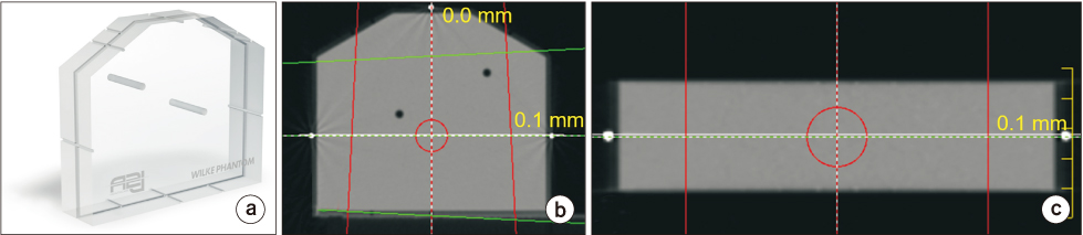
Fig. 2 (a) Wilke phantom, (b) an axial slice, and (c) a coronal slice. The deviations between laser position and groove position in the images are marked.
Cited by 1 articles
-
Basic Physical Principles and Clinical Applications of Computed Tomography
Haijo Jung
Prog Med Phys. 2021;32(1):1-17. doi: 10.14316/pmp.2021.32.1.1.
Reference
-
1. Geise RA, McCullough EC. The use of CT scanners in megavoltage photon-beam therapy planning. Radiology. 1977; 124:133–141.
Article2. Dobbs HJ, Parker RP, Hodson NJ, Hobday P, Husband JE. The use of CT in radiotherapy treatment planning. Radiother Oncol. 1983; 1:133–141.3. Parker RP, Hobday PA, Cassell KJ. The direct use of CT numbers in radiotherapy dosage calculations for inhomogeneous media. Phys Med Biol. 1979; 24:802–809.
Article4. Garcia-Ramirez JL, Mutic S, Dempsey JF, Low DA, Purdy JA. Performance evaluation of an 85-cm-bore X-ray computed tomography scanner designed for radiation oncology and comparison with current diagnostic CT scanners. Int J Radiat Oncol Biol Phys. 2002; 52:1123–1131.
Article5. Mutic S, Palta JR, Butker EK, Das IJ, Huq MS, Loo LN, et al. Quality assurance for computed-tomography simulators and the computed-tomography-simulation process: report of the AAPM Radiation Therapy Committee Task Group No. 66. Med Phys. 2003; 30:2762–2792.
Article6. Bissonnette JP, Balter PA, Dong L, Langen KM, Lovelock DM, Miften M, et al. Quality assurance for image-guided radiation therapy utilizing CT-based technologies: a report of the AAPM TG-179. Med Phys. 2012; 39:1946–1963.
Article7. American College of Radiology. CT accreditation program requirements. American College of Radiology;2019.8. European Commission. European guidelines on quality criteria for computed tomography. European Commission. 1999.9. Park HJ, Jung SE, Lee YJ, Cho WI, Do KH, Kim SH, et al. The relationship between subjective and objective parameters in CT phantom image evaluation. Korean J Radiol. 2009; 10:490–495.
Article10. Deak PD, Smal Y, Kalender WA. Multisection CT protocols: sex- and age-specific conversion factors used to determine effective dose from dose-length product. Radiology. 2010; 257:158–166.
Article11. McCollough CH, Leng S, Yu L, Cody DD, Boone JM, Mc-Nitt-Gray MF. CT dose index and patient dose: they are not the same thing. Radiology. 2011; 259:311–316.12. Chun M, Choi YH, Kim JH. Automated measurement of CT noise in patient images with a novel structure coherence feature. Phys Med Biol. 2015; 60:9107–9122.
Article13. Schuhbaeck A, Schaefer M, Marwan M, Gauss S, Muschiol G, Lell M, et al. Patient-specific predictors of image noise in coronary CT angiography. J Cardiovasc Comput Tomogr. 2013; 7:39–45.
Article14. Tomic N, Papaconstadopoulos P, Aldelaijan S, Rajala J, Seuntjens J, Devic S. Image quality for radiotherapy CT simulators with different scanner bore size. Phys Med. 2018; 45:65–71.
Article15. Seibert JA, Boone JM, Wootton-Gorges SL, Lamba R. Dose is not always what it seems: where very misleading values can result from volume CT dose index and dose length product. J Am Coll Radiol. 2014; 11:233–237.
Article16. Boone JM, Strauss KJ, Cody DD, McCollough CH, McNitt-Gray MF, Toth TL. Size-specific dose estimates (SSDE) in pediatric and adult body CT examinations: the report of AAPM task group 204. Med Phys. 2011.
Article17. Mccollough C, Bakalyar DM, Bostani M, Brady S, Boedeker K, Boone JM. Use of water equivalent diameter for calculating patient size and size-specific dose estimates (SSDE) in CT: the report of AAPM task group 220. Med Phys. 2014.
Article18. Eck BL, Fahmi R, Brown KM, Zabic S, Raihani N, Miao J, et al. Computational and human observer image quality evaluation of low dose, knowledge-based CT iterative reconstruction. Med Phys. 2015; 42:6098–6111.
Article19. Verdun FR, Racine D, Ott JG, Tapiovaara MJ, Toroi P, Bochud FO, et al. Image quality in CT: from physical measurements to model observers. Phys Med. 2015; 31:823–843.
Article20. Battista JJ, Bronskill MJ. Compton scatter imaging of transverse sections: an overall appraisal and evaluation for radiotherapy planning. Phys Med Biol. 1981; 26:81–99.
Article21. IAEA TRS Report 430. Commissioning and quality assurance of computerized planning systems for radiation treatment of cancer. Austria: International Atomic Energy Agency;2004. p. 430.22. Davis AT, Palmer AL, Nisbet A. Can CT scan protocols used for radiotherapy treatment planning be adjusted to optimize image quality and patient dose? A systematic review. Br J Radiol. 2017; 90:20160406.
Article
- Full Text Links
-
- Actions
-
Cited
- CITED
-
- Close
- Share
-
- Similar articles
-
- Commissioning and Validation of a Dedicated Scanning Nozzle at Samsung Proton Therapy Center
- The Broad-beam CT Image Reconstruction from Simulator Images
- Guideline on Acceptance Test and Commissioning of High-Precision External Radiation Therapy Equipment
- Comments on ‘Development of colonic stent simulator using three-dimensional printing technique: a simulator development study in Korea’
- Proposal on Guideline for Quality Assurance of Radiation Treatment Planning System
