Prog Med Phys.
2019 Dec;30(4):89-93. 10.14316/pmp.2019.30.4.89.
Low Magnetic Field MRI Visibility of Rubber-Based Markers
- Affiliations
-
- 1Department of Radiation Oncology, Seoul National University Hospital, Seoul, Korea.
- 2Institute of Radiation Medicine, Seoul National University Medical Research Center, Seoul, Korea. smjung@snu.ac.kr
- 3Biomedical Research Institute, Seoul National University Hospital, Seoul, Korea.
- KMID: 2468169
- DOI: http://doi.org/10.14316/pmp.2019.30.4.89
Abstract
- PURPOSE
This study aims to develop new markers based on silicone rubber and urethane rubber to enhance visibility in low magnetic field magnetic resonance (MR) imaging.
METHODS
Four types of markers were fabricated using two different base materials. Two of the markers were composed of two different types of silicone rubber: DragonSkinâ„¢ 10 MEDIUM and BodyDoubleâ„¢ SILK. The other two markers were composed of types of urethane rubber: PMCâ„¢ 780 DRY and VytaFlexâ„¢ 20. Silicone oil (KF-96 1000cs) was added to the fabricated markers. The allocated amount of oil was 20% of the weight (wt%) of each respective marker. The MR images of the markers, with and without the silicone oil, were acquired using MRIdian with a low magnetic field of 0.35 T. The signal intensities of each MR image for the markers were analyzed using ImageJ software and the visibility for each was compared.
RESULTS
The highest signal intensity was observed in VytaFlexâ„¢ 20 (279.67±3.57). Large differences in the signal intensities (e.g., 627% in relative difference between BodyDoubleâ„¢ SILK and VytaFlexâ„¢ 2 0) among the markers were observed. However, the maximum difference between the signal intensities of the markers with the silicone oil showed only a 62% relative difference between PMCâ„¢ 780 DRY and DragonSkinâ„¢ 10 MEDIUM. An increase in the signal intensity of the markers with the silicone oil was observed in all markers.
CONCLUSION
New markers were successfully fabricated. Among the markers, DragonSkinâ„¢ 10 MEDIUM with silicone oil showed the highest MR signal intensity.
MeSH Terms
Figure
-
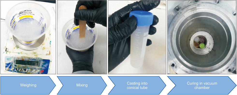
Fig. 1 Fabrication method of markers using DragonSkin™ MEDIUM, BodyDouble™ SILK, PMC™ 780 DRY, and VytaFlex™ 20 (Smooth-On Inc.).
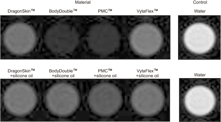
Fig. 2 Magnetic resonance images of markers with silicone oil (first row) and without silicone oil (second row).
Cited by 1 articles
-
Synthetic Computed Tomography Generation while Preserving Metallic Markers for Three-Dimensional Intracavitary Radiotherapy: Preliminary Study
Hyeongmin Jin, Seonghee Kang, Hyun-Cheol Kang, Chang Heon Choi
Prog Med Phys. 2021;32(4):172-178. doi: 10.14316/pmp.2021.32.4.172.
Reference
-
1. Kim JI, Park JM, Choi CH, An HJ, Kim YJ, Kim JH. Retrospective study comparing MR-guided radiation therapy (MRgRT) setup strategies for prostate treatment: repositioning vs. replanning. Radiat Oncol. 2019; 14:139.
Article2. Corradini S, Alongi F, Andratschke N, Belka C, Boldrini L, Cellini F, et al. MR-guidance in clinical reality: current treatment challenges and future perspectives. Radiat Oncol. 2019; 14:92.
Article3. Mutic S, Dempsey JF. The ViewRay system: magnetic resonance-guided and controlled radiotherapy. Semin Radiat Oncol. 2014; 24:196–199.
Article4. Park JM, Park S, Wu H, Kim J. Commissioning experience of tri-cobalt-60 MRI-guided radiation therapy system. Prog Med Phys. 2015; 26:193–200.
Article5. Klüter S. Technical design and concept of a 0.35 T MRLinac. Clin Transl Radiat Oncol. 2019; 18:98–101.
Article6. Bayouth JE, Low DA, Zaidi H. MRI-linac systems will replace conventional IGRT systems within 15 years. Med Phys. 2019; 46:3753–3756.7. Carrara M, Romanyukha A, Tenconi C, Mazzeo D, Cerrotta A, Borroni M, et al. Clinical application of MOSkin dosimeters to rectal wall in vivo dosimetry in gynecological HDR brachytherapy. Phys Med. 2017; 41:5–12.
Article8. Petoukhova A, Rüssel I, Nijst-Brouwers J, van Wingerden K, van Egmond J, Jacobs D, et al. In vivo dosimetry with MOSFETs and GAFCHROMIC films during electron IORT for accelerated partial breast irradiation. Phys Med. 2017; 44:26–33.
Article9. AAPM Report 87. Diode in vivo dosimetry for patients receiving external beam radiation therapy. AAPM Report. Bethesda: AAPM;2005. p. 87.10. Woulfe P, Sullivan FJ, O'Keeffe S. Optical fibre sensors: their role in in vivo dosimetry for prostate cancer radiotherapy. Cancer Nanotechnol. 2016; 7:7.
Article11. O'Neill AG, Jain S, Hounsell AR, O'Sullivan JM. Fiducial marker guided prostate radiotherapy: a review. Br J Radiol. 2016; 89:20160296.12. Shcherbakova Y, Bartels LW, Mandija S, Beld E, Seevinck PR, van der, et al. Visualization of gold fiducial markers in the prostate using phase-cycled bSSFP imaging for MRI-only radiotherapy. Phys Med Biol. 2019; 64:185001.
Article13. van den Ende RPJ, Rigter LS, Kerkhof EM, van Persijn van Meerten EL, Rijkmans EC, Lambregts DMJ, et al. MRI visibility of gold fiducial markers for image-guided radiotherapy of rectal cancer. Radiother Oncol. 2019; 132:93–99.
Article14. Smooth on Inc. Material Categories. Macungie: Smooth on Inc;cited 2019 December 13. Available from: https://www.smooth-on.com/products/.15. Lin AY. Fabrication and aeroelastic analysis of silicone membrane micro air vehicle wings [Master's thesis]. Florida: University of Florida;2009.16. Alqathami M. Novel 3D radiochromic dosimeters for advanced radiotherapy techniques [dissertation]. Melbourne: RMIT University;2013.17. An HJ, Kim MS, Kim JS, Son JM, Choi CH, Min J. Geometric evaluation of patient-specific 3D bolus from 3D printed mold and casting method for radiation therapy. Prog Med Phys. 2019; 30:32–38.
Article18. Smooth on Inc. Body double™ silk. Macungie: Smooth on Inc;cited 2019 December 13. Available from: https://www.smooth-on.com/tb/files/BODY_DOUBLE_SILK_TB.pdf.19. Jursinic PA. Characterization of optically stimulated luminescent dosimeters, OSLDs, for clinical dosimetric measurements. Med Phys. 2007; 34:4594–4604.
Article20. Joint Head and Neck MRI-Radiotherapy Development Cooperative. Kiser K, Meheissen MAM, Mohamed ASR, Kamal M, Ng SP, et al. Prospective quantitative quality assurance and deformation estimation of MRI-CT image registration in simulation of head and neck radiotherapy patients. Clin Transl Radiat Oncol. 2019; 18:120–127.
Article
- Full Text Links
-
- Actions
-
Cited
- CITED
-
- Close
- Share
-
- Similar articles
-
- Portable Low-Cost MRI System Based on Permanent Magnets/Magnet Arrays
- Water-Fat Imaging with Automatic Field Inhomogeneity Correction Using Joint Phase Magnitude Density Function at Low Field MRI
- The Survey of Magnetic Resonance Imaging Quality according to Magnetic Field Strength in Korea
- Dimensional stability and wettability of rubber impression materials
- Introduction to high field strength magnetic resonance imaging
