Chonnam Med J.
2020 Jan;56(1):85-86. 10.4068/cmj.2020.56.1.85.
Giant Sacral Schwannoma: A Case Report
- Affiliations
-
- 1Department of Neurosurgery, Chonnam National Univertisty Hospital & Medical School, Gwangju, Korea. leejk0261@hanmail.net
- 2Department of Urology, Chonnam National Univertisty Hospital & Medical School, Gwangju, Korea.
- KMID: 2468155
- DOI: http://doi.org/10.4068/cmj.2020.56.1.85
Abstract
- No abstract available.
MeSH Terms
Figure
-
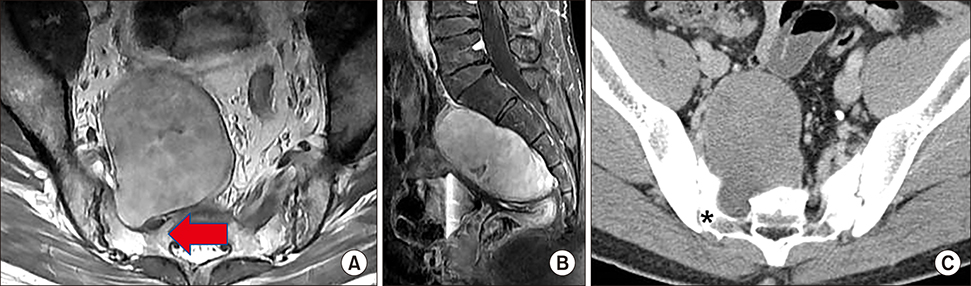
FIG. 1 Pre-operative lumbar spine MRI and CT. (A) Axial T1 enhanced MRI showed 10 cm sized homogenously enhanced mass arose from S1 foramen with S1 nerve root compression (arrow), (B) sagittal T1 enhanced MRI showed homogenously enhanced mass along ventral aspect of sacrum, (C) axial abdomen CT showed bony erosion of right S1 foramen (asteric).

FIG. 2 (A) Giant sacral schwannoma before resection. (B) Giant sacral schwannoma after resection measuring 10 cm.
Reference
-
1. Khan UA, Ismayl G, Malik I. Giant sacral schwannoma treated with a 360 approach: a rare case and systematic review of the literature. World Neurosurg. 2018; 115:65–72.
Article2. Lee BH, Hyun SJ, Park JH, Kim KJ. single stage posterior approach for total resection of presacral giant schwannoma: a technical case report. Korean J Spine. 2017; 14:89–92.
Article
- Full Text Links
-
- Actions
-
Cited
- CITED
-
- Close
- Share
-
- Similar articles
-
- Pre-sacral Giant Schwannoma: Removal by a Combined Anterior and Posterior Approach: A Case Report
- Giant Benign Schwannoma Involving Sacral Bone
- Cystic Giant Sacral Schwannoma Mimicking Aneurysmal Bone Cyst : A Case Report and Review of Literatures
- Giant Invasive Sacral Schwannoma Showing Chromosomal Numerical Aberrations [-14,+18,+22]
- Giant Presacral Schwannoma: A Case Report
