Height of elevated fetal buttock for prediction of successful external cephalic version
- Affiliations
-
- 1Department of Obstetrics and Gynecology, KonKuk University Medical Center, Konkuk University School of Medicine, Seoul, Korea.
- 2Department of Obstetrics and Gynecology, Seoul National University Bundang Hospital, Seoul National University College of Medicine, Seongnam, Korea.
- 3Department of Obstetrics and Gynecology, CHA Gangnam Medical Center, CHA University, Seoul, Korea.
- 4Department of Obstetrics and Gynecology, Gangseo Mizmedi Hospital, Seoul, Korea.
- 5Department of Obstetrics and Gynecology, CHA Bundang Medical Center, CHA University, Seongnam, Korea.
- 6Department of Obstetrics and Gynecology, National Medical Center, Seoul, Korea. hanjungyeol055@gmail.com
- KMID: 2467927
- DOI: http://doi.org/10.5468/ogs.2020.63.1.13
Abstract
OBJECTIVE
To increase the rate of successful external cephalic version (ECV) and to minimize the complications, it is important to identify the predictors of success. Therefore, the purpose of this study was to investigate whether the height of the elevated fetal buttock (HOB) is a valuable predictor of successful ECV or not.
METHODS
This prospective study was conducted from August 2016 to June 2018. A total of 139 pregnant women with breech presentation were enrolled in the study. HOB from the maternal pubic symphysis was measured on ultrasonography. The predictability and cut-off value of HOB for successful ECV were evaluated.
RESULTS
Among the 139 patients, 114 (82%) had successful ECV. The adjusted odds ratio for multiparity, amniotic fluid index (AFI) >14 cm, and HOB >7.8 cm were 10.80 (95% confidence interval [CI], 1.57-74.94), 5.26 (95% CI, 1.06-26.19), and 10.50 (95% CI, 1.03-107.12), respectively. Areas under the curve (AUCs) for AFI, HOB, and parity were 0.66 (95% CI, 0.54-0.78), 0.74 (95% CI, 0.64-0.85), and 0.69 (95% CI, 0.62-0.76), respectively. HOB had the largest AUC, but there were no significant differences among the AUCs of other factors. The cut-off value of HOB was 6 cm.
CONCLUSION
This study showed that the AUC of HOB was greater than that of parity and AFI, although it was not statistically significant. As HOB is a noninvasive and comprehensive marker to predict successful ECV, consideration of HOB would be helpful before conducting ECV. Further studies are needed.
MeSH Terms
Figure
-
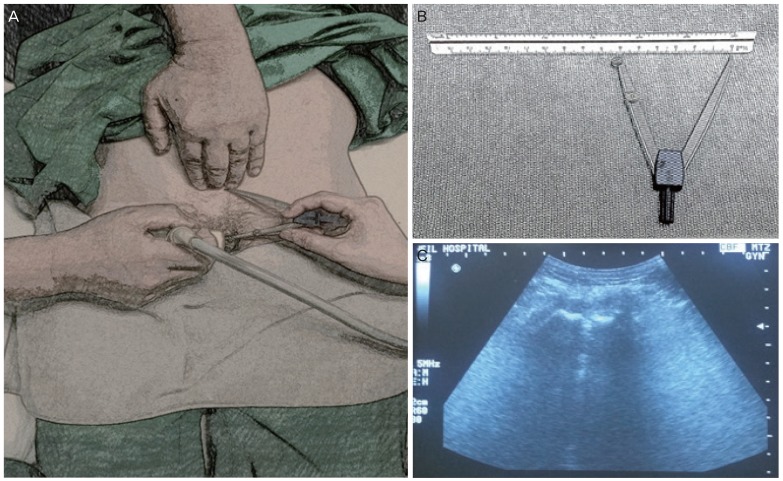
Fig. 1 Measurement of height of the elevated fetal buttock (HOB). (A) HOB by an operator's hand was measured from the maternal symphysis pubis under ultrasonography. (B) A compass and a ruler for measurement. (C) Symphysis pubis seen on ultrasonography.
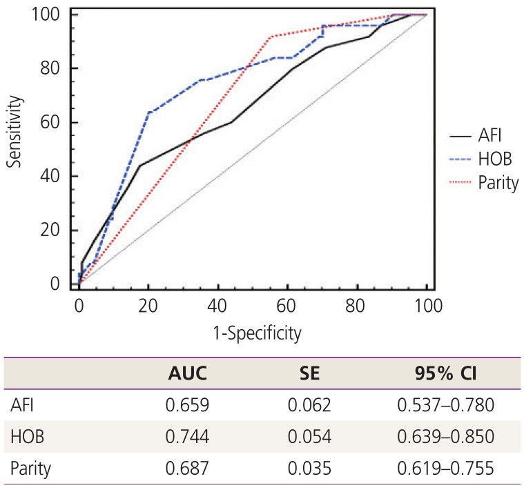
Fig. 2 Receiver operating characteristics curve and AUC for validity test with HOB, parity, AFI for prediction of successful external cephalic version.AUC, areas under the curve; SE, standard error; CI, confidence interval; AFI, amniotic fluid index; HOB, height of elevated fetal buttock.
Cited by 1 articles
-
Ritodrine in external cephalic version: is it effective and safe?
Sin Ae Kim, Eun-Hwan Cha, Kyoung-Chul Chun, Young Ah Kim, Jae-Whoan Koh, Jung Yeol Han, Jong Hee Hwang
Obstet Gynecol Sci. 2022;65(5):420-429. doi: 10.5468/ogs.22106.
Reference
-
1. Hannah ME, Hannah WJ, Hewson SA, Hodnett ED, Saigal S, Willan AR. Planned caesarean section versus planned vaginal birth for breech presentation at term: a randomised multicentre trial. Term Breech Trial Collaborative Group. Lancet. 2000; 356:1375–1383. PMID: 11052579.2. Kim MY, Park MY, Kim GJ. External cephalic version experiences in Korea. Obstet Gynecol Sci. 2016; 59:85–90. PMID: 27004197.
Article3. Hofmeyr GJ, Kulier R, West HM. External cephalic version for breech presentation at term. Cochrane Database Syst Rev. 2015; 4:CD000083.
Article4. Morgan ER, Hu AE, Brezak AMV, Rowley SS, Littman AJ, Hawes SE. Predictors of a successful external cephalic version: a population-based study of Washington state births. Women Birth. 2019; 32:e421–6. PMID: 30150151.
Article5. ACOG Committee opinion No. 745: mode of term singleton breech delivery. Obstet Gynecol. 2018; 132:e60–e63. PMID: 30045211.6. Burgos J, Melchor JC, Pijoán JI, Cobos P, Fernández-Llebrez L, Martínez-Astorquiza T. A prospective study of the factors associated with the success rate of external cephalic version for breech presentation at term. Int J Gynaecol Obstet. 2011; 112:48–51. PMID: 20870233.
Article7. Kim SY, Han JY, Chang EH, Kwak DW, Ahn HK, Ryu HM, et al. Evaluation of the learning curve for external cephalic version using cumulative sum analysis. Obstet Gynecol Sci. 2017; 60:343–349. PMID: 28791265.
Article8. Kok M, van der Steeg JW, van der Post JA, Mol BW. Prediction of success of external cephalic version after 36 weeks. Am J Perinatol. 2011; 28:103–110. PMID: 20661845.
Article
- Full Text Links
-
- Actions
-
Cited
- CITED
-
- Close
- Share
-
- Similar articles
-
- External Cephalic Version Attempted under Epidural Anesthesia : Case reports
- External cephalic version experiences in Korea
- Evaluation of the Success Rate Following Amnioinfusion in Pregnant Women Undergoing External Cephalic Version after Initial Failure
- Ritodrine in external cephalic version: is it effective and safe?
- Reviving external cephalic version: a review of its efficacy, safety, and technical aspects
